Categories
AAA AIOD aortoiliac occlusive disease (AIOD) bypass Commentary EndoRE EVAR imaging kidney transplant remote endarterectromy techniques

When both iliac systems are occluded below an abdominal aortic aneurysm: hybrid techniques on the cutting edge

preop CTA EVAR-ENDORE.jpg
AAA with iliac arterial occlusion -arrows point to right external iliac and left common iliac arterial occlusions

The patient is an 70 year old man referred for evaluation of claudication that occurred at under a block of walking. He reported no rest pain or tissue loss. He smoked heavily up to a pack a day, with congestive heart failure with an ejection fraction of 40%, prior history of myocardial infarction treated with PTCA, and pacemaker, and moderate dyspnea on exertion.

On examination, patient had a flaccid abdomen through which the AAA could be palpated, and he had no palpable femoral artery pulse bilaterally, nor anything below. He had a cardiac murmur and moderate bilateral edema. Preoperative risk evaluation placed him in the high risk category because of his heart failure, coronary artery disease, and his mild to moderate pulmonary disease.
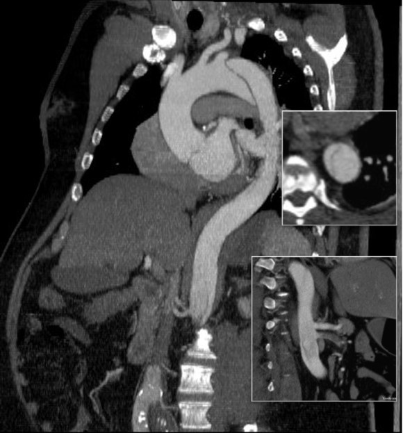
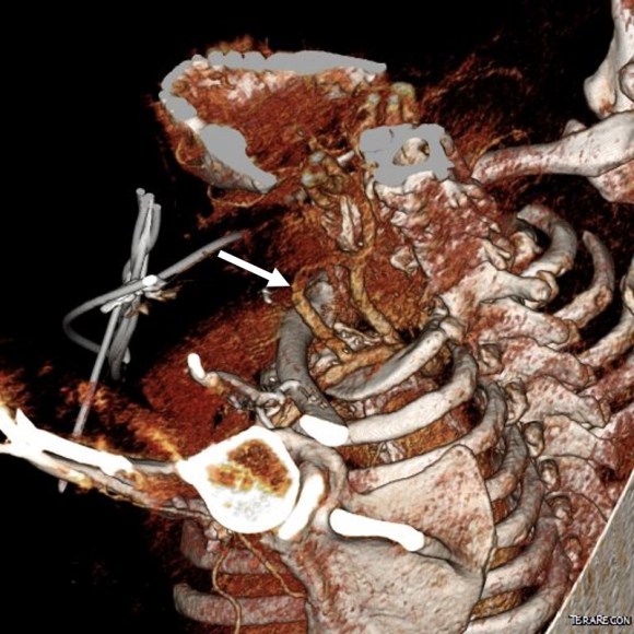
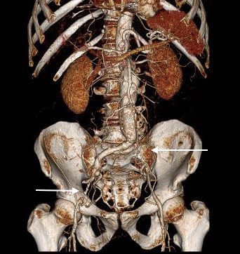
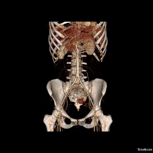
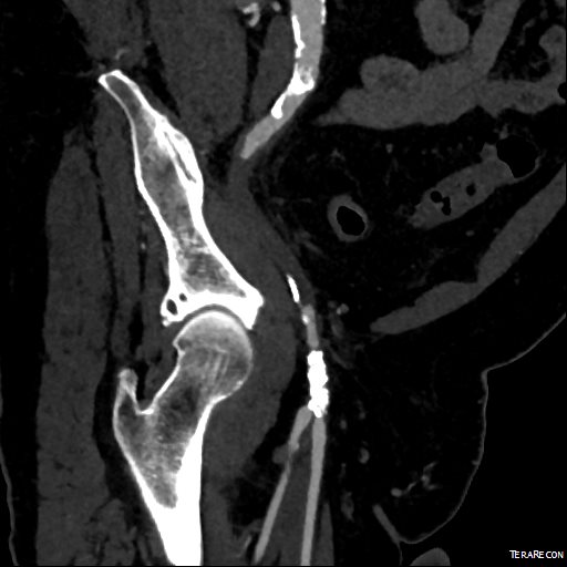
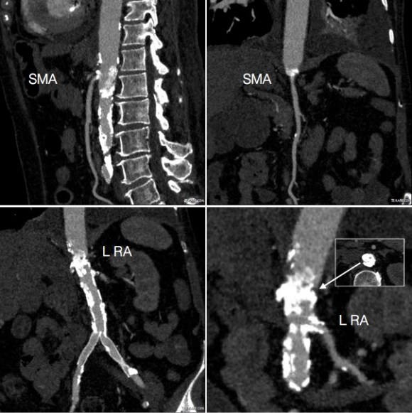
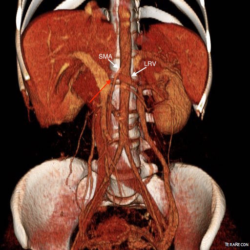
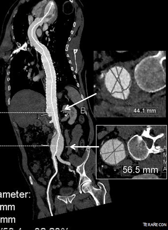
CTA (pictured above and below) showed a 5.1cm infrarenal AAA with an hourglass shaped neck with moderate atherosclerosis in the neck, an occluded left common iliac artery with external iliac artery reconstitution via internal iliac artery collaterals, and a right external iliac artery occlusion with common femoral artery reconstitution. There was calcified right common femoral artery plaque.

Preop left and right centerlines EVAR-ENDORE.jpg

Treatment options included open surgical aortobifemoral bypass with exclusion of the AAA, total endovascular repair with some form of endo-conduit revascularization of the occluded segments of iliac artery, or a hybrid repair.

Open aortic repair in patients with heart failure and moderate COPD can be performed safely (ref 1). Dr. Hollier et al, in the golden age of open repair, reported a 5.7% mortality rate operating on 106 patients with severe category of heart, lung, kidney, or liver disease.

Typically, the hybrid repair involves sewing in a conduit to deliver the main body of a bifurcated or unibody stent graft when endovascular access is not possible. Despite techniques to stay minimally invasive -largely by staying retroperitoneal, this is not a benign procedure (ref 2). Nzara et al reviewed 15,082 patients from the NSQIP database breaking out 1% of patients who had conduit or direct puncture access.

Matched analyses of comorbidities revealed that patients requiring [conduit or direct access] had higher perioperative mortality (6.8% vs. 2.3%, P = 0.008), cardiac (4.8% vs. 1%, P = 0.004), pulmonary (8.8% vs. 3.4%, P = 0.006), and bleeding complications (10.2% vs. 4.6%, P = 0.016).

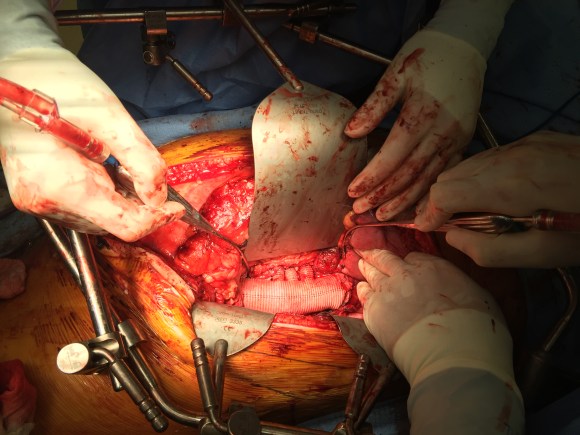
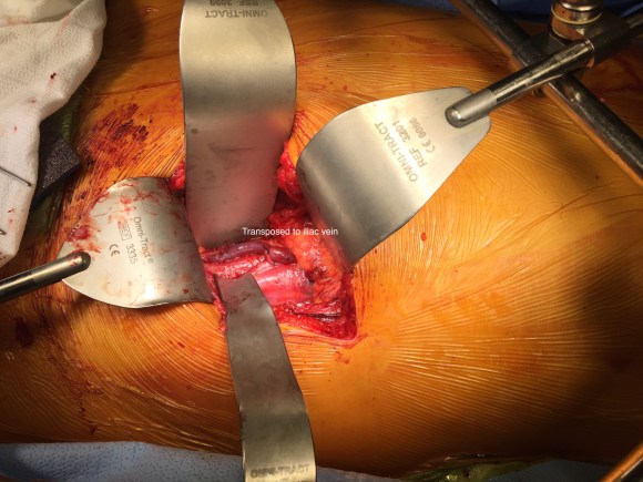
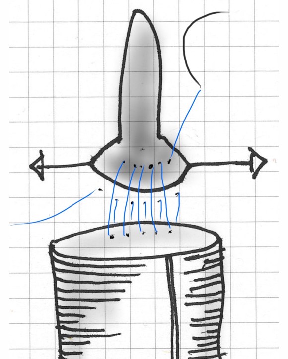
Despite these risks, I have performed AUI-FEM-FEM with good results with the modification of deploying the terminus of the stent graft across an end to end anastomosis of the conduit graft to the iliac artery (below), resulting in seal and avoiding the problems of bleeding from the usually heavily diseased artery

AUI fem fem.jpg
Aorto-uni-iliac stent graft across end to end conduit anastomosis to fem-fem bypass

The iliac limbs of some stent graft systems will have proximal flares and can be used in a telescoping manner to create an aorto-uni-iliac (AUI) configuration in occlusive disease. The Cook RENU converter has a 22mm tall sealing zone designed for deployment inside another stent graft and would conform poorly to this kind of neck as a primary  AUI endograft which this was not designed to act as. The Endurant II AUI converter has a suprarenal stent which I preferred to avoid in this patient as the juxtarenal neck likely was aneurysmal and might require future interventions

I chose to perform a right sided common femoral cutdown and from that exposure, perform an iliofemoral remote endarterectomy of the right external iliac to common femoral artery. This in my experience is a well tolerated and highly durable procedure (personal data). Kavanagh et al (ref 3) presented their experience with iliofemoral EndoRE and shared their techniques. This would create the lumenal diameter necessary to pass an 18F sheath to deliver an endograft. I chose the Gore Excluder which would achieve seal in the hourglass shaped neck and allow for future visceral segment intervention if necessary without having a suprarenal stent in the way. I planned on managing the left common iliac artery via a percutaneous recanalization.

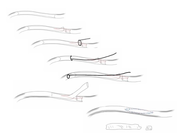
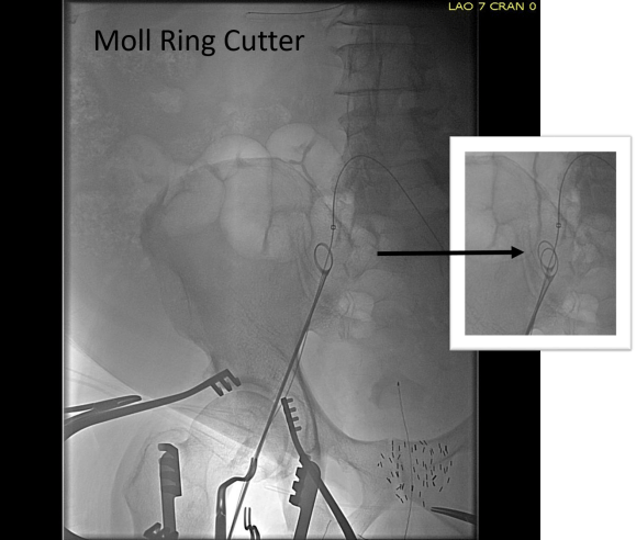
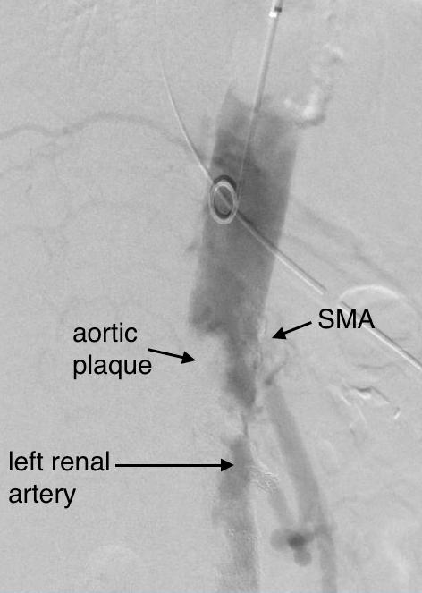
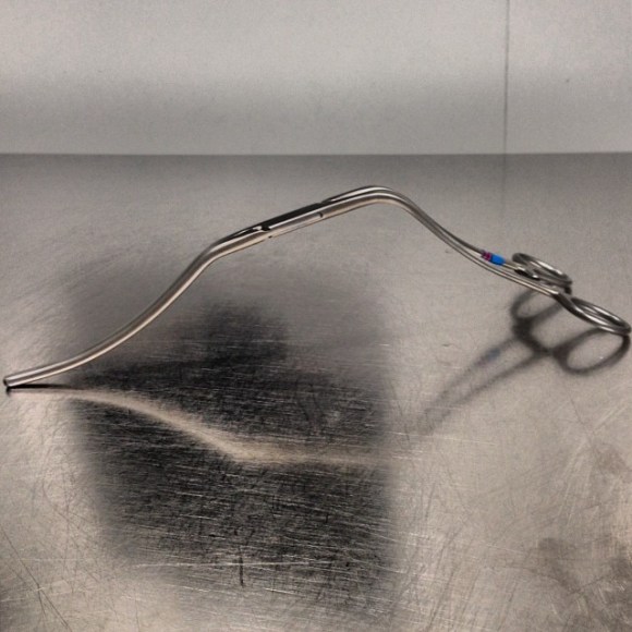
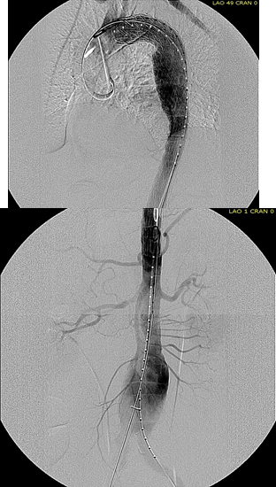
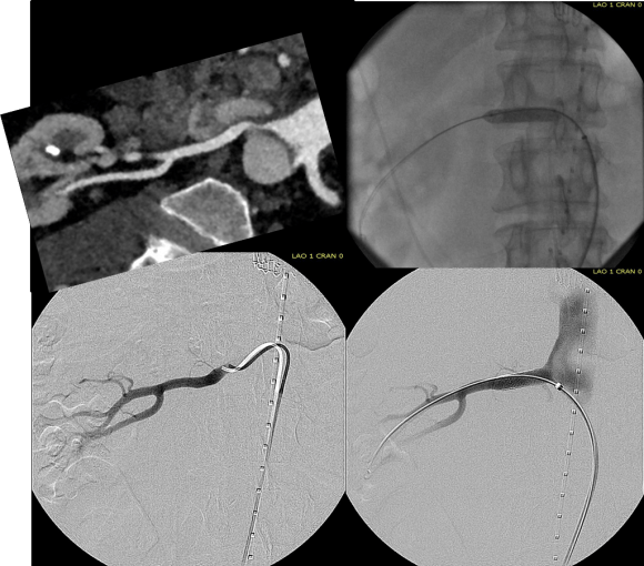
The patient’s right common femoral artery was exposed in the usual manner. Wire access across the occluded external iliac artery was achieved from a puncture of the common femoral artery. Remote endarterectomy (EndoRE) was performed over a wire from the common femoral artery to the external iliac artery origin (pictures below).

File Mar 31, 13 41 31.jpeg
External iliac to common femoral artery plaque removed with Moll ring cutter (LeMaitre Vascular) over a wire

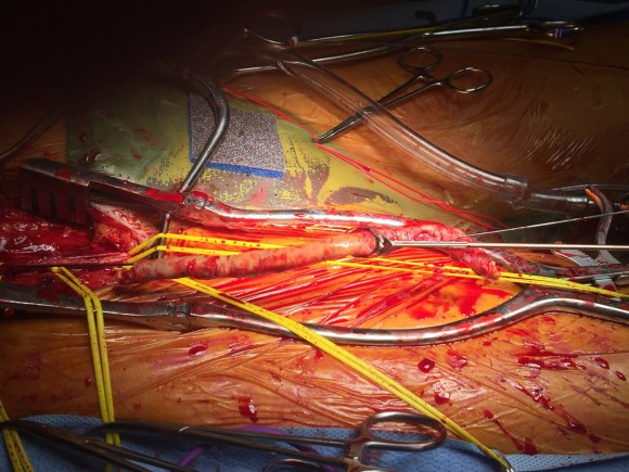
The 18F sheath went up with minimal resistance, and the EVAR was performed in the usual manner. The left common iliac artery occlusion was managed percutaneously from a left brachial access. The stent graft on the left was terminated above the iliac bifurcation and a self expanding stent was used to extend across the iliac bifurcation which had a persistent stenosis after recanalization.

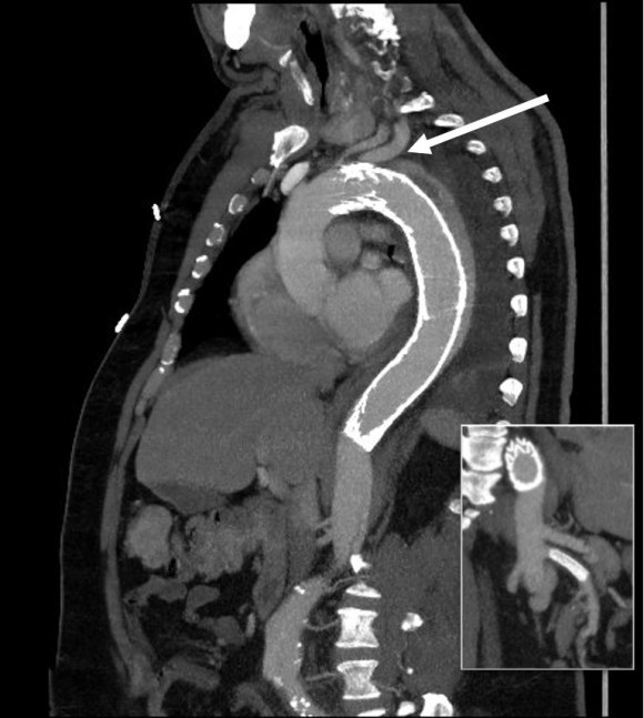
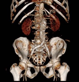
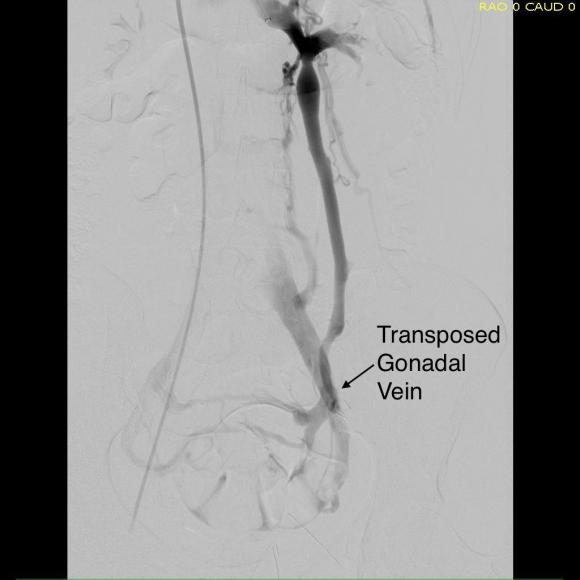
The patient recovered well and was sent home several days postprocedure. He returned a month later with healed wounds and palpable peripheral pulses. He no longer had claudication and CTA showed the aneurysm sac to have no endoleak (figures below).

post CTA EVAR-ENDORE

postop centerline EVAR-ENDORE
Composite imaging showing normal appearing right iliofemoral segment (EIA + CFA) and patent left common iliac artery.

Discussion
I have previously posted on using EndoRE (remote endarterectomy) for both occlusive disease and as an adjunct in EVAR. Iliofemoral EndoRE has excellent patency in the short and midterm, and in my experience has superior patency compared to the femoropopliteal segment where EndoRE is traditionally used. This case illustrates both scenarios. While the common iliac artery occlusions can be expected to have acceptable patencies with percutaneous interventions, the external iliac lesions typically fail when managed percutaneously especially when the stents are extended across the inguinal ligament. The external iliac artery is quite mobile and biologically, in my opinon, behaves much as the popliteal artery and not like the common iliac. Also, the common femoral arterial plaque is contiguous with the external iliac plaque, making in my mind, imperative to clear out all the plaque rather than what can just be seen through a groin exposure.

On microscopy, the external iliac artery is restored to a normal patent artery -I have sent arterial biopsies several months after endarterectomy and the artery felt and sewed like a normal artery and had normal structure on pathology. This implies that the external iliac can be restored to a near normal status and patients that are turned down for living related donor transplantation of kidneys can become excellent recipients. In this case, this hybrid approach effectively treated his claudication but also sealed off his moderate sized AAA while not precluding future visceral segment surgery or intervention with a large suprarenal stent.


Reference

  1. Hollier LH et al. J Vasc Surg 1986; 3:712-7.
  2. Nzara R et al. Ann Vasc Surg. 2015 Nov;29(8):1548-53
  3. Kavanagh CM et al. J Vasc Surg 2016;64:1327-34
Categories
common iliac artery aneurysm CTA Endoleak EVAR iliac artery aneurysm techniques

The Geometry of Parallel Grafts in the Iliac Arteries

The development of metachronous common iliac artery aneurysm, or progression of them, after prior treatment with EVAR (endovascular aneurysm repair), particularly with “bell bottoming” is typically treated with coil embolization of the internal iliac artery and extension of the stent graft into the external iliac artery. While CH-EVAR has been in the news with the recent results from the PERICLES registry, I have never been entirely convinced of its durability. That is different in the case of building parallel grafts in an iliac limb of an EVAR graft (reference).

Here, the geometries, thrombosis, and forces combine to make gutter flow and endoleak unlikely. Choosing the right size of stent grafts to channel to the external and internal iliacs seems to be a challenge, but is easily solved by this scheme -which I can’t claim as my own, but was thought up by a surgeon in upstate New York who choses to remain anonymous.*

The diameter of the stent graft to be sealed to is measured and an area calculated. The sum of the areas of the two grafts to be placed need to equal or slightly exceed the area of this inflow stent graft. If you have decided the size of the external iliac graft, for example, then the diameter of other graft is merely a few geometric formulas away.

Here is a table that can be helpful in avoiding those formulas.
diameter area table.jpegThe inflow graft area is taken from its measured diameter. Then usually one or the other artery has an obligate size -a size the graft has to be while the other has more “wiggle room.” The other thing that comes from experience is that the AFX graft’s iliac limb extension don’t get the B-infolding that can affect an oversized stent graft placed in a small artery and it accomodates a neighbor well.measurement_3

For example, take this patient who after EVAR of aortic aneurysm with AFX developed metachronous dilatation of the common iliac artery to 3.9cm with abdominal pain. The average diameter is 18.5mm. From the table, that rounds to 19mm corresponding to 283.53 square mm. If the internal iliac artery requires a 13mm graft, that is 132.73 square mm, the difference being 150.80 square mm. That corresponds to a 14mm diameter graft, but a slightly larger graft is preferred for oversizing. The external iliac artery is 8mm, and putting a 13mm Viabahn (largest available) in that would result in the B-infolding in the 8mm external iliac. Here, I bailed myself out by simply placing a 20mm AFX iliac limb extension, which by virtue of its design is resistent to infolding and tolerant of parallel grafts laid alongside in constricted channels. I found that the AFX iliac limb, a 20-13mm x 88mm length extension well suited for this.

Image-24.jpg

The AFX graft limb seems to adapt to the presence of the parallel “sandwich” graft which is deployed second and ballooned last. In followup, there was shrinkage of the common iliac artery aneurysm sac and no endoleak.

postop_1

postop_2.jpg

Compared to my other parallel graft case treating a metachronous saccular common iliac aneurysm years after an EVAR with a Gore endograft (link), which by table calculation, resulted in 8% oversize in calculated areas, this particular technique with a large AFX graft and an appropriately sized Viabahn seemed to work well the setting of a previously placed AFX graft. It allows one to avoid hypogastric occlusion.

The final option of a femoral or external iliac to internal iliac bypass after extension across the bifurcation to the external iliac artery is still a reasonable choice, although it seems to be receding into history.

Reference

Smith, Mitchell T. et al. “Preservation of Internal Iliac Arterial Flow during Endovascular Aortic Aneurysm Repair Using the ‘Sandwich’ Technique.” Seminars in Interventional Radiology 30.1 (2013): 82–86. PMC. Web. 9 Dec. 2016.

*While these grafts are not FDA approved for use in this manner, many times, with a prior endograft or graft in place, using the currently available and approved Gore Iliac Branch Endoprosthesis (IBE) in this common scenario would still be off label usage of an approved device, and only if it is feasible, which most times is not. For nonmedical readers, many commonly available devices and medications are used off-label, such as aspirin for blood thinning.

Categories
chronic mesenteric ischemia MALS median arcuate ligament syndrome Takayasu's Arteritis techniques

Takayasu’s Arteritis Driven Median Arcuate Ligament Syndrome: Unusual Symptoms Demand Unusual Solutions

precta_4

The patient is a young woman from overseas who was referred to the clinic for abdominal and chest pain that persisted after diagnosis and treatment of Takayasu’s Arteritis affecting her visceral (middle) aorta. A year prior to presentation, she had been having severe abdominal pain, 30 pound weight loss, and weakness. Laboratory findings included elevated inflammatory markers. CT scan showed inflammation around her celiac axis and superior mesenteric artery. She was treated with prednisone but only responded ultimately to immunosuppressives. Her pain remitted for a while and she regained weight and strength, but eventually in the months prior to consultation, symptoms of postprandial abdominal pain and nausea set in with concomitant constant midsternal chest pain which at times was incapacitating.

Examination was remarkable for a well nourished young woman in distress with epigastric tenderness. Inflammatory markers were normal and she was on methotrexate and tocilizumab. CTA (above) and duplex showed severe stenoses affecting the origins of the celiac axis and superior mesenteric artery. A composite of the centerlines through these showed the arteries to be critically narrowed at their origins (below), along with a mild to moderate stenosis of the aorta (above). No active inflammation could be seen.

precenterlineCA and SMA.png

The anterior view of the 3DVR images of her CT showed an absence of collateralization via the inferior mesenteric artery and Arc of Riolan.

preCTA_3.jpg

This could account for her postprandial abdominal pain which was midabdominal, but the midsternal chest pain was difficult to explain. A cardiac cause had been ruled out at her home institution. It was on the lower part of the sternum and bordered the epigastrium. Through my work with median arcuate ligament syndrome, it was not unusual to have chest pain be part of the pain syndrome which comes about through the compression of the celiac plexus by the median arcuate ligament (reference), and treated effectively with the division of the median arcuate ligament and celiac plexus neurolysis.

I did feel that revascularizing the SMA was likely to improve her postprandial symptoms, given the paucity of circulation to the gut. I had a discussion about her chest pain and the thought that this was a celiac plexus neuropathy as the result of compression of the celiac plexus by her Takayasu’s disease. Typically, for younger people, I perform a bifurcated graft to the celiac axis and SMA from the distal descending thoracic aorta for younger patients, but I had no intention of replacing her aorta at this time, and wanted to reserve any definitive revascularization of her visceral vessels for a later time if it became necessary. Her pulses were full in the legs and she had no hypertension, renal insufficiency, or claudication. I therefore planned a ilio-mesenteric bypass, as it would preserve planes for a later more definitive operation if necessary, and would address her mesenteric ischemia.

I also proposed taking down the median arcuate ligament and lysing her celiac plexus to treat her chest pain symptoms. While I knew this would be fraught with some hazard because of the inflammation that had been there, it would be the best chance at treating this symptom that was debilitating her. I did not think the constant chest/epigastric pain had a vascular etiology. Normally, I would test this with a celiac plexus block, but given the likely inflammation involved, a failed block would not be helpful, and a positive one achieved with some difficulty.

The patient thought this was reasonable and agreed. She underwent a midline laparotomy and I exposed both the SMA and right common iliac artery in the retroperitoneum and tunneled a PTFE graft in a C-pattern in the retroperitoneum. The SMA was diseased proximally under the pancreas and affected by inflammatory scar tissue, and the vessel was thickened as well. The iliac pulse was normal and full and provided excellent inflow.

Going into the lesser sac, the crurae of the diaphragm were heavily scarred near the celiac axis. Division of these fiber was taken slowly and with the aid of both loupe magnification and a hook cautery (borrowed from laparoscopic surgery). The aorta was cleared of tissue first -it was also encased in scar tissue. The dissection going distally was made difficult by inflammatory scar tissue as well, but several large nerve trunks were encountered and divided. The celiac axis was atretic and small within this scar tissue, but was released down to the trifurcation.

The recovery was notable for remission of her chest pain and her ability to resume eating without pain or nausea. A CTA done before discharge showed a patent bypass.

post-cta_1

Followup communication has revealed continued remission of her presenting symptoms months after her operation.

We are working on presenting a followup to our recent paper on MALS. Increasingly, it is apparent that consideration of celiac plexus compression as an etiology of epigastric abdominal pain unexplained by more common gastrointestinal causes provides solutions for patients given no explanation for their debilitating pain. This pain can also affect the chest, and flanks and back in the distal thoracic dermatomes, and mimic the some of the symptoms of mesenteric ischemia. In those with recurrent MALS after a successful ligament release and interval of symptom remission, even in the absence of celiac axis compression, a positive response to a celiac plexus block points to the presence of either remnant plexus fibers or compression by scar tissue of the cut nerve endings (neuromata) and reoperation has been successful. Extrapolating this experience to this patient with inflammatory compression of the celiac plexus secondary to Takayasu’s Arteritis made sense and celiac neurolysis appears effective.

Reference

Weber JM, Boules M, Fong K, Abraham B, Bena J, El-Hayek K, Kroh M, Park WM. Median arcuate ligament syndrome is not a vascular disease. Ann Vasc Surg. 2016 Jan;30:22-7.

Categories
EndoRE EVAR kidney transplant remote endarterectromy techniques TEVAR

External iliac remote endarterectomy restores the artery to normal, opening the way for EVAR, TAVR, TEVAR, and transplant: alternate applications of EndoRE

preop-cta

One of the nice things  about practicing at the Clinic is being able to offer unique solutions. A severely diseased or occluded external iliac artery (EIA) can be a vexing problem, particularly if bilateral, in this endovascular era. Many cardiovascular devices require femoral access that has to traverse compromised iliac arteries -those with large (>16F) delivery systems require a sufficiently wide path to get the devices to the heart and aorta. Also, living related donor kidney transplantation is predicated on minimizing risk to maximize results and having significant iliac plaque negates one as a recipient for this high stakes elective procedure. In situations where the EIA is too small to accommodate devices because of atherosclerotic plaque, the typical solution is placement of a conduit to the common iliac artery or the aorta. The practice of “endopaving” with a covered stent graft and ballooning is also described, but its long term outcomes are not reported and the internal iliac artery is usually sacrificed in this maneuver.

This patient presents with lifestyle limiting claudication and an absent right femoral pulse. ABI is moderately reduced on the right to 0.57, and he had no rest pain. CTA at our clinic revealed an occluded EIA bracketed by severely calcified and nearly occlusive plaque of the common iliac artery (CIA) and common femoral artery (CFA).

cta-preop_7
Centerline Projection

The patient was amenable to operation. Traditionally, this would have been treated with some form of bypass -aortofemoral or femorofemoral with a common femoral endarterectomy. While endovascular therapy of the occluded segment is available, one should not expect the patencies to be any better than that of occlusive lesions (CTO’s) in other arteries. Hybrid open/endovascular therapy is an option as well with CFA endarterecotmy and crossing CIA to EIA stents, but I have a better solution.

The common femoral endarterectomy rarely ends at the inguinal ligament, and is uniquely suitable for remote endarterectomy, a procedure from the early to mid twentieth century.

endoRE graphic
Steps in Remote Endarterectomy


The addition of modern fluoroscopic imaging and combining with endovascular techniques makes this a safe and durable operation.

pre intervention.png

The patient was operated on in a hybrid endovascular OR suite. A right groin incision was made to expose the common femoral artery for endarterectomy and left common femoral access was achieved for angiographic access, but also to place a wire across the occlusion into the common femoral artery.

All actions on the external iliac artery plaque are done with an up-and-over wire, allowing for swift action in the instance that arterial perforation or rupture occur. This event is exceedingly rare when the operation is well planned. With this kind of access, an occlusive balloon or repairing stent graft can be rapidly delivered.

The common femoral endarterectomy is done from its distal most point and the Vollmer ring is used to mobilize the plaque. A Moll Ring Cutter (LeMaitre Vascular) is then used to cut the plaque.

Ring dissection.png

Cutter.png

The plaque is extracted and re-establishes patency of the EIA.

plaque.png
Plaque Specimen

The plaque end point is typically treated with a stent -in this  case, the common iliac plaque was also treated.

result.png

What is nice about this approach is that this artery has been restored to nearly its original condition. I have taken biopsies of the artery several months after the procedure in the process of using the artery as inflow for a cross femoral bypass, and the artery clamped and sewed like a normal artery and the pathology returned normal artery.

This has several advantages over conduit creation which can be a morbid and high risk procedure in patients who require minimally invasive approach. A graft is avoided. The artery is over 8mm in diameter where with stenting up to 8mm with an occlusive plaque, the danger of rupture is present, and often ballooning is restricted to 6mm-7mm. This is insufficient for many TEVAR grafts and TAVR valves.

For patients being worked up for living related donor transplants who are turned down because of the presence of aortoiliac plaque, those with the right anatomy can undergo this procedure and potentially become candidates after a period of healing.


Categories
acute mesenteric ischemia AIOD aortoiliac occlusive disease (AIOD) chronic mesenteric ischemia CTA PAD techniques visceral malperfusion

When good enough is better than perfect: a case of end stage visceral segment aortic occlusive disease

The patient is a woman in her 60’s who self referred for complaint of abdominal pain, weight loss, and rest pain of the lower extremities. She is a 40 pack year smoker and had severe COPD, hypertension, congestive heart failure with mitral regurgitation, chronic kidney disease stage IV, and ischemic rest pain of the legs. She had a 30 pound weight loss due to severe postprandial abdominal pain. She had bloody stools. Her kidney function was worsening, and dialysis was being planned for likely renal failure but she was against dialysis. She had consulted several regional centers but was felt to be too high risk for surgery and with her refusal of dialysis, would be a high risk for renal failure and death with intervention. Physical examination revealed weakened upper extremity pulses, and nonpalpable lower extremity pulses and a tender abdomen. In clinic, she developed hypoxia and dyspnea and was admitted directly to the ICU.

preop-cta

CTA (above) revealed severely calcified atherosclerotic plaque of her visceral segment aorta occluding flow to her mesenteric and renal arteries and to her leg. The right kidney was atrophic. The left kidney had a prior stent which looked crushed. The infrarenal aorta was severely diseased but patent and there were patent aortic and bilateral iliac stents.

Echocardiography revealed a normal ejection fraction of 60%, diastolic failure,  +2 to+3 mitral regurgitation, and pulmonary artery hypertension. She did respond to diuresis and stabilized in the ICU. Intervention was planned.

Options that I considered were an extranatomic bypass to her legs and revascularization from below. I have come across reports of axillo-mesenteric bypass, and I have performed ascending and descending thoracic aorta to distal bypass for severe disease, but concluded, as did the outside centers, that she was a formidable operative risk. Also, there was a high likelihood of great vessel occlusive disease. Looking at her CTA, I felt that she needed just a little improvement in flow -not perfect but good enough. The analogy is like drilling an airline through a cave-in. Also, her left kidney gave a clue -it was normal sized and survived the stress test of a contrast bolus for the CTA without dying. A discussion with the patient green lighted an attempt -she understood the cost of failure but did not wish to linger with this abdominal pain.

Access for intervention was via the left brachial artery. Aortography showed the severe stenosis at the origin of the SMA and the nearly occlusive plaque in the visceral segment aorta.

preintervention-aortogram

The plaque was typical of the coral reef type, and had an eccentric channel that allowed passage of a Glidewire. Access into the left renal artery was achieved. Its stent was patent but proximally and distally there were stenoses; this was treated with a balloon expandable stent. The path to it was opened with a balloon expandable stent to 8 mm from femoral access. This was the improvement the renal needed. A large nitinol stent was placed from this access in the infrarenal aorta when severe disease above the iliac stents was encountered.  The SMA was then accessed and treated with a bare metal stent.

post intervention.jpg
Renal stent was reaccessed and ballooned in this pentultimate angiogram

Her creatinine improved, as did her intestinal angina. She was discharged home. She later returned a month after the procedure with complaints of nausea and vomiting and right lower quadrant abdominal pain and was discovered to have an ischemic stricture of her small bowel. This was removed laparoscopically and she recovered well. She recovered her lost weight and now a year and a half later, remains patent and symptom free.

Discussion: Dr. Jack Wiley includes in the preface to his atlas of vascular surgery the words of Dr. Joao Cid Dos Santos, the pioneer of endarterectomy techniques, “Vascular surgery is the surgery of ruins.” And in that context, good enough is sufficient.


Categories
pseudoaneurysm techniques ultrasound vascular lab venous intervention

Complex femoral pseudoaneurysm with arteriovenous fistula and large hematoma treated with novel hybrid therapy

wide avf and pseudo

The patient is a middle aged man who after an interventional procedure was referred to my clinic with an expanding hematoma due to a pseudoaneurysm complicated by an arteriovenous fistula. He was a week out from his procedure and had grown a hematoma roughly the size of a hard boiled egg in his left groin which caused him pain. A duplex scan showed a pseudoaneurysm (below) with fistula flow.

preduplex12

On examination, he had this well circumscribed indurated hematoma of hard boiled egg size with tenderness. There was a bruit on auscultation. Duplex showed a small chamber of flow adjacent to the proximal superficial femoral artery emptying into the femoral vein. Doppler in the common femoral vein showed relatively high fistula flow, and this is reported to be associated with failure of thrombin injection. CTA (top) demonstrated flow of contrast from femoral artery to vein through a pseudoaneurysm chamber that laid between. Angulation to an axial orientation showed this better (below).

axial AVF
Contrast flows from femoral artery (right) to the fistula chamber, then into the femoral vein.

axial CTA avf pseudo
Axial MPR
Operation was planned, but in the days leading up to the operation, I had a thought -the primary reason why ultrasound guided thrombin injection would fail is the AVF. It would be simple to fluoroscopically guide an angioplasty balloon on the arterial side to occlude the fistula inflow. The next step would be to get access to the pseudoaneurysm with a needle under ultrasound guidance, confirm location with a contrast injection. Once confirmed, the balloon is inflated and a small volume of thrombin would be injected. I discussed this with the patient in detail and he was enthusiastic about trying this before proceeding with an open repair.


pseudoaneurysm avf procedure sketch
Schematic of procedure
The procedure went as planned. Ultrasound guided access is aided with dual live display of B-mode and color flow (below)

Arteriography showed much of the contrast from injection of the pseudoaneurysm to preferentially go to the artery which made me worry less about creating a DVT/PE. With balloon inflated (below), thrombin was injected and balloon inflation held for about 30 seconds.

angio procedure31

There was resolution of flow in the pseudoaneurysm and in the fistula. Before and after duplexes are composited below.

prepost pseudo chamber

Repeat duplex on the following day showed resolution of the pseudoaneurysm and arteriovenous fistula.

In the days before ultrasound guided thrombin injection of pseudoaneurysms, open surgical repair of these was fraught with complications. First, these patients typically had cardiac disease. Second, they were usually anticoagulated often with multiple agents. And finally, they were  many times obese, making not only the operation fraught with complexity, but the ultimate wound healing a delicate and rare phenomena. Even now, we get emergency repairs when access hemostasis fails, and these patients are typically high risk. With hematoma evacuation, inflammation, lymph leaks, and infections may follow; the patient was correct in his enthusiasm for agreeing to proceed with a minimally invasive effort.

As to the techniques, they are all well established in the vascular surgeon’s toolbox. Ultrasound guided access of the pseudoaneursm should be obtained before arterial occlusion. This was  facilitated by general anesthesia which kept the patient from moving. Having access to excellent ultrasound and angiographic imaging made this possible. The patient felt much better and was discharged home the next day after his confirmatory duplex.

Categories
Nutcracker Syndrome techniques Venous venous intervention

Nutcracker Syndrome: A Simplified Approach With Gonadal Vein Transposition

CTV_1

The patient is a young woman in her twenties who developed severe right sided abdominal and back pain about 4 months prior to presentation associated with bouts of bloody urine. Activity and standing exacerbated her pain and inactivity and recumbency relieved it. She gained 15 pounds because of her inactivity. Examination was significant for tenderness over her left kidney. Urinanalysis showed positive proteinuria and hemaglobinuria.

Prior to consultation with me she had had an MR venogram showing compression of her left renal vein by the superior mesenteric artery (nutcracker phenomena). The presence of hematuria, proteinuria, and pain (albeit atypically right sided) made it nutcracker syndrome.

MRV color_Image006
Dilated left gonadal vein and pelvic varices indicate left renal vein (LRV) ouflow obstruction by the superior mesenteric artery (SMA)

I ordered a renal duplex and a CT venogram for procedural planning.

Duplex7680

On the duplex, the proximal left renal vein (LRV) was not visualized. The right kidney had normal parenchymal appearance and blood flows, while the left, the kidney appeared distended and had flows consistent with outflow obstruction.

spectral kidneys
Spectral Doppler flows show respirophasicity in right renal vein(RRV), outflow obstruction on left renal vein (LRV)
Duplex kidneys bmode
The left kidney is swollen and tender.

CT Venography showed the gonadal vein to be an important outflow vessel to the left renal vein with dilated proximal segment and reflux into pelvic varices.

CTV_1

CTA processed_5CTA processed_4

A left gonadal vein to iliac vein transposition was planned via a left lower quadrant retroperitoneal exposure. On the table, a venogram was performed with selective access of the left renal vein.

Venogram13

The injection from the LRV showed severe compression of the LRV with a channel only slightly larger than the sheath and avid reflux into the gonadal vein. Selective access into the gonadal vein and venography from a confluence in the pelvis showed that flow was one way from the LRV into the gonadal vein and this filled a large region of pelvic varices.

Venogram19

Venogram23

The gonadal vein was large caliber and refluxed into two large veins in the pelvis. The one that fed the varices was not selected for transposition, but rather the longer straighter tributary. A catheter was left for easier identification during the dissection.

A left lower quadrant incision was made and a retroperitoneal dissection performed exposing the gonadal vein and iliac vein.

IMG_0417

IMG_0420


Prior to ligation of the tributaries, a sheath was inserted and through this a LeMaitre valvulotome was brought up to the left renal vein and carefully deployed and pulled back, cutting the valves. This greatly increased the outflow from the vein as evidenced by the height of the blood spout from the vein when the sheath was removed. The varices were ligated at their root -treating them definitively. Transposition was to the external iliac vein, and I could see the feasibility of a laparoscopic or robotic approach to this operation (ref 3).

Completion venography showed excellent flow from the LRV down the gonadal vein into the iliac venous system.

Venogram41

The patient lost less than 10mL of blood and was discharged on postop day 2. Gratifyingly, all of her preoperative pain resolved and her UA showed no more hemoglobinuria or proteinuria.

Discussion

The described treatment options for nutcracker syndrome include (ref 1):

  1. Medical therapy aimed at decreasing renal venous hypertension (for hematuria)
  2. Renal autotransplantation
  3. Left renal vein transposition
  4. Left renal vein to vena cava bypass (autologous or PTFE)
  5. SMA transposition
  6. Nephrectomy
  7. Gonadal vein to IVC bypass
  8. Exovascular stenting (wrap of renal vein with ringed PTFE graft)
  9. Endovascular stenting

Many of the operations are of historic interest. Stenting deserves some comment. The patient self referred because she had read multiple reports of cardioembolization on internet support group comments. The largest nitinol stent (self expanding) available is 14mm. Wall stents in larger diameters are available, but are stiff, poorly conformable, and will elongate if constrained by a non-dilating stenosis like the external compression by the SMA. While acceptable results have been reported, the long term results (20-70 years) for younger patients is unknown. Migration is highly morbid, and usually to the heart, requiring sternotomy and cardiotomy to retrieve the stent. Optimally, a conforming 16-28mm self expanding stent should eventually become available, but conformability is typically inversely proportional to radial strength, and it is the less conformable stents that migrate. Work is ongoing to bring larger diameter nitinol stents for venous indications. The difference between May-Thurner Syndrome and Nutcracker syndrome isn’t merely the size of the veins and stents. The iliocaval confluence is relatively static with some movement of the lumbosacral joints and well suited for treatment with the relatively nonconforming Wall Stents. The left renal vein under the SMA is a very dynamic environment with motion of the SMA and the kidneys with respiration, ambulation, and activity leaving stents vulnerable early to migration and later to fracture.

The left renal vein transposition to the IVC is a nice operation with a good track record (ref 2). The downside is the long midline incision required with transperitoneal exposure. There is bleeding risk and postoperative complications of ileus, wound infection, and small bowel obstruction. Looking at the CTV, it seems obvious that the gonadal vein crosses over the iliac vein in the pelvis and would be a straightforward, less morbid, less invasive option. A review of the literature reveals only a single reference discussing three cases of left renal vein transposition (ref 3), and it was done with a surgical robot. I think that a laparoscopic approach would be simpler and less invasive and will consider developing this if volumes justify it. That said, the open retroperitoneal approach is very straightforward and well used exposure. Using venography to set up and then confirm the results of the transposition was helpful. I don’t think that measuring pressures and diameters and taking calipers to calculate stenoses is all that useful and in some instances a harmful method of justifying endovascular treatment of nutcracker phenomena in the absence of serious symptoms and a careful deliberate workup which includes a good history and physical, a UA, a duplex and CTV.

Intervening on the gonadal vein to iliac vein anastomosis should be straightforward from a groin or thigh venous access on the ipsilateral side. This operation doesn’t preclude any future interventions on the LRV. The pelvic varices were treated with direct ligation. The patient’s pain was successfully relieved in the short term.

Conclusion: Open retroperitoneal left gonadal vein to iliac vein transposition with gonadal vein valvulotomy is effecting in treating nutcracker syndrome.

References

  1. Kurklinsky AK, Rooke TW. Mayo Clin Proc. 2010 Jun; 85(6): 552–559.
  2. Reed NR et al. J Vasc Surg. 2009 Feb;49(2):386-93;
  3. White JV et al. J Vasc Surg Venous Lymphat Disord. 2016 Jan;4(1):114-8.


Categories
AAA techniques

Avoiding Aortic Exoleaks: principles of the proximal aortic anastomosis

IMG_9846

The proximal anastomosis is the most critical portion of an open aortic aneurysm repair. Several concepts are central to creating an anastomosis that doesn’t bleed at unclamping: managing narrow spaces, overcoming distortion, and using just enough tackle.

Narrow Spaces

The transabdominal exposure is a narrow space. The work is done under the overhanging left renal vein, the transverse colonic mesentery, the liver, the rib cage. Extra lateral space can be made by eviscerating the bowel, but at the cost of higher rates of ileus, and doesn’t solve the first problem. The standard DeBakey aortic clamp and straight Fogarty clamps stand nearly straight up, limiting the space above the incision at the aortic neck. My goto clamp is the Cherry Supra Celiac Aortic Clamp, designed by my mentor Ken Cherry. 1606988_10203082426724504_324421715_n

It hugs the contour of the mesentery and liver overhang, and the handles stay out of the way above the wound. It will also tilt up the aorta because of the weight balance. The other option is to apply a transverse clamp, which I will discuss in a later post. The transverse clamp leaves the suprarenal space free of clamp, but can be difficult if not hazardous to apply. The clamp has to be hemostatic and this can be challenging with atherosclerotic plaque -preoperative planning must include planning for safe clamp sites. A suprarenal clamp may be limited by the presence of the terminal insertions of the diaphragmatic crurae. I have recently found that dividing these crurae with a Maryland tipped Ligasure, a laparoscopic instrument I use to dissect the retroperitoneum, makes short order of what can sometimes be an awkward exposure in this tight space. Finally, endarterectomy of the neck should be done carefully to let needles pass without difficulty.

Distortion

The proximal anastomosis is ideally just another end to end anastomosis -attaching a circle to a circle, but clamping narrows and distorts the circular aortic neck (top illustration). To envision this, imagine the aortic neck being a clock face:

FullSizeRender

Lets say the suture should be applied at each of the hours and half hours. You get ready to sew your first aortic neck and after endarterectomizing some plaque, you get this:

IMG_9886

The important point is that you still have to apply the original plan of applying sutures evenly and at an appropriate frequency (about 3mm apart), to avoid gathers and gaps, especially on the posterior wall. One way is to apply outward tension with a Wheatlander retractor in the aortic sac.

IMG_9873
The posterior wall of the aorta should be distracted to avoid distortions that create uneven gaps between passes of the needle. 
The bites on the aorta should be generous, on the graft, less so. As long as the gaps between the sutures is the same on both aorta and graft, you shouldn’t get leaks.  The other principle to guide you is the needles should pass pointing to the center of the clock -this is challenging in the Dali clock, but if you pivot your shoulder, your suturing won’t be bullied by the distortions and the narrow space.

The Right Tackle

You don’t go after panfish with a deep sea tackle. Pictured below are a spinning lure for trolling with a large hook and a small dry fly with narrow guage hook for comparison.

IMG_9889

Which hook creates the larger hole in the fish’s mouth? In fact, both lures can be used to catch the same large trout, but in different situations.

OLYMPUS DIGITAL CAMERA
On a lake, large hook, on a stream, small hook for same fish.
One of the techniques which I have borrowed from our partners in cardiac surgery here at the clinic is using smaller guage monofilament and needles. I once co-scrubbed an aortic arch case with Eric Roselli, and was bemused to see him sew graft to the fearsome ascending aorta with 5-0 monofilament suture. Then I saw no needle hole bleeding and was sold. Up to then, I had switched from my 3-0 on SH needles to 2-0 on MH needles -basically the largest vascular needle, and saw posterior aortic wall break down from the needle trauma. Unfortunately, 5-0 suture doesn’t come long enough, the CV needles aren’t big enough to sew posterior wall. I now use 4-0 on SH with 48cm length suture. Ideally, we’d have a 5-0 monofilament on a 60cm suture with a SH shaped and sized needle with the narrow guage of the CV needle.

Also, because the needles are finer and I favor supersized Castro needle holders. The needle holes which represent rents in the aortic wall are far easier to deal with using a smaller needle. Right tackle.


Categories
AAA aortic dissection Commentary taaa tbad techniques TEVAR thoracabdominal aortic aneurysm visceral malperfusion

Moneyballing a Type II Thoracoabdominal Aortic Aneurysm


The innovation of sabremetrics in baseball management and finance as described in Michael Lewis’ wonderful book Moneyball wasn’t just the ability to quantify skill to predict outcomes, it was the ability to assemble that skill without overpaying. For a baseball team on a budget, spending all your payroll on a superstar makes no sense when you can get equivalent quants of skill in a statistical aggregate of no-name players with proven metrics. Rather than pay for an A-Rod, you can recruit, and pay for, 5 players that in aggregate, statistically achieve what you would get with a healthy A-Rod, so the thinking goes. How does this translate into vascular surgery? Can we arbitrage complication rates?

The open repair of type II thoracoabdominal aortic aneurysms is a heroic endeavor, putatively best done by surgeons wearing cowboy boots, and classically comes with sobering complication rates that exceed 20% for death and paralysis. Is it possible to reduce this risk by subdividing this most enormous of cardiovascular operations into component parts?

The patient is a middle aged man in his 50’s who presented with a type B aortic dissection. His dissection flap spanned from his left subclavian artery to the infrarenal aorta. He was a long time smoker and had hypertension that was difficult to control, made much worse after his dissection. He had a moderate dilatation of his thoracic aorta, maximally 36mm and tapering to 35mm in visceral segment. There was a 4.9cm infrarenal AAA where the dissection terminated.

CTA at presentation

His chest pain resolved with blood pressure control and he was discharged, but in followup his thoracic aortic segment grew and his blood pressure worsened, never getting below a 150mmHg systolic despite multiple agents. CTA two months after presentation, showed growth of his TAA to 44mm from  36mm in two months  and the visceral segment showed that his dissection flap impinged on flow to the right renal artery. His AAA remained the same. He continued to have bouts of chest pain related to hypertension.

CTA at 2mo post presentation

Twenty years ago, the board answer would have been to replace the whole aorta. In young, otherwise healthy man who had been working in road construction up to the dissection, he would have been considered a candidate for a direct open repair of the type II thoracoabdominal aortic aneurysm. From the landmark paper out of Houston by Dr. Svensson in 1993, open type II TAAA repair was associated with about a 10% death rate and 30 percent paralysis rate. Waiting a few months for the aneurysms to grow further in this patient, in the 90’s this patient would probably have ended up with an open TAAA repair. Good thing we have better options.

The goals of modern therapy are to treat the urgent indication while holding off repair of less critical segments of the aorta, and to do so in a way that each operation builds on the previous one.

This patient needed a left subclavian artery debranching and then TEVAR of his dissecting thoracic aortic aneurysm, and intervention on his right renal artery. We did this in one setting performing first a left carotid subclavian artery transposition and then percutaneous TEVAR from the left common carotid artery origin to the supraceliac abdominal aorta.

TEVAR with carotid SCA transposition

completion TEVAR.png

The completion aortography showed good deployment of the CTAG device from the left common carotid artery origin to the celiac axis origin. The false lumen was no longer visualized. The right renal artery which was narrowed was treated with a balloon expandable stent.

The distal thoracic aorta, the true lumen was constrained by a chronic dissection flap. It is here I gently dilate the distal thoracic stent graft with the hopes of eliminating the distal false lumen. This is different from the acute dissection where I rarely balloon.

The TEVAR was done percutaneously, minimizing the overall time in the operating room. The technical details of the transposition can be found in the excellent paper by Dr. Mark Morasch.


renal PTAS

When I do this procedure for acute dissection, I quote the patient a general risk of stroke, paralysis of about 2-5% and death of 1-2 percent for someone with low cardiopulmonary risk like this patient had. He recovered rapidly and went home post op day 5.


Followup post TEVAR

He at 6 month post TEVAR followup, CTA showed stablility in his thoracic aorta. in infrarenal AAA grew from 5.0 to 5.7cm between the 1 month CT and the 6 month CT.

6 month CTA imaging

The terminus of the stent graft excluded the false lumen in the thoracic aorta but also resulted in filling and pressurization of the false lumen beyond and can be seen as a 44mm lateral dilation of the visceral segment of the aorta which had developed in the 6 month interval since the TEVAR.
The infrarenal neck continued the dissection and had dilated to about 36mm, but was parallel for a good length above the AAA. I decided to treat the inrarenal aorta with direct transabdominal repair. This would allow me to fenestrate the aorta, and possibly prevent further growth of the viseral segment while reserving the retroperitoneum for the visceral segment repair if it came to it. The neck diameter was 36mm.

tube graft
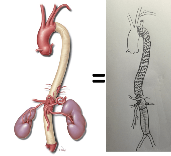
His operation was performed via an anterior approach with the patient supine. A tube graft repair was performed expeditiously and included resecting the dissection flap up to the clamp. Care was taken to avoid injury to the renal stent. The proximal anastomosis went well – the dilated aorta yet had strong tissue strength. A felt strip was used to buttress the aortic side of the anastomosis. The estimated risk of paralysis was less than 1% and risk of death was less than 2%. The patient recovered uneventfully and went home on POD 5.

He did well in subsequent followup, having successfully quit smoking. He retired early on disability and was becoming more active, but the visceral segment dilatation was concerning. At 6 months post infrarenal AAA repair, he underwent CTA and it showed patent thoracic stent graft and infrarenal abdominal graft. The intervening visceral segment continued to enlarge and was now 46mm. The decision was to wait another interval 9 months to see if this would stabilize. The segment grew some more and was 49mm. He wanted to give it another 6 months and at that time, CTA showed further growth over 5cm, and he had developed some abdominal discomfort. He was taken to the operating room.

IMG_8659

A four branch repair of the visceral segment thoracoabdominal aortic aneurysm was performed. The diaphragm was taken down and the stent graft was clamped as was the infrarenal tube graft. A premade Coselli graft was used to bypass to the right renal, SMA, celiac, and left renal in those order. The patient had a CSF drain for the case which was removed on postoperative day 2. He recovered rapidly and went home on postoperative day 6. His estimated risk of paralysis was about 2-5%, mitigated by a protocol centered on CSF drainage and blood pressure control. His risk of death was 5%. Telephone followup reveals the he is pain free at a month out and functional nearly at baseline.

This illustrates the notion that three smaller operations in an aggregate over three years achieved the equivalent of the single big open type II TAAA repair.

equivalence
Illustration on left from Svensson et al.

The idea is to make each step achievable -like coming down a mountain taking three days on well marked paths rather than base jumping off the summit.

Clearly, the patient was younger and a fast healer, and credit must also be given to the anesthesia/critical care team who see high acuity cases in volume every day and not every patient can expect to have such short stays and excellent outcome, but these are far more likely if operations are planned out in such a manner.

Reference

Svensson LG, Crawford ES, Hess KR, Coselli JS, Safi HJ. Experience with 1509 patients undergoing thoracoabdominal aortic operations.  J Vasc Surg 1993;17(2):357-36.


Categories
complications CTA tbad techniques TEVAR type b aortic dissection visceral malperfusion

Reversing paralysis with a bypass

Dissection CTA

The patient is middle aged and had a type B thoracic aortic dissection (TBAD) as a consequence of recreational substances that acutely raised his blood pressure. At the outside hospital, he had a CTA showing the dissection extending from his left subclavian artery and causing occlusion of his superior mesenteric artery (SMA). He developed abdominal pain and was swiftly transported to our acute aortic syndrome unit. He was taken to the operating room and underwent a TEVAR of the dissection and stenting of his SMA -this is similar to other cases I have discussed in prior posts so I am omitting the technical details. The stent covered the left subclavian artery origin to exclude the origin of the dissection. The stent was extended to the distal thoracic aorta but did not go to the celiac origin. 

TBAD post stent

Post procedure, his lactate never rose and he was maintained on the usual post procedure protocol of keeping MAP’s (mean arterial pressure) above 80mmHg. His left subclavian artery was covered but I do not routinely bypass, especially when the left vertebral artery is at least equal in size to the contralateral one. I don’t often place spinal drains for urgent/emergent cases particularly in patients who have never had infrarenal aortic surgery and patent hypogastric arteries. He was kept sedated overnight and awoke in the morning unable to move his legs to command. He had no pain sensation up to his umbilicus.

A spinal drain was emergently placed and his blood pressure was raised to MAPs of 90+, but these failed to reverse his paralysis. After discussion among my world class partners, I chose to take the patient back for a carotid subclavian bypass which was done through a single incision with a dacron bypass graft.

Carotid subclavian bypass CTA

His paralysis resolved. He was discharged home, ambulating without assistance. Spinal cord complications are reported to occur between 1-5 percent of patients undergoing TEVAR for complicated TBAD. They were seen in 2 of 72 patients in the TEVAR arm of the INSTEAD trial (Circulation, 2009 vol. 120(25) pp. 2519-28), and was permanent in 1. While there are some who routinely place prophylactic drains, it is unclear to me that they have a significant effect if placed unselectively. I will place a Preop drain in the instance of infra renal graft, hypogastric arterial occlusive disease. In the instance of a dominant left vertebral, I will perform concomitant bypass, but just as often not. This is a gratifying and rare outcome of paralysis reversed with a carotid subclavian bypass when spinal drain and permissive hypertension did not.