Categories
common iliac artery aneurysm CTA Endoleak EVAR iliac artery aneurysm techniques

The Geometry of Parallel Grafts in the Iliac Arteries

The development of metachronous common iliac artery aneurysm, or progression of them, after prior treatment with EVAR (endovascular aneurysm repair), particularly with “bell bottoming” is typically treated with coil embolization of the internal iliac artery and extension of the stent graft into the external iliac artery. While CH-EVAR has been in the news with the recent results from the PERICLES registry, I have never been entirely convinced of its durability. That is different in the case of building parallel grafts in an iliac limb of an EVAR graft (reference).

Here, the geometries, thrombosis, and forces combine to make gutter flow and endoleak unlikely. Choosing the right size of stent grafts to channel to the external and internal iliacs seems to be a challenge, but is easily solved by this scheme -which I can’t claim as my own, but was thought up by a surgeon in upstate New York who choses to remain anonymous.*

The diameter of the stent graft to be sealed to is measured and an area calculated. The sum of the areas of the two grafts to be placed need to equal or slightly exceed the area of this inflow stent graft. If you have decided the size of the external iliac graft, for example, then the diameter of other graft is merely a few geometric formulas away.

Here is a table that can be helpful in avoiding those formulas.
diameter area table.jpegThe inflow graft area is taken from its measured diameter. Then usually one or the other artery has an obligate size -a size the graft has to be while the other has more “wiggle room.” The other thing that comes from experience is that the AFX graft’s iliac limb extension don’t get the B-infolding that can affect an oversized stent graft placed in a small artery and it accomodates a neighbor well.measurement_3

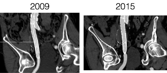
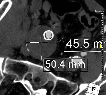
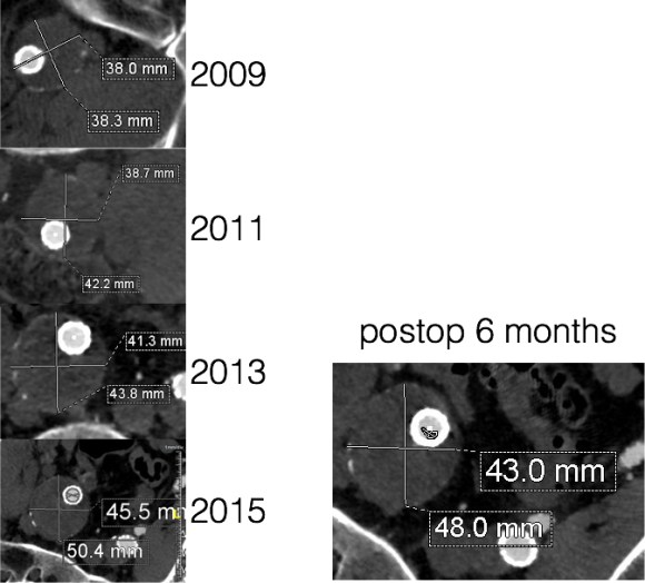
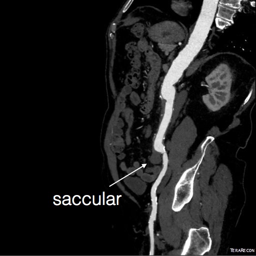
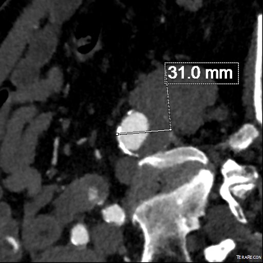
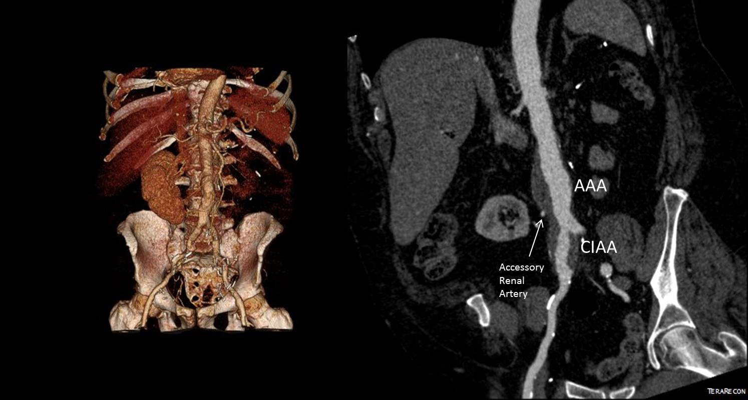
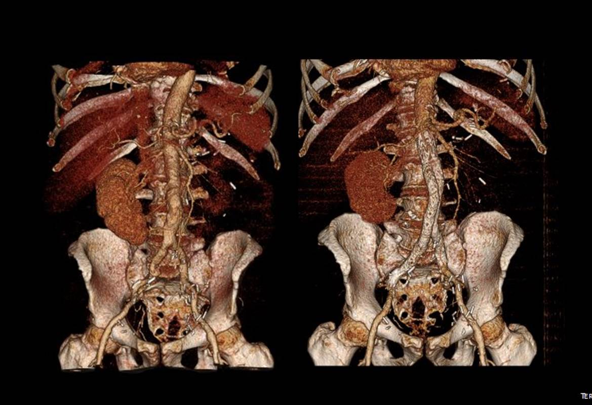
For example, take this patient who after EVAR of aortic aneurysm with AFX developed metachronous dilatation of the common iliac artery to 3.9cm with abdominal pain. The average diameter is 18.5mm. From the table, that rounds to 19mm corresponding to 283.53 square mm. If the internal iliac artery requires a 13mm graft, that is 132.73 square mm, the difference being 150.80 square mm. That corresponds to a 14mm diameter graft, but a slightly larger graft is preferred for oversizing. The external iliac artery is 8mm, and putting a 13mm Viabahn (largest available) in that would result in the B-infolding in the 8mm external iliac. Here, I bailed myself out by simply placing a 20mm AFX iliac limb extension, which by virtue of its design is resistent to infolding and tolerant of parallel grafts laid alongside in constricted channels. I found that the AFX iliac limb, a 20-13mm x 88mm length extension well suited for this.

Image-24.jpg

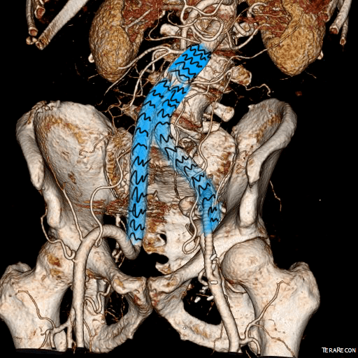
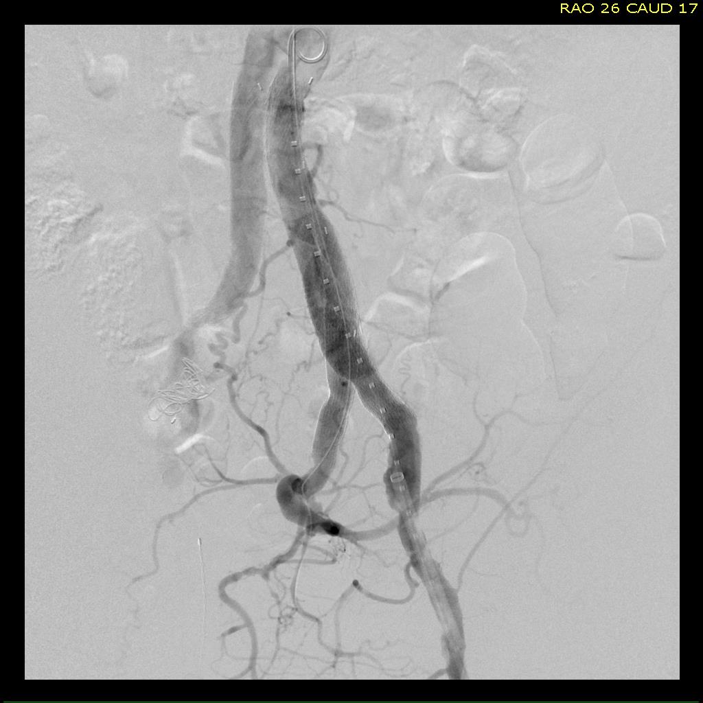
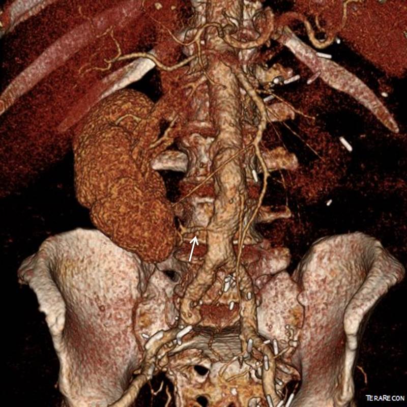
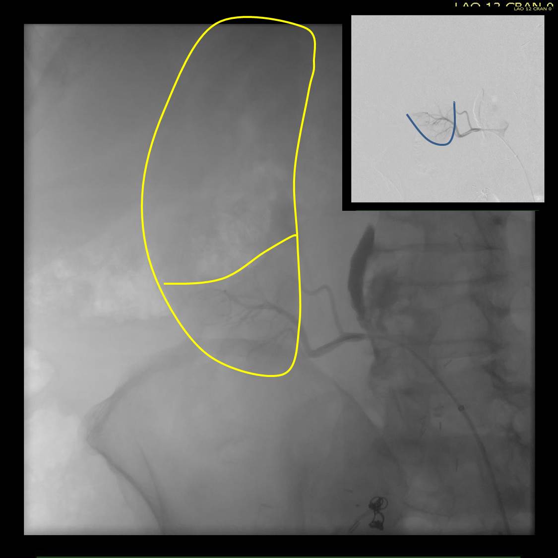
The AFX graft limb seems to adapt to the presence of the parallel “sandwich” graft which is deployed second and ballooned last. In followup, there was shrinkage of the common iliac artery aneurysm sac and no endoleak.

postop_1

postop_2.jpg

Compared to my other parallel graft case treating a metachronous saccular common iliac aneurysm years after an EVAR with a Gore endograft (link), which by table calculation, resulted in 8% oversize in calculated areas, this particular technique with a large AFX graft and an appropriately sized Viabahn seemed to work well the setting of a previously placed AFX graft. It allows one to avoid hypogastric occlusion.

The final option of a femoral or external iliac to internal iliac bypass after extension across the bifurcation to the external iliac artery is still a reasonable choice, although it seems to be receding into history.

Reference

Smith, Mitchell T. et al. “Preservation of Internal Iliac Arterial Flow during Endovascular Aortic Aneurysm Repair Using the ‘Sandwich’ Technique.” Seminars in Interventional Radiology 30.1 (2013): 82–86. PMC. Web. 9 Dec. 2016.

*While these grafts are not FDA approved for use in this manner, many times, with a prior endograft or graft in place, using the currently available and approved Gore Iliac Branch Endoprosthesis (IBE) in this common scenario would still be off label usage of an approved device, and only if it is feasible, which most times is not. For nonmedical readers, many commonly available devices and medications are used off-label, such as aspirin for blood thinning.

Categories
AAA common iliac artery aneurysm EVAR iliac artery aneurysm techniques

The Interrupted Natural History of Aortic and Iliac Artery Aneurysms

graphic

The patient, now in his 90’s, found out about his aortic and iliac artery aneurysms in his early 80’s, had been offered repair, but had refused. Several years ago, one of my partners emergently repaired his ruptured AAA (abdominal aortic aneurysm) via a retroperitoneal approach using a tube graft. At the time of the repair of the AAA, the common iliac artery aneurysms (CIAA’s) were not ruptured and would have added risky time to the repair. He survived and had a postop CT done about two years ago which showed his CIAA’s.

CT 5cm L CIAA 2014.png
Two years ago
The patient chose not to pursue repair of these aneurysms, I assume figuring that at his age, he’d again take the chance that he would pass on without the hassle of another procedure.

He was recently admitted for treatment of another condition, when his physicians noted a large visible pulsatile mass on his lower abdomen.

CIAA

A CT scan was performed. It showed a 13 cm left common iliac artery aneurysm which was responsible for the visible puslatile mass and a large right common iliac artery aneurysm. The left internal iliac artery was thrombosed. His right common iliac artery aneurysm was over 5cm in size.

CT 13cm L CIAA preop

My partner, Dr. Ezequiel Parodi, was consulted for this case. He performed a percutaneous EVAR. The procedure was made difficult by tortuosity in iliac artery and the tube graft in the aorta requiring a secondary access from the arm to straighten out and advance the stent graft (aka body floss).

Dr. Ezequiel Parodi
 
In followup, the aneurysms decreased in size and showed no endoleak around a patent stent graft.

CT postop L CIAA (1)

Common iliac artery aneurysms expand at a rate proportional to their starting size and have increased rates of expansion in those with prior aortic aneurysm expansion (ref 1). Rupture probably signals a tendency to expand rapidly. There is evidence that iliac ectasia and aneurysms left over after tube graft repair (aorto-aortic) of AAA is benign and can be safely observed (ref 2), but these were all small at the start.

I had been trained at the dusk of the open surgical era and the dictum was aortobi-iliac bypasses to avoid future problems with the iliac arteries. With EVAR, and soon bifurcated iliac branched stent-grafts (currently on trial), staged repair of metachronous iliac aneurysms after tube graft repair of AAA has not only an appeal, but some logic as open bypass to iliac bifurcations, particularly in large men, is challenging and potentially morbid. This is a case of a patient who had a large iliac aneurysm that was not repaired initially due to the exigencies of ruptured AAA and had refused planned staged repair. His aneurysm grew from over 5cm to 13cm in 2 years time without rupturing. Such good fortune is very rare.

Vascular surgeons like to collect large aneurysm stories like fishermen talk about big fish. This is the largest unruptured common iliac artery aneurysm I have seen. While it is baffling to many who are in healthcare, it is important to understand noncompliance is common. Denial is a powerful urge, and a uniquely human one.

2014-03-25


References

  1. J Vasc Surg. 2009 Apr;49(4):881-5
  2. Surgery. 2008 Nov;144(5):822-6.
Categories
AAA CTA EVAR open aneurysm surgery techniques training Uncategorized

Never Stop Following Stent Grafts -Type IV endoleak causing slow growth in 12 year old stent graft

Centerline


The patient had undergone EVAR for bilateral common iliac artery aneurysm with the original Gore Excluder stent graft a dozen years before with coil embolization and extension to the external iliac on the larger side and femoral to internal iliac artery bypass on the other side. A coagulopathy, one of the clotting factor deficiencies, had made him high risk for bleeding with major open surgery. His aneurysms never shrank but remained stable and without visible endoleak by CT for a long time resulting in ever longer intervals between followup.

2013_1
2013

Between 2009 and 2013, there was subtle enlargement on the embolized side without a type I or type III leak, and the patient was brought back a year and a half later, with further growth of the sac.

1-2015_3
2015

This was a relatively rare type IV endoleak that was causing sac enlargement due to excessive graft porosity of the original Excluder’s graft material. Its treatment is either explantation or relining. We chose to reline the graft with an Excluder aortic cuff at the top and two Excluder iliac limbs.

2015-11-26 13_25_23

This was done percutaneously and in short followup, there has been stabilization and even some reduction in the aneurysm circumference.

CT Scans


It was long known that a certain percentage of PTFE grafts “back in the day” would sweat ultrafiltrated plasma. The relative porosity of the grafts allowed for transudation of a protein rich fluid.

Slide2
Tanski W, Fillinger M. J Vasc Surg 2007;45(2):243-249.


This results in a hygroma formation. I remember seeing this in AV graft fistulae back in the 90’s -after flow was introduced, the grafts would start sweating! The newer grafts are lower porosity and this is seen very infrequently. Drs. Morasch and Makaroun published a paper in 2006 comparing parallel series of patients who received the original Gore Excluder (OGE), the currently available Excluder Low-Permeability Device (ELPD), and the Zenith device (ZEN). Sac enlargement occurred in equal measure between OGE and ZEN but zero was reported for the ELPD.

Slide3
Haider S et al. J Vasc Surg 2006;44(4):694-700.

The ELPD had higher rates of sac shrinkage than the OGE, and equal rates of sac shrinkage compared to ZEN.

Slide4
Haider S et al. J Vasc Surg 2006;44(4):694-700.

The diagnosis in my patient’s case came about through serial followup through a decade. While I doubt that the aneurysm would have ruptured in the same way as in a Type I, II, or III endoleak, I am sure it would have progressed to developing symptoms from aneurysmal distension or local pelvic compression.

Is it possible to visualize this kind of endoleak at the time it is suspected? I came across a case series from the Netherlands using Gadofosveset trisodium which takes longer to clear than the usual Gd-based MR contrasts and they successfully visualized transudative leaks in 3 serial patients with the original Excluder graft.

Slide2
Cornelissen SA et al. J Vasc Surg 2008;47(4):861-864.

The problem is that Gd-based contrasts have toxicity, especially for patients with poor renal function. The protocol is time consuming. And I suspect that ten years out, a lot of grafts will have positive findings, especially cloth based grafts that are sutured to their supporting stents, without clinical basis for treatment as their sacs size are likely stable on a year to year basis.

That said, as we are well into the second decade of commercially available stent grafts, it is even more important than ever to continue lifelong followup even for what is assumed stable, patent grafts and anatomy.

Categories
EVAR peripheral aneurysm techniques

Parallel Grafts in the iliac bifurcation -an option at least until branched grafts become commercially available

CTA_1

This patient had developed metachronous common iliac artery aneurysms after aorto-bi-iliac graft placement of a AAA a decade ago. This is not infrequent occurence in a significant number of patients with aneurysmal degeneration seen in the thoracic or visceral segment abdominal aorta, iliac arteries, and popliteal arteries, years after a primary AAA repair. The patients are often older than they were at the original repair, with concomitant risk factors, and so a minimally invasive option is preferred.

Right CIAA -vulnerable tissue

saccular r ciaa

The teaching during my fellowship was that aorto-iliac bypasses for aneurysmal disease were to be taken to the iliac bifurcation to go around vulnerable tissues. These tissues vulnerable to aneurysmal degeneration were infrarenal aorta up to the renal artery origins, common iliac and internal iliac arteries, and popliteal arteries. An anastomosis to the iliac bifurcation however normal appearing may degenerate given enough time. This patient developed a saccular aneurysm on the right iliac bifurcation and a small internal iliac artery aneurysm (1.5cm).

CTA_2

This was treated with coil embolization and stent graft from the right iliac limb to the external iliac artery.

RCIAA treatment

This is the standard endovascular therapy for common iliac artery aneurysms, and acceptable in the setting of unilateral disease, and in a staged fashion has been considered acceptable for bilateral disease, acknowledging there is a 10-40% incidence of buttock claudication and when the contralateral hypogastric is occluded or when the patient is diabetic, the risk of buttock or colorectal necrosis is not insignificant. The patient had transiently some buttock claudication and hip and thigh neuralgia with walking but this improved in the weeks leading up to treating his left common iliac artery aneurysm.

Left CIAA

The left common iliac artery bifurcation is sometimes challenging to access from a midline incision and exposure requiring a separate sigmoid mobilization. In men, the narrow pelvis can increase the challenge, so it is without fault that sometimes common iliac artery is left behind. This is what became aneurysmal, developing into a 3.0cm fusiform aneurysm beyond the left limb of the graft.

CTA_6

The internal iliac artery had a moderate 50-75% stenosis at its origin but was not aneurysmal, and I chose to revascularize this. The patient was sexually active and walked for exercise. My options included proceeding with left hypogastric embolization and stent grafting, mirroring the right but with a significant risk for buttock claudication, sexual dysfunction, and a small risk for colorectal ischemia. Other option is an external iliac or common femoral to internal iliac artery bypass which is an excellent option in good risk patients.

Endovascular options

Iliac branched stent grafts are undergoing trial. My center is participating in both available industrial FDA approval trials (disclosure, I am site PI for the Gore trial), but this patient’s presentation and anatomy exclude him from the trials. The final option is placing a parallel stent grafts -one to the internal iliac artery and the other to the external iliac artery from a large common iliac stent graft. While not ideal, until branched grafts become available, this remains a viable option. The principle is to size the grafts to minimize potential gutters between the grafts, and have long seal zones to minimize the impact of the gutters. Access from two points is required to get two grafts into position. With the acute angle of the aortobi-iliac graft, up and over access is generally not possible. The 10mm Viabahn graft that I chose to place in the hypogastric requires a 12Fr sheath, which cannot be placed from the brachial artery, so I prepped for an axillary cutdown. The left common femoral access was percutaneous.

Image-17

The left CFA access allowed placement of a 16mmx10cm Excluder iliac graft limb to cover the aneurysm down to the iliac bifurcation. The left axillary arterial cutdown access allowed placement of a 12Fr sheath (Flexor) to allow access of the left internal iliac artery and safe delivery of a 10mm Viabahn stent graft. The left external iliac artery was sealed with a 13mm Viabahn stent graft that was deployed simultaneously. Ballooning was performed to both.

Completion
Completion

No leak was seen. The axillary access was repaired directly and the CFA access was repaired with two Perclose S devices.

Discussion

Despite initial acceptance of bilateral hypogastric occlusion, even staged, it can be the cause of significant disability aside from buttock claudication, which sometimes does not remit with exercise. Ischemia of the pelvis can drive a plexopathy that can result in motor and sensory neuropathy and disability. Death can occur. Preserving one of the hypogastrics can go a long way to preventing these complications, and everyone eagerly awaits adding iliac branched grafts to the armamentarium.

Categories
peripheral aneurysm techniques

Open repair is preferred for younger patients

  
The patient had an isolated 3.0cm common iliac artery aneurysm. Patient is in his fifties and wants to avoid the need for annual CT scans, buttock claudication. He had also read about neurological complications with open aortic surgery like retrograde ejaculation. 

  
An older patient may be well served with hypogastric artery embolization and iliac stent grafting. In the absence of an aortic or contralateral common iliac artery aneurysm, it would be hard to justify placing a bifurcated aortic stent graft to then accessorize with snorkels. He was not a candidate for the branched iliac stent graft trial (disclosure: I am a site PI for the Gore iliac branched trial and the Cook iliac branched device is also available on trial) and he was not enthusiastic about the follow up -neither was I, when we discussed other endovascular options. 

When I broached open surgery, there was a pause because he had read about all the endovascular procedures that were possible, but truthfully, he had never had an honest discussion about open repair. 

In the current set up of care and training, there would be opinions favoring a purely endovascular approach. Ironically, in another time, the approach we chose would have been considered minimally invasive. The operation was planned with a left lower quadrant retroperitoneal pelvic exposure. The plan was to replace only the aneurysm and revascularize both the internal and external iliac arteries. The internal was revascularized with an end to side anastomosis to a 12mm graft and the common iliac to external iliac revascularization was end to end. 


The patient recovered and was discharged in two days. The good thing is that he won’t face buttock claudication and has a low risk of neurologic complications (primarily retrograde ejaculation). Future endovascular options were maintained in the way the graft was tailored -particularly in the creation of a generous landing zone for any future aortic endograft. The patient won’t need to come back for surveillance on the same rigorous schedule as an endograft. 

Categories
AAA EVAR techniques training

A Troublesome Accessory Renal Artery Complicating a Complicated Patient

Preop Figure

The patient is an 65 year old man with a growing right common iliac artery aneurysm of 3.7cm, a small AAA, and severe COPD (not oxygen dependent, FEV 1.5L) . He had a prior left nephrectomy for cancer as well as a bladder resection and prostatectomy with an ileal conduit (Indiana pouch or neobladder), with complex abdominal wall closure complicated by infection of Marlex in the past, and prior operations for small bowel obstruction. He is morbidly obese. His kidney function was stable with a Cr 1.5dL/mL, calculated GFR or 44mL/min. His nuclear cardiac stress test (pharmacologic) was normal.

A magnified view of the accessory renal artery is shows below with the arrow

mag preop CT

He needed to have his right CIAA treated but the issues were what to do with his accessory renal artery. Vascular surgery is all about making the right decisions with fall back plans. As with most complicated patients, the options are numerous.

  1. Direct transabdominal open repair
  2. Open retroperitoneal repair –Left sided approach.
  3. Open retroperitoneal repair –Right sided approach
  4. Open debranching right accessory renal artery and EVAR
  5. Parallel graft to right accessory renal artery and EVAR
  6. Coil embolization right accessory renal artery, anticipate worst case postop GFR 20ml/min
  7. Medical management

I informally polled my partners and found an absence of consensus except for rejecting #1, 2, and 7. The first two options were not optimal because of his prior operation and because of the location of his disease. The third option had its proponents, but I felt that the kidney and pouch were in jeopardy from dissection in that area. The open debranching had its appeal for others, but for the same reasons that I rejected #3, I rejected #4 –potential harm to the kidney. #5 may be an option, but in my experience, I have seen too many patients referred for failure of parallel grafts to feel secure about offering it.  #6 would be reasonable if the patient could avoid dialysis. With a calculated CGF of 44ml/min, losing half the remaining kidney would barely leave him off dialysis. By appearances though, the smart money was on losing less than 50% but more than 20%. A 30% loss would result in a GFR of 30mL/min or a Cr of 2.1 which made dialysis not likely. In my experience, the kidney does have some collateralization as evidenced by backbleeding of accessory renals with an infrarenal clamp so it may be that he might lose only 10-15%. I discussed all of these options and medical management with the patient who agreed to proceed with option 5 under my recommendation. My plan was to assess the flow from the accessory renal and proceed if it was small, with plan B being a parallel graft, plan C debranching.

nephrogram

In the OR, the right accessory renal artery was selectively catheterized and a nephrogram revealed that it supplied less than 20% of the kidney. The above diagram shows the extent of the total kidney and the area perfused by the accessory renal artery. I proceeded with coil embolization of it and the right hypogastric artery and EVAR of the AAA/R.CIAA.

post CT

In followup, the patient had a Cr of 1.7mg/dL, representing about 15% loss of kidney function. As the case was done percutaneously, he only had 1cm incision in both groins, and was pleased with his result. No endoleak was seen (CT above).

The telling lesson about this case is that at the time of initial consultation, my first instinct was to prepare the patient for open repair via a right retroperitoneal approach with debranching of the right accessory renal artery as a fallback position. Open surgery is my fallback as it was the foundation of my training. But experience has also taught me that patients with multiple comorbidities often struggle to recover from big operations even if one particular problem is not prohibitively severe. Finally, having smart partners to bounce ideas off of is a not only a luxury but a critical asset.