Categories
AAA techniques

Avoiding Aortic Exoleaks: principles of the proximal aortic anastomosis

IMG_9846

The proximal anastomosis is the most critical portion of an open aortic aneurysm repair. Several concepts are central to creating an anastomosis that doesn’t bleed at unclamping: managing narrow spaces, overcoming distortion, and using just enough tackle.

Narrow Spaces

The transabdominal exposure is a narrow space. The work is done under the overhanging left renal vein, the transverse colonic mesentery, the liver, the rib cage. Extra lateral space can be made by eviscerating the bowel, but at the cost of higher rates of ileus, and doesn’t solve the first problem. The standard DeBakey aortic clamp and straight Fogarty clamps stand nearly straight up, limiting the space above the incision at the aortic neck. My goto clamp is the Cherry Supra Celiac Aortic Clamp, designed by my mentor Ken Cherry. 1606988_10203082426724504_324421715_n

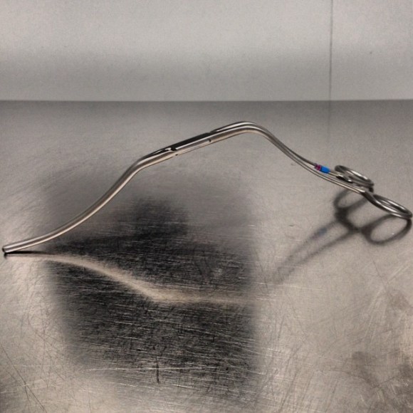
It hugs the contour of the mesentery and liver overhang, and the handles stay out of the way above the wound. It will also tilt up the aorta because of the weight balance. The other option is to apply a transverse clamp, which I will discuss in a later post. The transverse clamp leaves the suprarenal space free of clamp, but can be difficult if not hazardous to apply. The clamp has to be hemostatic and this can be challenging with atherosclerotic plaque -preoperative planning must include planning for safe clamp sites. A suprarenal clamp may be limited by the presence of the terminal insertions of the diaphragmatic crurae. I have recently found that dividing these crurae with a Maryland tipped Ligasure, a laparoscopic instrument I use to dissect the retroperitoneum, makes short order of what can sometimes be an awkward exposure in this tight space. Finally, endarterectomy of the neck should be done carefully to let needles pass without difficulty.

Distortion

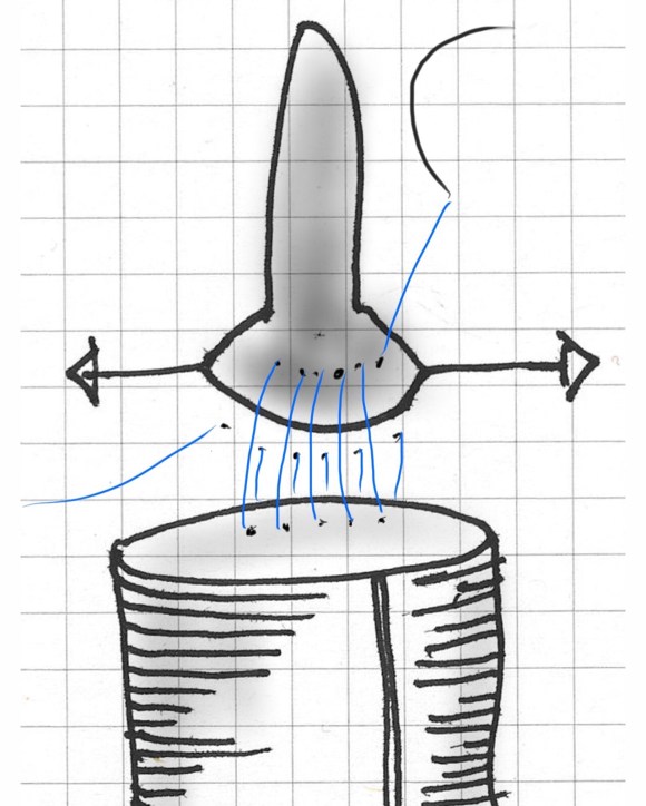
The proximal anastomosis is ideally just another end to end anastomosis -attaching a circle to a circle, but clamping narrows and distorts the circular aortic neck (top illustration). To envision this, imagine the aortic neck being a clock face:

FullSizeRender

Lets say the suture should be applied at each of the hours and half hours. You get ready to sew your first aortic neck and after endarterectomizing some plaque, you get this:

IMG_9886

The important point is that you still have to apply the original plan of applying sutures evenly and at an appropriate frequency (about 3mm apart), to avoid gathers and gaps, especially on the posterior wall. One way is to apply outward tension with a Wheatlander retractor in the aortic sac.

IMG_9873
The posterior wall of the aorta should be distracted to avoid distortions that create uneven gaps between passes of the needle. 
The bites on the aorta should be generous, on the graft, less so. As long as the gaps between the sutures is the same on both aorta and graft, you shouldn’t get leaks.  The other principle to guide you is the needles should pass pointing to the center of the clock -this is challenging in the Dali clock, but if you pivot your shoulder, your suturing won’t be bullied by the distortions and the narrow space.

The Right Tackle

You don’t go after panfish with a deep sea tackle. Pictured below are a spinning lure for trolling with a large hook and a small dry fly with narrow guage hook for comparison.

IMG_9889

Which hook creates the larger hole in the fish’s mouth? In fact, both lures can be used to catch the same large trout, but in different situations.

OLYMPUS DIGITAL CAMERA
On a lake, large hook, on a stream, small hook for same fish.
One of the techniques which I have borrowed from our partners in cardiac surgery here at the clinic is using smaller guage monofilament and needles. I once co-scrubbed an aortic arch case with Eric Roselli, and was bemused to see him sew graft to the fearsome ascending aorta with 5-0 monofilament suture. Then I saw no needle hole bleeding and was sold. Up to then, I had switched from my 3-0 on SH needles to 2-0 on MH needles -basically the largest vascular needle, and saw posterior aortic wall break down from the needle trauma. Unfortunately, 5-0 suture doesn’t come long enough, the CV needles aren’t big enough to sew posterior wall. I now use 4-0 on SH with 48cm length suture. Ideally, we’d have a 5-0 monofilament on a 60cm suture with a SH shaped and sized needle with the narrow guage of the CV needle.

Also, because the needles are finer and I favor supersized Castro needle holders. The needle holes which represent rents in the aortic wall are far easier to deal with using a smaller needle. Right tackle.


Categories
AAA EVAR techniques training

A Troublesome Accessory Renal Artery Complicating a Complicated Patient

Preop Figure

The patient is an 65 year old man with a growing right common iliac artery aneurysm of 3.7cm, a small AAA, and severe COPD (not oxygen dependent, FEV 1.5L) . He had a prior left nephrectomy for cancer as well as a bladder resection and prostatectomy with an ileal conduit (Indiana pouch or neobladder), with complex abdominal wall closure complicated by infection of Marlex in the past, and prior operations for small bowel obstruction. He is morbidly obese. His kidney function was stable with a Cr 1.5dL/mL, calculated GFR or 44mL/min. His nuclear cardiac stress test (pharmacologic) was normal.

A magnified view of the accessory renal artery is shows below with the arrow

mag preop CT

He needed to have his right CIAA treated but the issues were what to do with his accessory renal artery. Vascular surgery is all about making the right decisions with fall back plans. As with most complicated patients, the options are numerous.

  1. Direct transabdominal open repair
  2. Open retroperitoneal repair –Left sided approach.
  3. Open retroperitoneal repair –Right sided approach
  4. Open debranching right accessory renal artery and EVAR
  5. Parallel graft to right accessory renal artery and EVAR
  6. Coil embolization right accessory renal artery, anticipate worst case postop GFR 20ml/min
  7. Medical management

I informally polled my partners and found an absence of consensus except for rejecting #1, 2, and 7. The first two options were not optimal because of his prior operation and because of the location of his disease. The third option had its proponents, but I felt that the kidney and pouch were in jeopardy from dissection in that area. The open debranching had its appeal for others, but for the same reasons that I rejected #3, I rejected #4 –potential harm to the kidney. #5 may be an option, but in my experience, I have seen too many patients referred for failure of parallel grafts to feel secure about offering it.  #6 would be reasonable if the patient could avoid dialysis. With a calculated CGF of 44ml/min, losing half the remaining kidney would barely leave him off dialysis. By appearances though, the smart money was on losing less than 50% but more than 20%. A 30% loss would result in a GFR of 30mL/min or a Cr of 2.1 which made dialysis not likely. In my experience, the kidney does have some collateralization as evidenced by backbleeding of accessory renals with an infrarenal clamp so it may be that he might lose only 10-15%. I discussed all of these options and medical management with the patient who agreed to proceed with option 5 under my recommendation. My plan was to assess the flow from the accessory renal and proceed if it was small, with plan B being a parallel graft, plan C debranching.

nephrogram

In the OR, the right accessory renal artery was selectively catheterized and a nephrogram revealed that it supplied less than 20% of the kidney. The above diagram shows the extent of the total kidney and the area perfused by the accessory renal artery. I proceeded with coil embolization of it and the right hypogastric artery and EVAR of the AAA/R.CIAA.

post CT

In followup, the patient had a Cr of 1.7mg/dL, representing about 15% loss of kidney function. As the case was done percutaneously, he only had 1cm incision in both groins, and was pleased with his result. No endoleak was seen (CT above).

The telling lesson about this case is that at the time of initial consultation, my first instinct was to prepare the patient for open repair via a right retroperitoneal approach with debranching of the right accessory renal artery as a fallback position. Open surgery is my fallback as it was the foundation of my training. But experience has also taught me that patients with multiple comorbidities often struggle to recover from big operations even if one particular problem is not prohibitively severe. Finally, having smart partners to bounce ideas off of is a not only a luxury but a critical asset.