

The patient is a young woman in her twenties who developed severe right sided abdominal and back pain about 4 months prior to presentation associated with bouts of bloody urine. Activity and standing exacerbated her pain and inactivity and recumbency relieved it. She gained 15 pounds because of her inactivity. Examination was significant for tenderness over her left kidney. Urinanalysis showed positive proteinuria and hemaglobinuria.
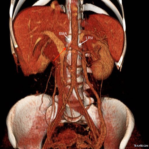
Prior to consultation with me she had had an MR venogram showing compression of her left renal vein by the superior mesenteric artery (nutcracker phenomena). The presence of hematuria, proteinuria, and pain (albeit atypically right sided) made it nutcracker syndrome.

I ordered a renal duplex and a CT venogram for procedural planning.

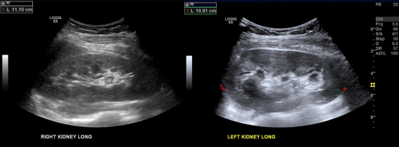
On the duplex, the proximal left renal vein (LRV) was not visualized. The right kidney had normal parenchymal appearance and blood flows, while the left, the kidney appeared distended and had flows consistent with outflow obstruction.

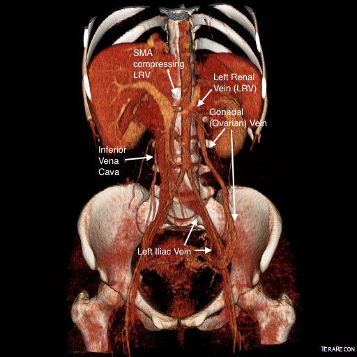
CT Venography showed the gonadal vein to be an important outflow vessel to the left renal vein with dilated proximal segment and reflux into pelvic varices.

A left gonadal vein to iliac vein transposition was planned via a left lower quadrant retroperitoneal exposure. On the table, a venogram was performed with selective access of the left renal vein.

The injection from the LRV showed severe compression of the LRV with a channel only slightly larger than the sheath and avid reflux into the gonadal vein. Selective access into the gonadal vein and venography from a confluence in the pelvis showed that flow was one way from the LRV into the gonadal vein and this filled a large region of pelvic varices.


The gonadal vein was large caliber and refluxed into two large veins in the pelvis. The one that fed the varices was not selected for transposition, but rather the longer straighter tributary. A catheter was left for easier identification during the dissection.
A left lower quadrant incision was made and a retroperitoneal dissection performed exposing the gonadal vein and iliac vein.

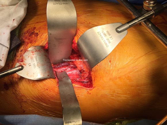
Prior to ligation of the tributaries, a sheath was inserted and through this a LeMaitre valvulotome was brought up to the left renal vein and carefully deployed and pulled back, cutting the valves. This greatly increased the outflow from the vein as evidenced by the height of the blood spout from the vein when the sheath was removed. The varices were ligated at their root -treating them definitively. Transposition was to the external iliac vein, and I could see the feasibility of a laparoscopic or robotic approach to this operation (ref 3).
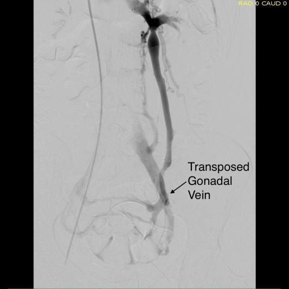
Completion venography showed excellent flow from the LRV down the gonadal vein into the iliac venous system.
The patient lost less than 10mL of blood and was discharged on postop day 2. Gratifyingly, all of her preoperative pain resolved and her UA showed no more hemoglobinuria or proteinuria.
Discussion
The described treatment options for nutcracker syndrome include (ref 1):
- Medical therapy aimed at decreasing renal venous hypertension (for hematuria)
- Renal autotransplantation
- Left renal vein transposition
- Left renal vein to vena cava bypass (autologous or PTFE)
- SMA transposition
- Nephrectomy
- Gonadal vein to IVC bypass
- Exovascular stenting (wrap of renal vein with ringed PTFE graft)
- Endovascular stenting
Many of the operations are of historic interest. Stenting deserves some comment. The patient self referred because she had read multiple reports of cardioembolization on internet support group comments. The largest nitinol stent (self expanding) available is 14mm. Wall stents in larger diameters are available, but are stiff, poorly conformable, and will elongate if constrained by a non-dilating stenosis like the external compression by the SMA. While acceptable results have been reported, the long term results (20-70 years) for younger patients is unknown. Migration is highly morbid, and usually to the heart, requiring sternotomy and cardiotomy to retrieve the stent. Optimally, a conforming 16-28mm self expanding stent should eventually become available, but conformability is typically inversely proportional to radial strength, and it is the less conformable stents that migrate. Work is ongoing to bring larger diameter nitinol stents for venous indications. The difference between May-Thurner Syndrome and Nutcracker syndrome isn’t merely the size of the veins and stents. The iliocaval confluence is relatively static with some movement of the lumbosacral joints and well suited for treatment with the relatively nonconforming Wall Stents. The left renal vein under the SMA is a very dynamic environment with motion of the SMA and the kidneys with respiration, ambulation, and activity leaving stents vulnerable early to migration and later to fracture.
The left renal vein transposition to the IVC is a nice operation with a good track record (ref 2). The downside is the long midline incision required with transperitoneal exposure. There is bleeding risk and postoperative complications of ileus, wound infection, and small bowel obstruction. Looking at the CTV, it seems obvious that the gonadal vein crosses over the iliac vein in the pelvis and would be a straightforward, less morbid, less invasive option. A review of the literature reveals only a single reference discussing three cases of left renal vein transposition (ref 3), and it was done with a surgical robot. I think that a laparoscopic approach would be simpler and less invasive and will consider developing this if volumes justify it. That said, the open retroperitoneal approach is very straightforward and well used exposure. Using venography to set up and then confirm the results of the transposition was helpful. I don’t think that measuring pressures and diameters and taking calipers to calculate stenoses is all that useful and in some instances a harmful method of justifying endovascular treatment of nutcracker phenomena in the absence of serious symptoms and a careful deliberate workup which includes a good history and physical, a UA, a duplex and CTV.
Intervening on the gonadal vein to iliac vein anastomosis should be straightforward from a groin or thigh venous access on the ipsilateral side. This operation doesn’t preclude any future interventions on the LRV. The pelvic varices were treated with direct ligation. The patient’s pain was successfully relieved in the short term.
Conclusion: Open retroperitoneal left gonadal vein to iliac vein transposition with gonadal vein valvulotomy is effecting in treating nutcracker syndrome.
References
- Kurklinsky AK, Rooke TW. Mayo Clin Proc. 2010 Jun; 85(6): 552–559.
- Reed NR et al. J Vasc Surg. 2009 Feb;49(2):386-93;
- White JV et al. J Vasc Surg Venous Lymphat Disord. 2016 Jan;4(1):114-8.
