Categories
AAA Commentary common iliac artery aneurysm EVAR iliac artery aneurysm imaging open aneurysm surgery

When You Pay Your Own Way, You Chose Value

abd angio  11836788149..jpg

The patient is an active man in his 60’s with a history of hypertension who had known about a right common iliac artery aneurysm for several years and had come for an opinion. He was asymptomatic of pain. He had a prior splenic artery aneurysm embolization about a decade prior to presentation.. CT scan showed a large eccentric aneurysm arising from a retrograde chronic dissection dilating the right common iliac artery to over 4cm. This is typically iatrogenic, but impossible to know for sure. The left common iliac artery was ectatic to 2cm as was the aorta to 3 cm and all were “wavy.” This sort of tortuosity is the result of remodeling in the axis of flow resulting in lengthening of the artery and is found in those with the substrate for aneurysmal degeneration (footnote). He did not smoke and he could climb stairs without dyspnea or chest pain.

abd angio  1605093750..jpg

On examination, he was a fit middle aged man with a slight paunch. His abdomen was soft and his peripheral pulses were present and normal. Laboratory results were normal, including creatinine. EKG and echocardiogram were also normal.Treatment options were discussed in detail. The patient was paying for the operation himself and wanted to understand in detail the possible options. These included

1. Open aortobi-iliac bypass with a jump bypass to the right internal iliac artery
2. Open aortobi-iliac bypass with ligation of right internal iliac artery
3. EVAR with right external iliac extension after embolization of right internal iliac artery
4. EVAR with parallel grafts to right external and internal iliac artery (off label)
5. EVAR with iliac branched graft to right internal and external iliac artery (off label)

People are known to react with emotions and to decide typically for near term gain over far term benefits. The offer of an operation involving laparotomy and a possible weeklong hospitalization with all the attendant risks of death, heart attack, stroke, ileus, wound infection, pneumonia, organ failure and so on provides a stark contrast to the appeal of endovascular repair which can be done percutaneously, with local anesthesia, and with a short hospital stay. The lifelong CT scans are in the murky future compared to the present which is sharply in focus. This is why few people save for retirement, why profligate grasshoppers far outnumber industrious ants. In the same vein, the offer of an “advanced minimally invasive” solution plays to several cognitive biases that exists in the mind of not only the patient but the health-care provider. These include this preference for short term gain over long term gain, but also viewing all innovation as being necessarily better than what was available.

After going over the operation in great detail, the patient cut me off when I mentioned the need for lifelong followup CT scanning. Because he lives in several countries, typically, he has to pay for his healthcare out of pocket and he balked at the notion of paying for an annual CT scan. He was also disdainful of the possibility of reintervention (quoted at 10%) and having to pay for it. Also, the stent grafts, which he would have to pay for, end up being as costly as a new luxury sedan based on local pricing.

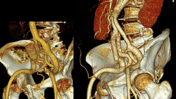
We chose open surgical repair via a midline laparotomy. An aortobi-iliac bypass was performed from proximal aorta to right internal iliac artery and left common iliac artery bifurcation with a jump bypass from the right graft branch to the right external iliac artery. Technically, this sequence was chosen for ease of access to the internal iliac artery with the external clear of graft. Operative time was 3 hours. He was in the ICU for one night. He went home after 10 days after contracting a UTI. In followup three weeks after discharge, because he had fevers, a CT scan was performed (figure). He was treated for an upper respiratory infection which cleared, and he has been doing well since. We are both happy that he will never need a followup CT scan.

Before and After

The literature supports this stance. The long term followup of the EVAR-1 Trial (Reference 1) is an example. This was the late followup of the prospective randomized study looking at open surgery versus EVAR in 1252 patients. The initial EVAR 1 findings are well known and put into question the long term benefits of EVAR as the initial mortality benefit of EVAR is lost within a few years of treatment. At a mean of 12.7 years of followup, there were more deaths from aortic aneurysm rupture and aneurysm related death in the EVAR group compared OPEN surgery (adjusted HR 5.82, p=.0064), with 13 AAA ruptures. I have mentioned before that the failure of a handful of pacemakers drove the Guidant company to recall over 20,000 of their devices. Plus, the cost of annual CT scans in the treatment group and cost of devices and cost of reintervention have driven the UK’s NICE Guidelines recommending against EVAR in elective AAA repair. The meta-analysis of EVAR-1, DREAM, OVER, and ACE trials comprising 2783 patients is confirmatory, that aneurysm related mortality was significantly higher after the initial mortality benefit of EVAR fades away, and that patients of marginal fitness gain no advantage from EVAR, particularly those with heart or renal disease, and those with PAD had lower mortality in the period of 6 months to 4 years (reference 2).

I post this case, because despite a complications, in this case UTI and URI, the patient did fine. So why is open aortic surgery considered a dying art? Why is there such pushback against the UK NICE Guidelines?During my residency in the 1990’s, in the heyday of open surgery, I observed a lot of mediocre vascular surgeons and a very few great ones. The average vascular surgeon would take 6-8 hours to perform open aortic surgery, and the patient would come out with a typical picture of oliguria, third spacing, SIRS, that would generate a 1-2 week stay that would even be described as the normal and expected course for AAA repair in surgical and critical care textbooks. The best surgeons back in the day did these operations under 3 hours with 100mL blood loss and the patients would spend a day in the ICU (often not needing it), and 3-7 days in the hospital, but they were the exception.A higher percentage of surgeons today can do EVAR well than surgeons 25 years ago could perform competent open aortic surgery. Most surgeons graduating from training rarely see or do open aortic surgery compared to the multitude of interventions. Capable open surgery basically is not available outside of a few centers, and most surgeons admit to not having equipoise to start a new trial with modern devices. Open aortic surgery is a lost art, like growing your own vegetables, dressing your own game, reading cursive script, and dialing a rotary telephone.

It is not for a lack of desire. I have several younger colleagues I have met or interacted with via social media who have an intense interest in gaining open vascular skills. They have organized open skills courses at major European vascular meetings, but I believe that is not enough.The need for exovascular fellowship, the running topic of conversation of older surgeons through the 2000’s, is never as critical as it is now as we see milennia of surgeon-years of experience retiring to golf courses and cottages. The recommendation for preferring open surgery in the younger and fitter patients is sadly out of reach for most patients and surgeons. The same passion in disseminating endovascular knowledge needs to be applied to repair the damage to vascular education by over-relying on and over-prescribing endovascular approaches.

Finally, and sadly, this patient is the exception. When given clear options and outcomes and costs, this patient made a rational decision, choosing value over convenience.

Reference
1. Lancet 2016;388: 2366-2374.
2. BJS 2017;104:166-178

Footnote:

Pearls for finding AAA:
1. Tortuosity of the internal carotid arteries including loops and hairpin turns found in patients particularly smokers implies the present of a AAA until proved otherwise
2. Palpating bounding popliteal or pedal pulses in an older smoker implies the presence of a AAA until proved otherwise. Especially if the medical student can feel these pulses.
3. African-American Females with Diabetes almost never get AAA.
4. Palpate their abdomen

Categories
common iliac artery aneurysm CTA Endoleak EVAR iliac artery aneurysm techniques

The Geometry of Parallel Grafts in the Iliac Arteries

The development of metachronous common iliac artery aneurysm, or progression of them, after prior treatment with EVAR (endovascular aneurysm repair), particularly with “bell bottoming” is typically treated with coil embolization of the internal iliac artery and extension of the stent graft into the external iliac artery. While CH-EVAR has been in the news with the recent results from the PERICLES registry, I have never been entirely convinced of its durability. That is different in the case of building parallel grafts in an iliac limb of an EVAR graft (reference).

Here, the geometries, thrombosis, and forces combine to make gutter flow and endoleak unlikely. Choosing the right size of stent grafts to channel to the external and internal iliacs seems to be a challenge, but is easily solved by this scheme -which I can’t claim as my own, but was thought up by a surgeon in upstate New York who choses to remain anonymous.*

The diameter of the stent graft to be sealed to is measured and an area calculated. The sum of the areas of the two grafts to be placed need to equal or slightly exceed the area of this inflow stent graft. If you have decided the size of the external iliac graft, for example, then the diameter of other graft is merely a few geometric formulas away.

Here is a table that can be helpful in avoiding those formulas.
diameter area table.jpegThe inflow graft area is taken from its measured diameter. Then usually one or the other artery has an obligate size -a size the graft has to be while the other has more “wiggle room.” The other thing that comes from experience is that the AFX graft’s iliac limb extension don’t get the B-infolding that can affect an oversized stent graft placed in a small artery and it accomodates a neighbor well.measurement_3

For example, take this patient who after EVAR of aortic aneurysm with AFX developed metachronous dilatation of the common iliac artery to 3.9cm with abdominal pain. The average diameter is 18.5mm. From the table, that rounds to 19mm corresponding to 283.53 square mm. If the internal iliac artery requires a 13mm graft, that is 132.73 square mm, the difference being 150.80 square mm. That corresponds to a 14mm diameter graft, but a slightly larger graft is preferred for oversizing. The external iliac artery is 8mm, and putting a 13mm Viabahn (largest available) in that would result in the B-infolding in the 8mm external iliac. Here, I bailed myself out by simply placing a 20mm AFX iliac limb extension, which by virtue of its design is resistent to infolding and tolerant of parallel grafts laid alongside in constricted channels. I found that the AFX iliac limb, a 20-13mm x 88mm length extension well suited for this.

Image-24.jpg

The AFX graft limb seems to adapt to the presence of the parallel “sandwich” graft which is deployed second and ballooned last. In followup, there was shrinkage of the common iliac artery aneurysm sac and no endoleak.

postop_1

postop_2.jpg

Compared to my other parallel graft case treating a metachronous saccular common iliac aneurysm years after an EVAR with a Gore endograft (link), which by table calculation, resulted in 8% oversize in calculated areas, this particular technique with a large AFX graft and an appropriately sized Viabahn seemed to work well the setting of a previously placed AFX graft. It allows one to avoid hypogastric occlusion.

The final option of a femoral or external iliac to internal iliac bypass after extension across the bifurcation to the external iliac artery is still a reasonable choice, although it seems to be receding into history.

Reference

Smith, Mitchell T. et al. “Preservation of Internal Iliac Arterial Flow during Endovascular Aortic Aneurysm Repair Using the ‘Sandwich’ Technique.” Seminars in Interventional Radiology 30.1 (2013): 82–86. PMC. Web. 9 Dec. 2016.

*While these grafts are not FDA approved for use in this manner, many times, with a prior endograft or graft in place, using the currently available and approved Gore Iliac Branch Endoprosthesis (IBE) in this common scenario would still be off label usage of an approved device, and only if it is feasible, which most times is not. For nonmedical readers, many commonly available devices and medications are used off-label, such as aspirin for blood thinning.

Categories
AAA common iliac artery aneurysm iliac artery aneurysm ruptured AAA training

If the odds are against the patient, who is for the patient?

IMG_1484

The first patient, a man in his late 70’s, ruptured in the emergency room at around four in the afternoon on a weekday, which was fortuitous, as the hospital was fully staffed, fully armed. The patient had arrived only a bit earlier with the complaint of severe abdominal pain, and soon after getting his CT, arrested. CPR commenced as I arrived by Dr. Timothy Ryan, our chief resident at that time. 

Ruptured CIAA with CPR 1 -_1
a rupture
The patient was wheeled upstairs with ongoing chest compressions. The anesthesia and operating room staff started a bucket brigade of blood -there was enough staff to start a symposium. Within 5 minutes of hitting the operating, I poured betadine on the chest and belly and took a blade and cut open the abdomen. Blood poured out onto our scrubs and to the floor and our shoes. I pushed my hand into the retroperitoneum, gently sweeping aside the torn tissues and blood clot to feel the hill of the aneurysm. I walked over the slope of the aneurysm and tweedled my fingers around the aorta above the aneurysm. The cross clamp rode my fingers into position around the aorta. The patient, so very dead minutes before in the ED, came back as I began to feel a pulse above the clamp. The patient lived through the operation and the night where grim data -pH of 6.8, lactates in the double digits, four figure LFT’s, kidney failure all predicted a bad outcome. And yet he survived, and a few days later, a second operation to washout and close his belly which had been left opened and packed occurred, and he recovered. We still talk about that day now three years out, and while he thanks me, I thank the whole hospital because I don’t remember speaking very much -the right things just happened around me as we worked, the whole hospital and me.

More recently, while I was finishing up two urgent cases, I got a call that the patient with the leaking aneurysm had arrived from across town and was becoming hypotensive.

ruptured AAA -_1
another rupture
Gratefully, one of my partners, Dr. Christopher Smolock, was rounding that Saturday and stepped in to finish up the last of the two cases while I ran down to the patient, a man in his late sixties, who had arrived in our acute aortic syndrome unit.

IMG_7195 (1)
Dr. Christopher Smolock
We conversed, the patient and I, and he understood what laid ahead. We rolled him up to the OR, and while we were prepping and draping, my fellow, Dr. Francisco Vargas, looked to me gravely and said with certitude, “I think he’s dead.

IMG_7197
Dr. Francisco Vargas
Chest compressions commenced and again, knife in hand, I cut him open from sterum to pubis and got the clamp on. It took 15 minutes of CPR to get a pulse back. I was very pessimistic as during the case, ridiculously bad lab data came back like a pH of 6.9, lactate above 10, no urine.

IMG_7234
Graft Repair of Rupture
The blood bank sent down coolers like the kind you take to tailgates, only filled with blood and plasma. The aneurysm had grown like a rotten apple on a stick and the graft we needed to repair it was surprisingly short. He too made it to the ICU, and after a long recovery which included dialysis, a tracheostomy, and a reboot of the brain -the brain takes a while to recover from the anoxia, but his went “bonnnnng” like a waking Mac after days of spinning beach balls, and he started to follow commands. The morning before he transferred to rehab, we talked about what he could have done to prevent the rupture. Not knowing about it, not much, I replied. People traditionally lived to about 20-30 years of age, I said, before dying of disease, violence, or predation. Longevity has meant wear and tear on irreplaceable parts. We agreed it was good to be alive.

Ruptured aortic aneurysms are the sine qua non of vascular surgical practice. As a junior resident back in the antedeluvian 90’s, I remember one of my chiefs, Dr. Eric Toschlog, now a trauma surgeon out East, running a patient upstairs from the ER with a rupture, and before the attending arrived by taxi, had the graft in. When it became my turn, as a fellow working on a patient who had been helicoptered in from the frozen wastes of Minnesota, I remember playing a trick with my mind -that the patient was proportionally the same size as the rabbits I was working with in the research lab, that I was really big and the patient’s aneurysm very small. This works to calm the heart, steady the hand. Nowadays, my mind is blank, and my hands working reflexively.

There has been a series of papers that create scores that allow prediction of odds for survival, and both of these patients, particularly with their prolonged CPR, have greater than 90% predicted mortality on these measures. In this month’s JVS, Broos et al, in the aptly named paper, “A ruptured abdominal aortic aneurysm that requires preoperative cardiopulmonary resuscitation is not necessarily lethal” describe a 38.5% survival rate among their series of patients with rupture who had CPR (ref 1).

Practically speaking, no one I know would use these scores to decide to not operate. While many series show better survival for emergency EVAR compared to open repair, several randomized control trial failed to show better results when these methods were directly compared. A retroperitoneal approach is preferred by some in our group, but having tried both closed chest CPR with the patient in right lateral decubitus position and open cardiac massage -(both died), I prefer supine.

There is no survival if there is no attempt.

Reference

  1. J Vasc Surg 2016;63:49-54.
Categories
AAA common iliac artery aneurysm EVAR iliac artery aneurysm techniques

The Interrupted Natural History of Aortic and Iliac Artery Aneurysms

graphic

The patient, now in his 90’s, found out about his aortic and iliac artery aneurysms in his early 80’s, had been offered repair, but had refused. Several years ago, one of my partners emergently repaired his ruptured AAA (abdominal aortic aneurysm) via a retroperitoneal approach using a tube graft. At the time of the repair of the AAA, the common iliac artery aneurysms (CIAA’s) were not ruptured and would have added risky time to the repair. He survived and had a postop CT done about two years ago which showed his CIAA’s.

CT 5cm L CIAA 2014.png
Two years ago
The patient chose not to pursue repair of these aneurysms, I assume figuring that at his age, he’d again take the chance that he would pass on without the hassle of another procedure.

He was recently admitted for treatment of another condition, when his physicians noted a large visible pulsatile mass on his lower abdomen.

CIAA

A CT scan was performed. It showed a 13 cm left common iliac artery aneurysm which was responsible for the visible puslatile mass and a large right common iliac artery aneurysm. The left internal iliac artery was thrombosed. His right common iliac artery aneurysm was over 5cm in size.

CT 13cm L CIAA preop

My partner, Dr. Ezequiel Parodi, was consulted for this case. He performed a percutaneous EVAR. The procedure was made difficult by tortuosity in iliac artery and the tube graft in the aorta requiring a secondary access from the arm to straighten out and advance the stent graft (aka body floss).

Dr. Ezequiel Parodi
 
In followup, the aneurysms decreased in size and showed no endoleak around a patent stent graft.

CT postop L CIAA (1)

Common iliac artery aneurysms expand at a rate proportional to their starting size and have increased rates of expansion in those with prior aortic aneurysm expansion (ref 1). Rupture probably signals a tendency to expand rapidly. There is evidence that iliac ectasia and aneurysms left over after tube graft repair (aorto-aortic) of AAA is benign and can be safely observed (ref 2), but these were all small at the start.

I had been trained at the dusk of the open surgical era and the dictum was aortobi-iliac bypasses to avoid future problems with the iliac arteries. With EVAR, and soon bifurcated iliac branched stent-grafts (currently on trial), staged repair of metachronous iliac aneurysms after tube graft repair of AAA has not only an appeal, but some logic as open bypass to iliac bifurcations, particularly in large men, is challenging and potentially morbid. This is a case of a patient who had a large iliac aneurysm that was not repaired initially due to the exigencies of ruptured AAA and had refused planned staged repair. His aneurysm grew from over 5cm to 13cm in 2 years time without rupturing. Such good fortune is very rare.

Vascular surgeons like to collect large aneurysm stories like fishermen talk about big fish. This is the largest unruptured common iliac artery aneurysm I have seen. While it is baffling to many who are in healthcare, it is important to understand noncompliance is common. Denial is a powerful urge, and a uniquely human one.

2014-03-25


References

  1. J Vasc Surg. 2009 Apr;49(4):881-5
  2. Surgery. 2008 Nov;144(5):822-6.