Categories
CTA imaging Uncategorized Venous venous aneurysm venous intervention

3D VR Images from CT Data Very Useful in Open Surgical Planning: Popliteal Venous Aneurysm

ct-3dvr-planning

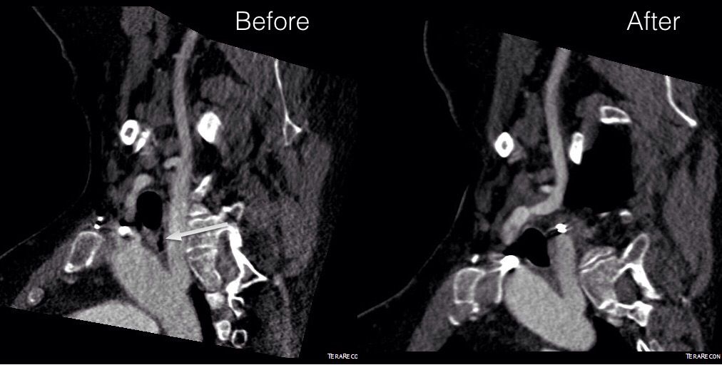
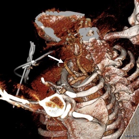
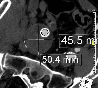
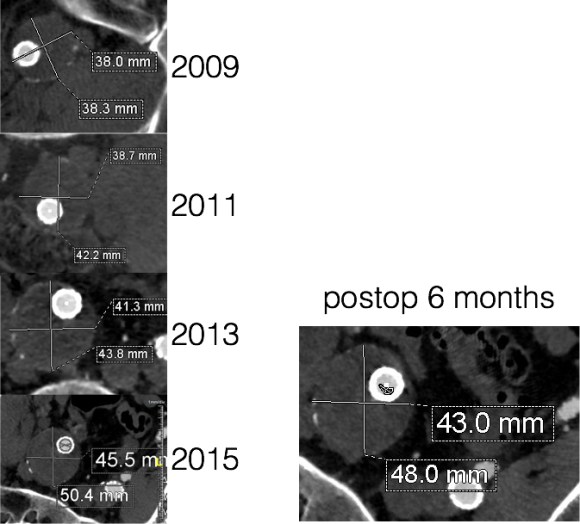
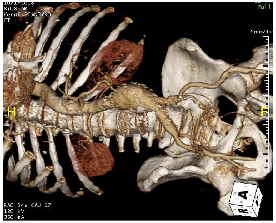
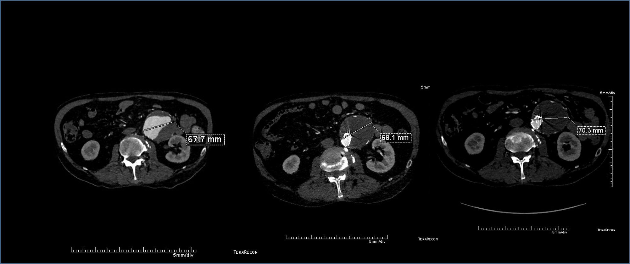
Patient is a middle aged man with history of DVT and PE who in preoperative workup for another operation was found to have a popliteal venous aneurysm affecting his right leg. Unlike the recently posted case (link) which was fusiform, this aneurysm was saccular (CT above, duplex below). Popliteal venous aneurysms have a high risk of pulmonary embolism because: they tend to form clot in areas of sluggish flow and once loaded with clot, will eject it when compressed during knee flexion.

preop-duplex

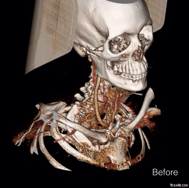
When I perform open vascular surgery, I tend to get a CTA not just because it is minimally invasive and convenient, but because it gives important information for operative planning. The volume rendering function, which takes the 3 dimensional data set from a spiral CT scan, and creates voxels (3 dimensional pixels) of density information and creates stunning images such as the one featured on the current September 2016 issue of the Journal of Vascular Surgery. But these are not just pretty pictures.

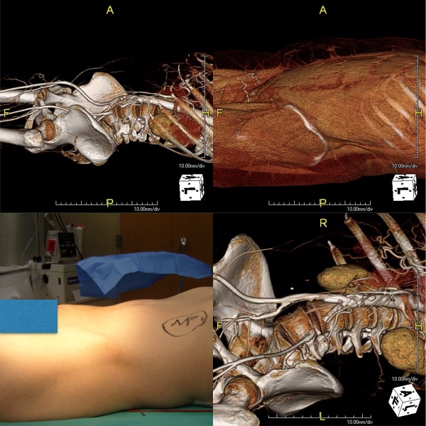
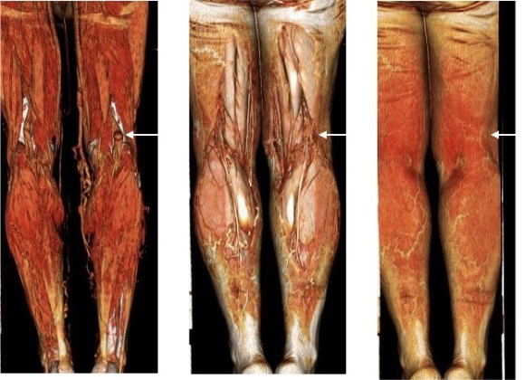
In fact, I use these images to plan open surgery, even to the location of incisions. Vital structures are seen in 3D and injuries are avoided. Take for example the CT Venogram on the panel below. By adjusting the window level, you have first the venographic information showing the saccular popliteal venous aneurysm on the left panel, you can also see where it is in reference to the muscles in the popliteal fossa. The greater saphenous vein and varicose veins below are well seen.

ct-3dvr-planning

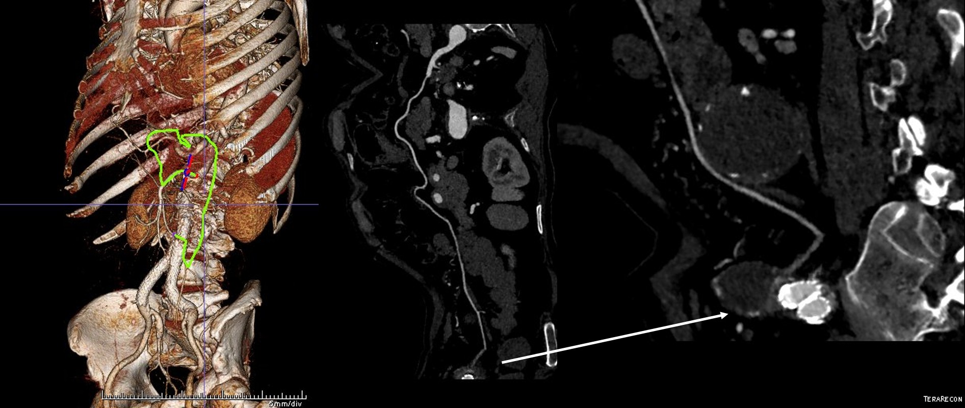
By adjusting the level, subcutaneous structures are better seen including the small saphenous vein which could be harvested to create a patch or a panel graft from a posterior approach. A final adjustment of the window level on the right shows the skin, and I can now plan the curvilinear incision.

By changing the orientation, I can also recreate the surgeon’s eye view of the leg in the prone position (below).

or-view_1

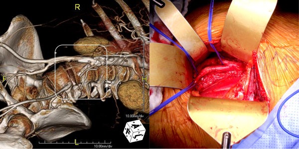
And you can see how well it matches up to the actual operation shown below:

Intraop Photo.png

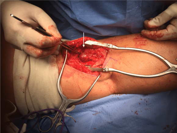
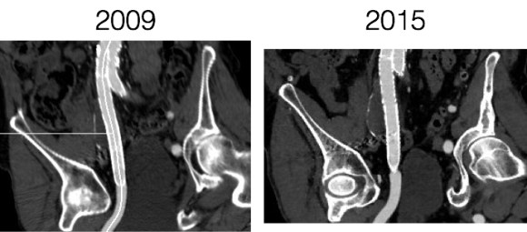
This was treated with plication of the saccular aneurysm and unlike the fusiform aneurysm, I did not sew over a mandrill (a large 24F foley) inserted through a transverse venotomy, but rather ran a Blalock type stitch under and over a clamp.

sketch15473555

The several weeks postoperatively showed no further trace of the saccular aneurysm.

postop-duplex

The volume rendering software grew out of the 3D gaming industry. The voxel data that paints flesh and bone on skeletons and costumes and weapons is far more complex than what is applied for the 3DVR packages that are available. The images shown for this post comes from TeraRecon/Aquarius, but they are also available as open source software from Osirix, Vitrea, and various software packages sold with CT scanners. While those that are tied to the scanners are often tied to dedicated workstations -limiting you to going to Radiology and taking over their workstation, many will work in the cloud for both the DICOM data and for virtual desktop access through mobile. Contrast is not necessary if the patient has kidney dysfunction -the vessels can be manually centerlined -ie. a line can be dropped in the center of the artery to illustrate its course when viewing the VR images.

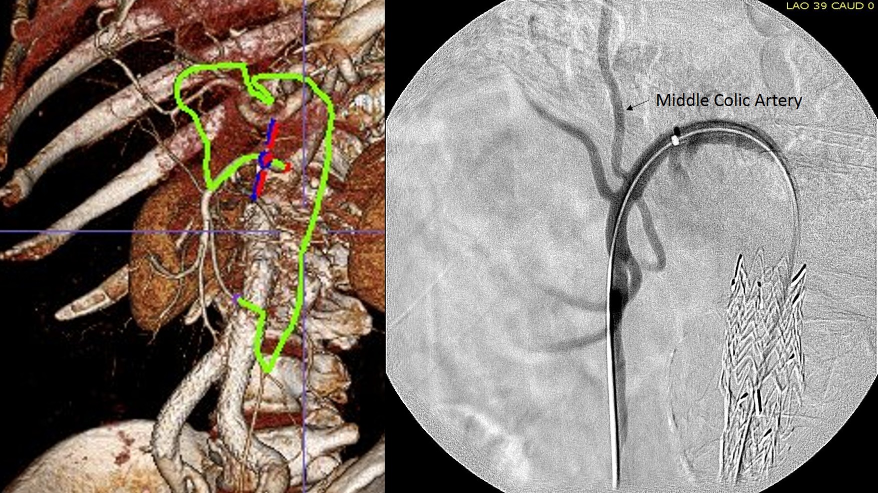
I will plan the surgery while in the clinic with the patient, actually tracing out the incisions and dissections necessary to achieve success. It is a wonderful teaching tool for trainees. But most critically, it helps me imagine the operation and its successful completion.

Categories
Aberrant right subclavian artery Carotid CTA Dysphagia lusoria imaging

Dysphagia Lusoria -a simplified approach

Arrow points to the esophagus. Tension is maintained by the tether on either side of the esophagus. By releasing one side, the tension is relieved.

The patient had been suffering with dysphagia for over a decade and had had extensive head and neck work up which found a goiter. Medical treatment of this goiter failed to relieve the lingering sensation of food getting stuck and the constant feelingof choking. It was only after a search for mediastinal sources of dysphagia that an aberrant right subclavian artery was found. 


One of the advantages of working at the Clinics (I was a fellow at the Mayo, and currently on staff the the Cleveland) is that the infrequent is common while the common is rare. Recently in clinic, I had not one but two patients with dysphagia lusoria. It was the observation of Dan Clair’s,  chairman emeritus, that by simply transposing the aberrant and yet nonaneurysmal right subclavian artery, the tension on the esophagus and trachea are relieved. Or as the dictum might go: it takes two hands to garrote someone


The question is then what to do with the stump? The natural history of the untreated stump is unknown but may be more benign than one might assume. It certainly doesn’t degenerate into an aneurysm all the time -chest CT’s are fairly common and when these are discovered, they are not usually aneurysmal like persistent sciatic arteries which present typically as aneurysms with thromboembolism. Perhaps because we don’t sit on the subclavian artery as we would on a persistent sciatic artery that these aberrant right subclavian arteries don’t degenerate. 


The old fashioned way I learned to treat these aneurysms (Kommerell Diverticula) was through a high thoracotomy and short graft repair of the aorta, replacing the origin of the diverticulum.  This is a dangerous operation for  an older, sicker, and often cachectic patient. The more recent reports involve a left carotid subclavian bypass or transposition and TEVAR after a right carotid subclavian revascularization. This second step may be unecessary if the non-aneurysmal stump proves to be benign. I don’t recommend coil embolization of the stump as mass effect of packed coils adjacent to the esophagus can cause dysphagia to recur, and this may necessitate an open resection and repair (observation, DC). 

The patient underwent a successful right carotid subclavian transposition and had immediate relief of her dysphagia for the first time in over a decade, especially because she had been told she may have been imagining the discomfort. Kudos to her physicians who ordered the CT of the chest that discovered her arch anomaly. Follow up at 6 weeks showed a stable subclavian stump and patent transposition (images above). My plan is for regular interval CT’s with increasing intervals as time passes. 

Categories
complications CTA tbad techniques TEVAR type b aortic dissection visceral malperfusion

Reversing paralysis with a bypass

Dissection CTA

The patient is middle aged and had a type B thoracic aortic dissection (TBAD) as a consequence of recreational substances that acutely raised his blood pressure. At the outside hospital, he had a CTA showing the dissection extending from his left subclavian artery and causing occlusion of his superior mesenteric artery (SMA). He developed abdominal pain and was swiftly transported to our acute aortic syndrome unit. He was taken to the operating room and underwent a TEVAR of the dissection and stenting of his SMA -this is similar to other cases I have discussed in prior posts so I am omitting the technical details. The stent covered the left subclavian artery origin to exclude the origin of the dissection. The stent was extended to the distal thoracic aorta but did not go to the celiac origin. 

TBAD post stent

Post procedure, his lactate never rose and he was maintained on the usual post procedure protocol of keeping MAP’s (mean arterial pressure) above 80mmHg. His left subclavian artery was covered but I do not routinely bypass, especially when the left vertebral artery is at least equal in size to the contralateral one. I don’t often place spinal drains for urgent/emergent cases particularly in patients who have never had infrarenal aortic surgery and patent hypogastric arteries. He was kept sedated overnight and awoke in the morning unable to move his legs to command. He had no pain sensation up to his umbilicus.

A spinal drain was emergently placed and his blood pressure was raised to MAPs of 90+, but these failed to reverse his paralysis. After discussion among my world class partners, I chose to take the patient back for a carotid subclavian bypass which was done through a single incision with a dacron bypass graft.

Carotid subclavian bypass CTA

His paralysis resolved. He was discharged home, ambulating without assistance. Spinal cord complications are reported to occur between 1-5 percent of patients undergoing TEVAR for complicated TBAD. They were seen in 2 of 72 patients in the TEVAR arm of the INSTEAD trial (Circulation, 2009 vol. 120(25) pp. 2519-28), and was permanent in 1. While there are some who routinely place prophylactic drains, it is unclear to me that they have a significant effect if placed unselectively. I will place a Preop drain in the instance of infra renal graft, hypogastric arterial occlusive disease. In the instance of a dominant left vertebral, I will perform concomitant bypass, but just as often not. This is a gratifying and rare outcome of paralysis reversed with a carotid subclavian bypass when spinal drain and permissive hypertension did not. 

Categories
AAA CTA EVAR open aneurysm surgery techniques training Uncategorized

Never Stop Following Stent Grafts -Type IV endoleak causing slow growth in 12 year old stent graft

Centerline


The patient had undergone EVAR for bilateral common iliac artery aneurysm with the original Gore Excluder stent graft a dozen years before with coil embolization and extension to the external iliac on the larger side and femoral to internal iliac artery bypass on the other side. A coagulopathy, one of the clotting factor deficiencies, had made him high risk for bleeding with major open surgery. His aneurysms never shrank but remained stable and without visible endoleak by CT for a long time resulting in ever longer intervals between followup.

2013_1
2013

Between 2009 and 2013, there was subtle enlargement on the embolized side without a type I or type III leak, and the patient was brought back a year and a half later, with further growth of the sac.

1-2015_3
2015

This was a relatively rare type IV endoleak that was causing sac enlargement due to excessive graft porosity of the original Excluder’s graft material. Its treatment is either explantation or relining. We chose to reline the graft with an Excluder aortic cuff at the top and two Excluder iliac limbs.

2015-11-26 13_25_23

This was done percutaneously and in short followup, there has been stabilization and even some reduction in the aneurysm circumference.

CT Scans


It was long known that a certain percentage of PTFE grafts “back in the day” would sweat ultrafiltrated plasma. The relative porosity of the grafts allowed for transudation of a protein rich fluid.

Slide2
Tanski W, Fillinger M. J Vasc Surg 2007;45(2):243-249.


This results in a hygroma formation. I remember seeing this in AV graft fistulae back in the 90’s -after flow was introduced, the grafts would start sweating! The newer grafts are lower porosity and this is seen very infrequently. Drs. Morasch and Makaroun published a paper in 2006 comparing parallel series of patients who received the original Gore Excluder (OGE), the currently available Excluder Low-Permeability Device (ELPD), and the Zenith device (ZEN). Sac enlargement occurred in equal measure between OGE and ZEN but zero was reported for the ELPD.

Slide3
Haider S et al. J Vasc Surg 2006;44(4):694-700.

The ELPD had higher rates of sac shrinkage than the OGE, and equal rates of sac shrinkage compared to ZEN.

Slide4
Haider S et al. J Vasc Surg 2006;44(4):694-700.

The diagnosis in my patient’s case came about through serial followup through a decade. While I doubt that the aneurysm would have ruptured in the same way as in a Type I, II, or III endoleak, I am sure it would have progressed to developing symptoms from aneurysmal distension or local pelvic compression.

Is it possible to visualize this kind of endoleak at the time it is suspected? I came across a case series from the Netherlands using Gadofosveset trisodium which takes longer to clear than the usual Gd-based MR contrasts and they successfully visualized transudative leaks in 3 serial patients with the original Excluder graft.

Slide2
Cornelissen SA et al. J Vasc Surg 2008;47(4):861-864.

The problem is that Gd-based contrasts have toxicity, especially for patients with poor renal function. The protocol is time consuming. And I suspect that ten years out, a lot of grafts will have positive findings, especially cloth based grafts that are sutured to their supporting stents, without clinical basis for treatment as their sacs size are likely stable on a year to year basis.

That said, as we are well into the second decade of commercially available stent grafts, it is even more important than ever to continue lifelong followup even for what is assumed stable, patent grafts and anatomy.

Categories
AIOD CTA imaging techniques

3DVR -Very Helpful in Planning Open Surgical Cases

3DVR CIA Endart

The images above show a patient with on isolated occlusion of his left common iliac artery. He was young, in his forties, but was a heavy smoker and suddenly developed claudication of his left leg which interfered with his work. He quit smoking and did not progress with exercise. Discussion involving possible stenting was made and initially offered but he turned it down because erroneously he assumed that his father’s coronary stents were the same as an iliac stent in terms of longevity. I do think that common iliac and aortoiliac occlusive disease is well treated with stents, but I felt it was possible to do a common iliac endarterectomy. We went over these images together and he settled on proceeding with endarterectomy.

The images show how well the 3D Volume Rendering, which I mentally call Virtual Reality, of CTA makes it possible to plan out operations and exposures virtually. The bottom left image shows the surgeon’s eye view of the exposed vessel.

Below, the virtual and the actual are juxtaposed.

3DVR CIA Endart Exposure

The outline on the virtual image (volume rendered) shows the areas of retraction -for the trainees, the retractor systems work to make quadrilaterals out of linear incisions, and as a rule, the incision should be twice the length of the square that you want to expose. The end points of the endarterectomy were at the aortic and iliac bifurcations.

SAMSUNG DIGITAL CAMERA

The arteriotomy was repaired with a patch at the iliac bifurcation -the common iliac was large and was repaired primarily.

SAMSUNG DIGITAL CAMERA

The specimen below was fibrocalcific. The thing about this disease is that the plaque truly has no endpoint -intimal thickening and mild plaque was present that could be taken all the way to the aortic root and to the feet on the other end!

SAMSUNG DIGITAL CAMERA

This patient did very well and had palpable pulses. He did not develop neointimal hyperplasia and successfully quit smoking.

One of the exciting developments is the ongoing development of wearable virtual reality and display solutions -particularly from the gaming industry. The gaming industry ironically drives all computer imaging because that is where the money is at. The advances in imaging trickle down to medicine -the VR images seen here are the result of the same algorithms that drive first person shooting games. It would be great to see this displayed intraop on a HoloLens, on a virtually positioned screen behind the assistant!

Categories
AIOD aortoiliac occlusive disease (AIOD) CTA EndoRE PAD remote endarterectromy techniques

EndoRE-ABF -an alternative to the EndoABF which is in turn an alternative to the ABF.

  
The patient is 70 year old woman with prior history of smoking who developed severe claudication and near rest pain. She was unable to walk more than 50 feet before having to stop due to severe leg pain. On exam, neither femoral artery pulses were palpable. PVR’s (pulse volume recordings) and ABI’s (ankle brachial index) are shown below.

PVR pre2

PVR’s demonstrate the presence of severe inflow (aortoiliac occlusive disease or AIOD). CTA was acquired and the findings were consistent with the PVR’s.

preop centerline CTA composite

There was diffuse bilateral iliac atherosclerotic plaque with occlusion of the right common femoral artery and left common and external iliac artery. The 3DVR (three dimensional virtual reality) reconstruction image below shows this as well as the abdominal and pelvic wall collaterals feeding the legs around the occluded iliofemoral system.

Pre CTA

Plans were made to perform a hybrid common femoral and profunda femoral endarterectomy, remote external iliac artery endarterectomy (EndoRE), and common iliac artery stenting. The specific challenges to this case was getting into and staying in the true lumen. Typically, this is easiest to achieve from a left arm access with wires being pushed antegrade, but in a smaller person, particularly woman, this increases the chances for access site complications. My plan was to expose both common femoral arteries and get control of the external iliac arteries at the inguinal ligament and the profunda femoral arteries at the point the proximal plaque dissipated -typically at the second branch point, and then get micropuncture access of the right iliac system by accessing from the common femoral plaque. This would give me true lumen access, and with a sheath and curved catheter (VCF in this case, but a similarly shaped OMNI Flush catheter would do as well), wire access up and across the occluded left iliac system could be achieved and the wire retrieved from the left common femoral artery. This up and over access with the wire allows for control of the aortic bifurcation and both iliac systems.

I perform EndoRE over this wire -this allows for quick access if the artery is ruptured. To minimize blood loss, I gain control of the common femoral artery in the following fashion -a 4cm segment of common femoral artery is left intact and looped above the inferior epigastrics -this loop is brought out in the lateral lower quadrant of the abdomen so that the loop doesn’t travel distally over the arteriotomy. The second loop adjacent to the arteriotomy is sent through periadventitial tissues behind the artery to keep the loop migrating over the arteriotomy. The arteriotomy is created from the distal CFA (common femoral artery) onto the profunda femoral artery (PFA) where the endarterectomy is started. A separate arteriotomy on the superficial femoral artery (SFA) allows me to divide the plaque and mobilize the proximal segment up to the SFA origin, freeing the CFA plaque in this manner. It also gives me the option to perform EndoRE of the SFA if warranted. The dissected plaque and system of loops which I call the blood lock is shown below:

  
The yellow loops are major control points (the blood lock loop is drawn in the picture above) and the red loops are around smaller branch arteries. At this point, micropuncture access through the plaque core was achieved into the true lumen of the yet patent EIA (external iliac artery, picture below).

 
The right EIA plaque was mobilized with a Vollmer ring dissector, and cut with a Moll ring cutter (LeMaitre).


This allowed for cutting and removal of the plaque. 

  
Up and over access and control of the wire from the contralateral (left) arteriotomy allowed for EndoRE on the other side. The occluded left common iliac plaque was ballooned and wire access into the aorta from the left was achieved. 


Kissing balloon angioplasty was performed with revascularization of the aortoiliac bifurcation and common iliac arteries. 


The stents were extended across the dissected end points of the external iliac artery origins. The arteriotomies were closed with bovine pericardial patches. Because the PFA were of small caliber, to avoid narrowing the distal end of the patch, the patches were sewn over Argyll shunts which also allowed perfusion of the legs during the suturing of the patches. The loops made this a straighforward maneuver. 

The completed CFA to PFA patch on the left is shown below:


Closure involved reapproximating the Scarpa’s type investing fascia of the femoral triangle and a running dermal layer of absorbable monofilament, dressed with a surgical glue. No drains were used, but if needed, they would be exited through the counter incisions created for the EIA loops. 

The patient recovered well. I always use cell salvage -sometimes, profundaplasties can be bloody, particularly if they are in reoperative fields. The ABI’s and PVR’s at the ankles improved significantly.

  The postoperative CTA shows good results as well. Below is the composite right and left centerline from aorta to the PFA’s. 

  
The 3DVR reconstruction images are shown below, with the comparison to preop shown in the first image of this blog entry:

  
The pre and postoperative images of the centerlines (composited) are shown below:

  
EndoABF is an established hybrid procedure involving an open endarterectomy of the common femoral and PFA/SFA with iliac balloon angioplasty and stenting, often taking the stents distally into the CFA and the patch to deal with complex distal EIA plaque. This procedure, which would be an EndoRE ABF, offers some advantages in eliminating the need for EIA stents which are often placed across the inguinal ligament and into the patch during EndoABF. In my experience, the EIA EndoRE performed as an extension of a CFA endarterectomy is safe, and made even safer by performing the EndoRE over a wire. Published results from Europe shows for TASC C and D disease, EIA EndoRE has excellent patency, and I would expect the same here. EndoRE and Endo ABF both offer advantages over traditional ABF, particularly in patients with medical comorbidities. 


Categories
AAA CTA techniques

Using TeraRecon for planning minimally invasive aortic surgery

CTA VR

From my notes

November 7, 2008 9:05 PM

Using TeraRecon for planning minimally invasive aortic surgery

Terarecon, Vitrea, Osirix, all allow for visualization of three dimensional CT data. The 3DVR (virtual reality) view, is often overlooked, but is an important feature of Terarecon. It is a synthesis of the axial data and does for you what you tried to do in your head back in the days of cut axial film -that is reconstruct a three dimensional picture from 2 dimensional sections.  This is a moderate risk patient, 65 years of age, with a 5.8cm AAA. The top image shows the standard 3DVR perspective with the surgeon standing on the patient’s left. By adjusting the levels, you can bring in the organs (not shown), and then the muscles (panel below).

CT SQ

You can then bring in the skin by manipulating the “window levels” -in TeraRecon this is done by pressing both left and right mouse buttons. This allowed me to plan the location of a skin incision (measuring 15cm) for a minimally invasive AAA repair.

CT S

While 15cm hardly qualifies as a mini-laparotomy, it is less than half the length of a “stem to stern” laparotomy.

OR vu

Dr. Jon Cohen et al. reviewed their experience with laparoscopic versus minilaparotomy averaging 8-10cm in length, and found that OR time, fluid given, and length of stay was superior in mini-laparotomy compared to open and laparoscopic assisted repair (ref).

Chart open lap

I would say that learning curve probably accounted for the difficulties with laparoscopic-assisted. In this patient the tube graft AAA took 2.5hrs, and patient was extubated post op and went home in 4 days. TeraRecon made short work of planning out the location of incision and was predictive of the viewing perspectives.

Addendum 11/30/2014

Using the 3DVR perspectives in thoracoabdominal aortic aneurysms is indispensable for planning retroperitoneal thoracoabdominal exposures, and I will post an example.

reference

J Vasc Surg 1999;30:977-84

Categories
AAA CTA EVAR techniques training

Type II endoleak from IMA treated via SMA -concept of building the intervention machine

Picture1

The patient had a successful EVAR or an eccentric infrarenal AAA which in followup grew due to the presence of a type II endoleak from the inferior mesenteric artery. This was seen on the CTA and duplex ultrasound. Planning for assessment and treatment involved analyzing the CTA in centerline, tracking the source of the arterial blood flow into the sac.

Picture2

The centerline from the SMA into the middle colic artery shows a meandering but patent path via the Arc of Riolan to the left colic artery to the inferior mesenteric artery. In my experience this is straightforward to access selectively from the femoral approach, but it illustrates for the trainees the concept of building up access which I refer to as building the intervention machine.

The first step in the access involves getting stable footing in the SMA. Selective access can be performed with a shaped catheter, and once accessed, a Rosen wire is used to track in a curved long sheath. Parking this sheath in the proximal SMA forms the foundation of this machine. The next step is access into the middle colic artery.

Picture3

The CTA is particularly helpful in identifying the middle colic on the 3DVR projection. Selection of this is straightforward with a an angle catheter which I place a Tuohy Borst connector. This is the second stage of the machine, because further access with 0.35guage wires and catheters could result in spasm. This second sheath access (the Tuohy turns the catheter into a sheath) of the middle colic allows for selective 0.18 gauge catheters and wires to make the final step to the IMA and the AAA.

Picture4
Selective access of middle colic artery (left) and later phase showing IMA and endoleak (right)
Picture5
6F Ansel Sheath in SMA, Angled Glide Catheter into Arc of Riolan, 0.18 Glidewire and catheter in IMA
Picture6
NBCA glue used to seal endoleak and IMA

The embolization with NBCA sealed the IMA and the cavity in the AAA sac. This was checked with intraoperative duplex, done with a transabdominal aortic probe.

Picture7
Before embolization

Picture8

Transabdominal aortic duplex is easier on sleeping patient and potentially gives more information than arteriography alone.  The patient in followup had no endoleak and demonstrated sac shrinkage.

The access machine concept is important for planning interventions. Every major branch or turn needs to be crossed by your ultimate access sheath, if you want to avoid having to arduously reaccess those points, and building up a telescoping layer of sheaths is very handy. Every interventional case is done at some distance away from the access point on the skin, and so some though has to be given to how you will build that machine.

With this example, I have shown that you can readily access the AAA sac from the SMA. An earlier article showed iliofemoral access via the hypogastric artery (link). I will give in an upcoming post how this can be done laparoscopically in under 20 minutes.

Categories
CTA imaging PAD techniques training

Intuition Aquarius (TeraRecon) Trick -Applying Virtual Reality to Operative Planning

I have used many different flavors of image post processing software including Osiris, Vitrea, and now Aquarius, aka TeraRecon. But I notice that outside of endovascular planning, people rarely use the virtual 3D reconstructed images (the pretty pictures) for anything other than posting images for publication in JVS, and even there I think we have reached saturation.

I have found 3D reconstruction to be especially useful for open surgical planning, and that is by doing two things. First, on viewing the 3DVR data, I reorient and center on the surgeon’s perspective, using left button to rotate the picture around the zero at the center of the screen, and the right mouse button to grab the whole image and recenter as necessary.

Window Leveling.001
Surgeon’s eye OR view

I then window-level in tissue density -this is done by pressing both the right and left mouse buttons, but you can choose this off the menu.

Window Leveling.002

I can plan the incisions and exposures from any angle -in this case, I can see the saphenous vein and its relative proximity to the CFA to perform an in site bypass to the AK POP. And I see the loci of the tributaries that I may need to ligate.

Window Leveling.004

This is a powerful tool that is often overlooked.