
From my notes
November 7, 2008 9:05 PM
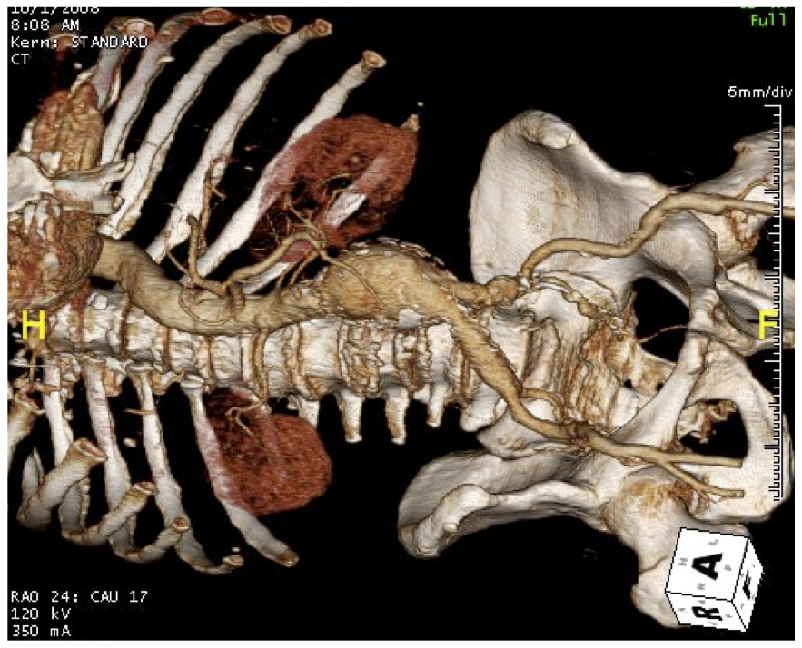
Using TeraRecon for planning minimally invasive aortic surgery
Terarecon, Vitrea, Osirix, all allow for visualization of three dimensional CT data. The 3DVR (virtual reality) view, is often overlooked, but is an important feature of Terarecon. It is a synthesis of the axial data and does for you what you tried to do in your head back in the days of cut axial film -that is reconstruct a three dimensional picture from 2 dimensional sections. This is a moderate risk patient, 65 years of age, with a 5.8cm AAA. The top image shows the standard 3DVR perspective with the surgeon standing on the patient’s left. By adjusting the levels, you can bring in the organs (not shown), and then the muscles (panel below).

You can then bring in the skin by manipulating the “window levels” -in TeraRecon this is done by pressing both left and right mouse buttons. This allowed me to plan the location of a skin incision (measuring 15cm) for a minimally invasive AAA repair.

While 15cm hardly qualifies as a mini-laparotomy, it is less than half the length of a “stem to stern” laparotomy.

Dr. Jon Cohen et al. reviewed their experience with laparoscopic versus minilaparotomy averaging 8-10cm in length, and found that OR time, fluid given, and length of stay was superior in mini-laparotomy compared to open and laparoscopic assisted repair (ref).

I would say that learning curve probably accounted for the difficulties with laparoscopic-assisted. In this patient the tube graft AAA took 2.5hrs, and patient was extubated post op and went home in 4 days. TeraRecon made short work of planning out the location of incision and was predictive of the viewing perspectives.
Addendum 11/30/2014
Using the 3DVR perspectives in thoracoabdominal aortic aneurysms is indispensable for planning retroperitoneal thoracoabdominal exposures, and I will post an example.
reference
J Vasc Surg 1999;30:977-84
