Categories
AIOD aortoiliac occlusive disease (AIOD) BKA bypass CTA EndoRE EVAR graft infection imaging remote endarterectromy

The Unclampable: Strategies for Managing a Heavily Calcified Infrarenal Aorta

Leriche Syndrome -one of those disease names that adds to our work in a way that an ICD codes and even the “aortoiliac occlusive disease” fails to describe. When I hear someone described as having Leriche Syndrome, I think about a sad, chain smoking man, unmanned, complaining of legs that cramp up at fifty feet, pulseless.

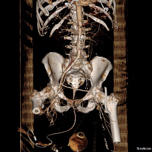
The CT scan will occasionally show an aorta ringed by calcium in the usual places that are targetrs for clamping below and above the level of the renal arteries. Even without the circumferential calcium, a bulky posterior plaque presages the inability to safely clamp the aorta. Woe to the surgeon who blithely clamps a calcified lesion and finds that the rocky fragments have broken the aorta underneath the clamp! The first way to deal with this is to look for ways not to clamp the aorta, by planning an endovascular procedure, but circumstances may necessitate the need to control the aorta despite the unclampability.

The traditional methods of avoiding clamping the calcifed peri-renal aorta are extra-anatomic bypasses including femorofemoral bypass and axillo-femoral bypass. I propose these following options for the consideration when the patient needs a more durable solution while avoiding a heavily diseased aorta.

Not Clamping I:

EndoABF.png
An EndoABF (actually EndoRE-ABF)

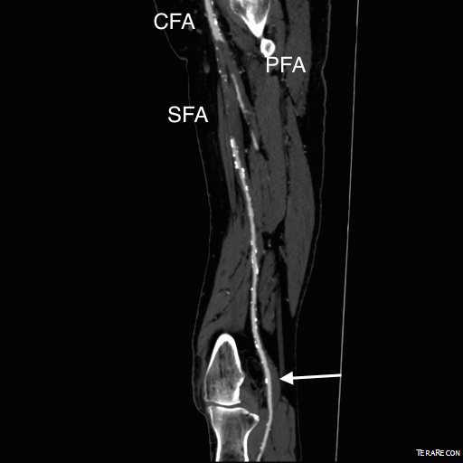
EndoABF does work to avoid clamping -these are common femoral endarterectomies supplemented by stenting of the aortoiliac segment, including in those with appropriate anatomy, a bifurcated aortic stent graft. This is often not possible to treat both sides, but one side is usually more accessible. Often, people will compromise and perform an AUI-FEM-FEM, but I have found the fem-fem bypass to be the weak link, as you are drawing flow for the lower half of the body through a diseased external iliac artery. The orientation of the proximal anastomosis is unfavorable and in the instance of highly laminar or organized flow, the bypass is vulnerable to competitive flow on the target leg, leading to thrombosis.

auifemfem.png
AUI prior to fem-fem bypass for acute aortoiliac occlusion causing critical limb ischemia

The femorofemoral bypass is the option of patients whose options have largely run out. It is made worse when fed by an axillofemoral bypass. Sometimes, you have no choice, but in the more elective circumstance, you do.


Not Clamping II:

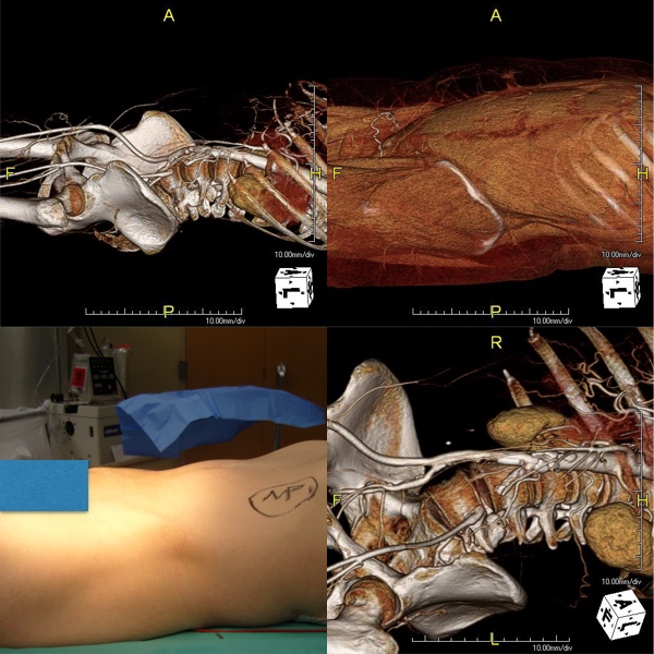
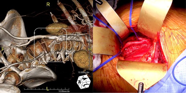
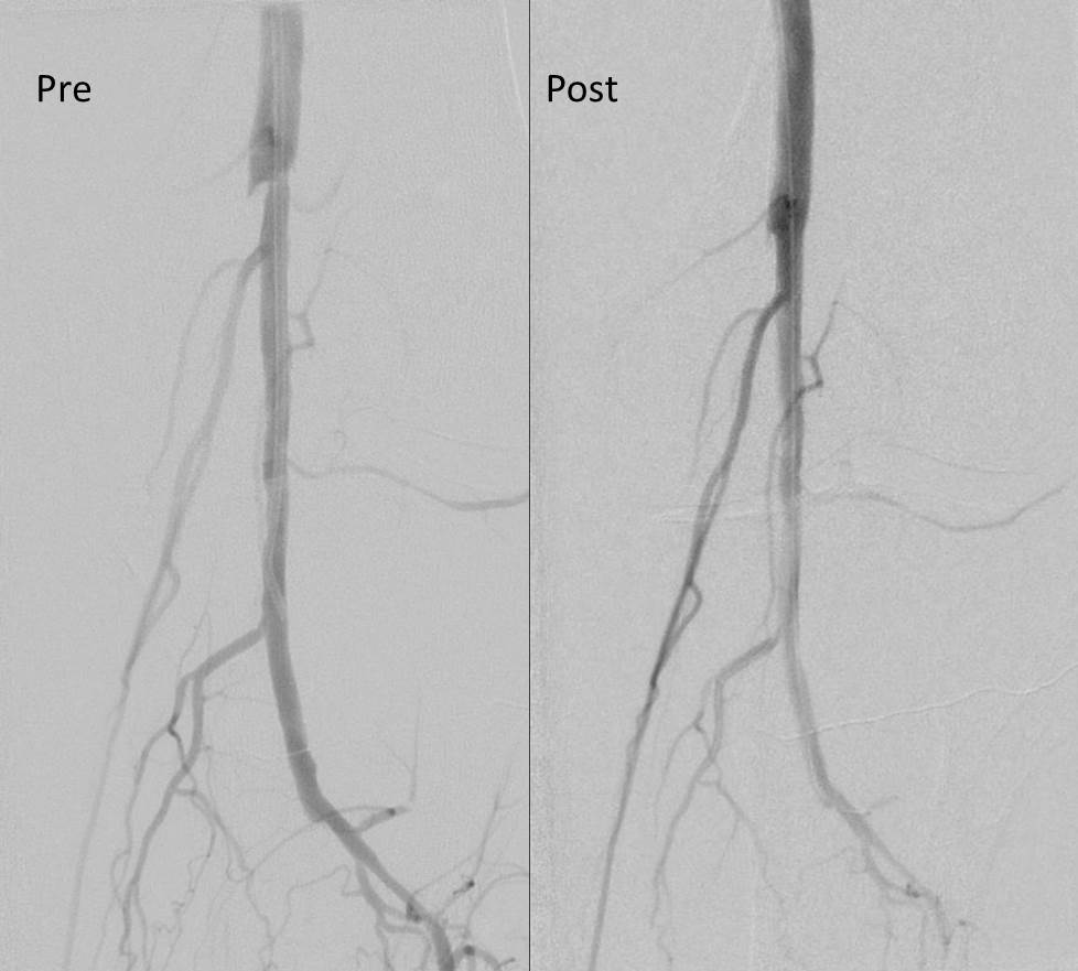
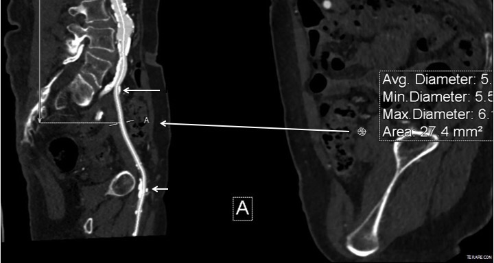
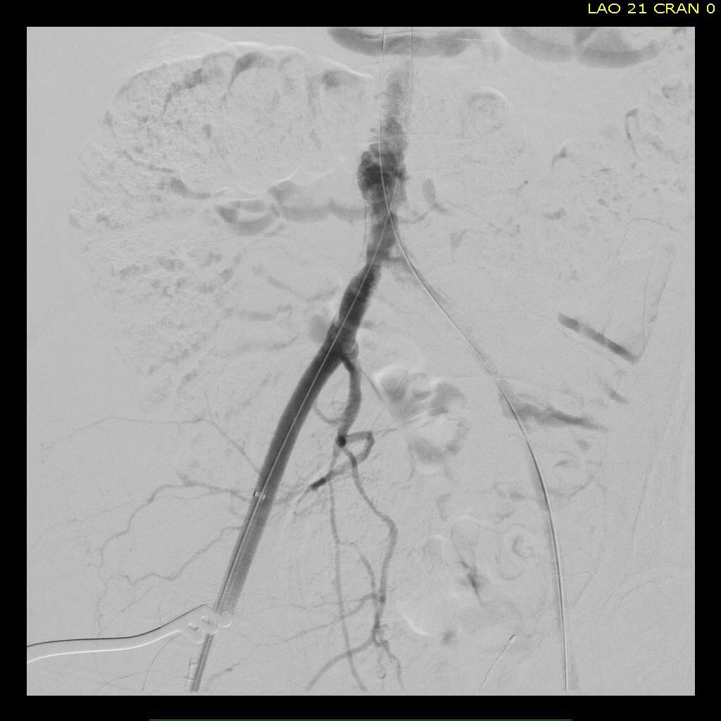
The second method is performing a aorto-uni-iliac stent graft into a conduit sewn end to end to the common iliac aftery, oversewing the distal iliac bifurcation.

conduit.png

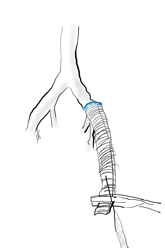
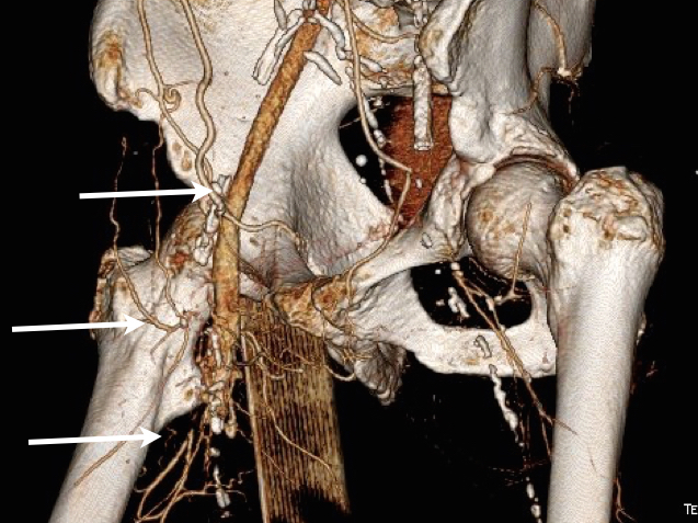
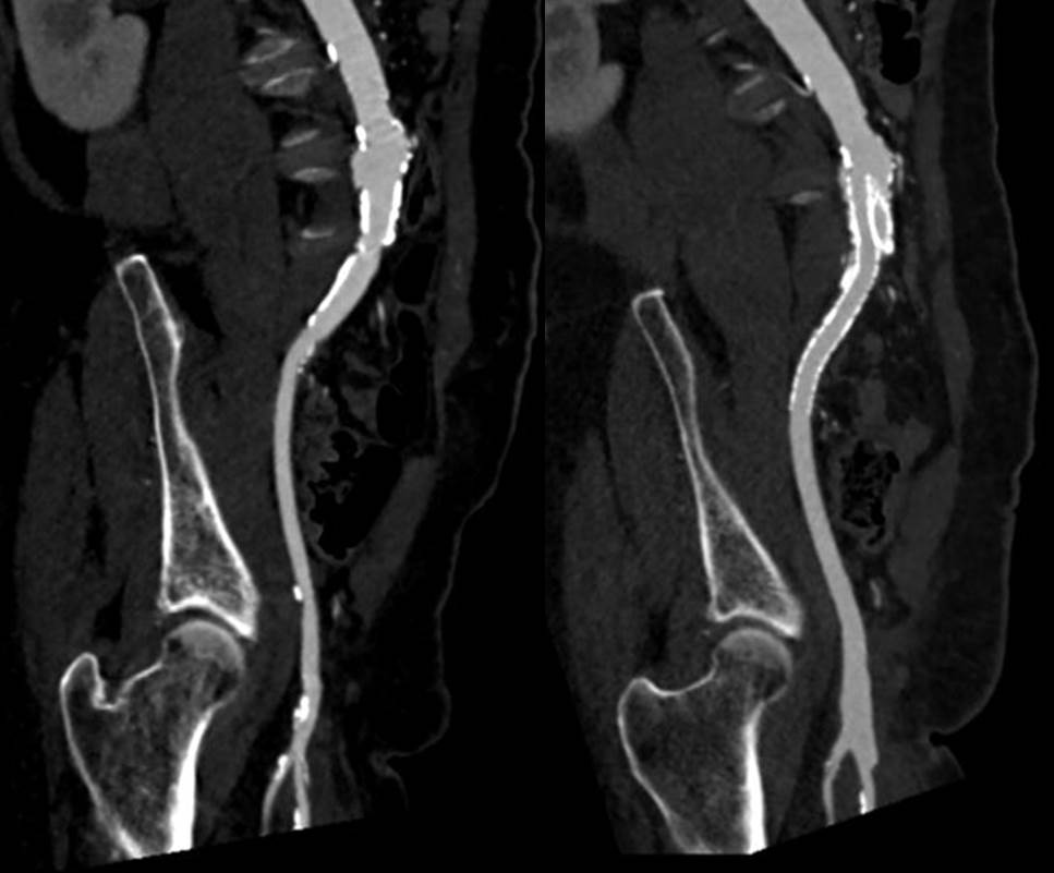
The conduit is 12mm in diameter, the key is to deliver the stent graft across the anastomosis, sealing it. The conduit is then sewn to the side of a fem-fem bypass in the pelvis, maintaining antegrade flow to both legs. The other option is to sew the conduit to a 14×7 bifurcated graft. Illustrated above is this 12mm conduit sewn end to end to the diseased common iliac artery with wire access into the aorta and a aorto-uni-iliac device. Typically, a small AUI converter (Cook, Medtronic) can be used, but the aorta is often too small even for a 24mm device, and an iliac limb with a generous sized docking segment (Gore) ending in a 12mm diameter fits nicely. Below is a CTA from such a case, where the stent graft is deployed across the anastomosis, sealing it off from anastomotic leaks (exoleaks).

AUI fem fem.jpg

Not Clamping III:

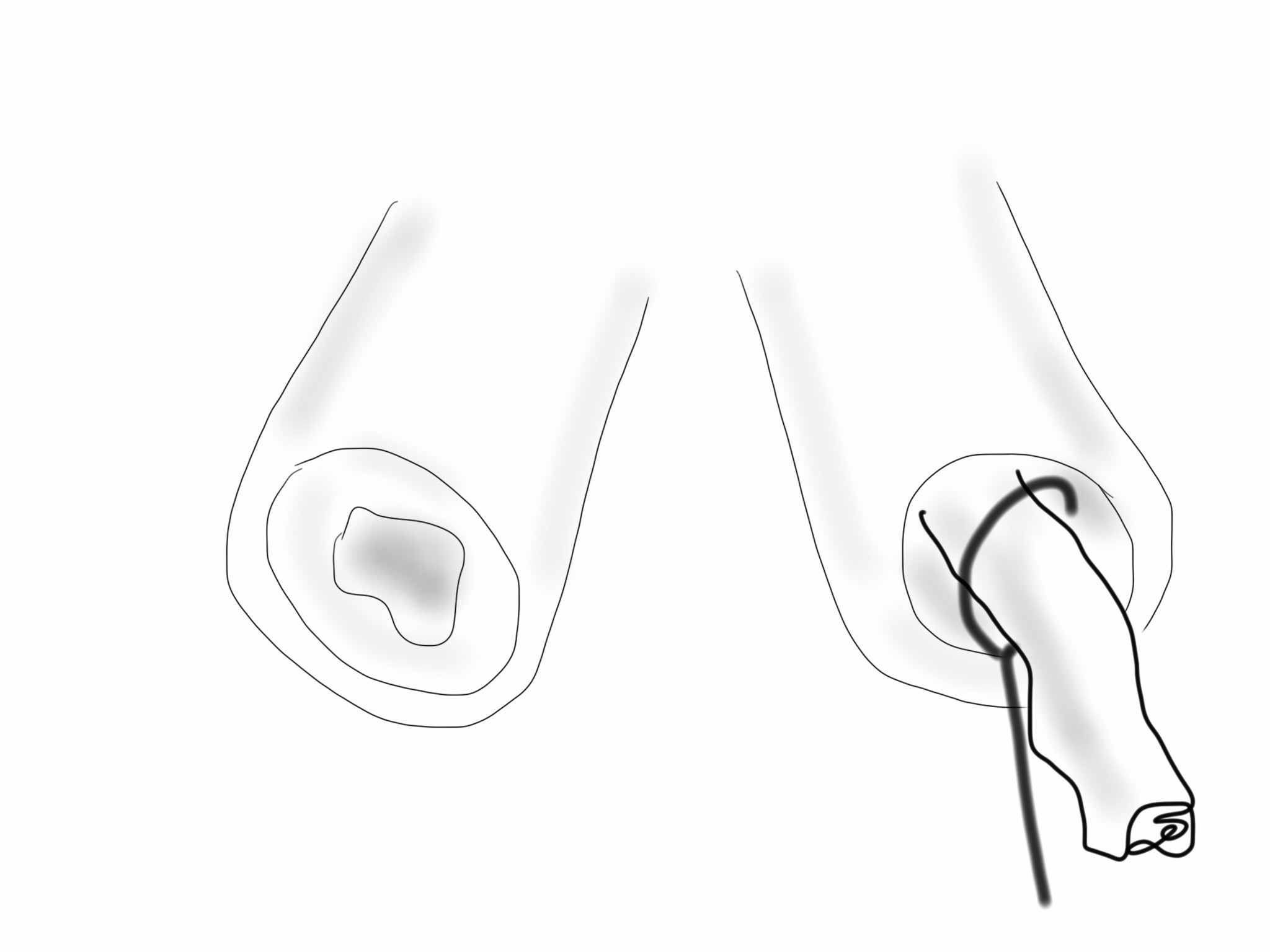
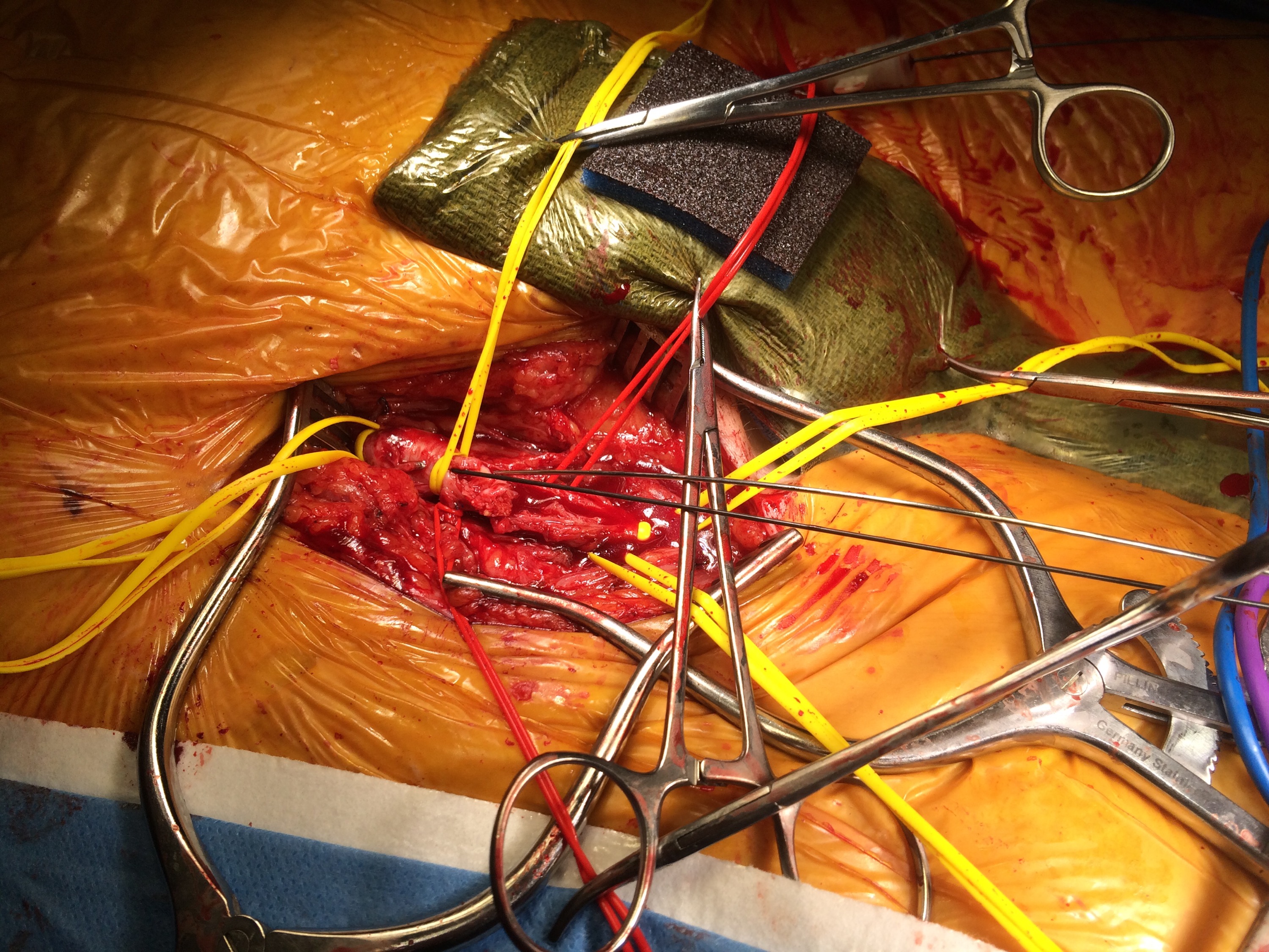
Often, the infrarenal aorta is soft anteriorly and affected only by posterior plaque at the level of the renal arteries. While a clamp is still not entirely safe (I prefer clamping transversely in the same orientation as the plaque with a DeBakey sidewinder clamp), a balloon is possible. I do this by nicking the aorta -simple application of a finger is sufficient to stop the bleeding if you have ever poked the ascending aorta to place cardioplegia line.

conduit1.png

A Foley catheter is inserted and inflated. The Foley’s are more durable and resist puncture better than a large Fogarty. This is usually sufficient for control, although supraceliac control prior to doing this step is advised. The aorta can be endarterectomized and sewn to the graft quite easily with this non-clamp. conduit2.png

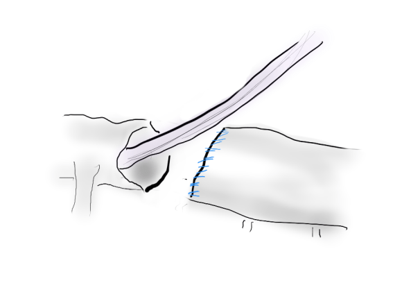
This has worked well, Although pictured above with an end-to end anastomosis planned, it works just as well end-to-side. I actually prefer end to side whenever possible because it preserves the occluded native vessels for future intervention in line.

The Non-Thoraco-Bi-Femoral Bypass

The typical board answer for the non-clampable aorta is taking the inflow from the thoracic aorta or from the axillary artery -neither of which are good options. The first because the patient is positioned in right lateral decubitus and tunneling is not trivial. The second because of long term durability. The supraceliac aorta, technically it is the thoracic aorta, is often spared from severe plaque and clampable. Retropancreatic tunelling is straightforward, and a 12 or 14mm straight graft can be tunelled in this fashion from the lesser sac to the infrarenal retroperitoneum. It then sewn to the supraceliac aorta and then anastomosed to a 12x6mm or 14x7mm bifurcated aorto-bifemoral bypass, of which limbs are tunneled to the groins.

Sketch134215736.jpg

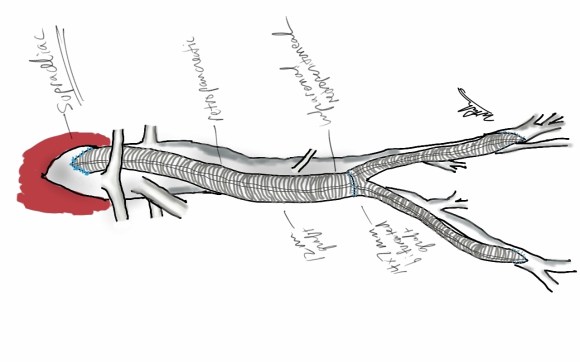
This worked very well recently, allowing a middle aged patient with severe medical problems, occluded aorta and iliac arteries, with critical limb ischemia, survive with minimal blood loss and home under 5 days. It delivers excellent flow to both legs in an antegrade fashion. Dr. Lew Schwartz gave me a list of references showing that this is not novel, but represents a rediscovery as the papers were published in the 80’s [reference], and buttresses the principle that innovations in open vascular surgery are exceedingly rare, largely because we have been preceeded by smart people. 

Conclusion: All of these come about through application of some common sense and surgical principles. The most important this is that the aorta is the best inflow source and reconstructing it with the normal forward flow of down each leg and not reversing directions as in a fem-fem bypass gives each of these options a hemodynamic advantage.


References for Supraceliac Aorta to Lower Extremity Bypass

  1. Surgery [Surgery] 1987 Mar; Vol. 101 (3), pp. 323-8.
  2. Annals of Vascular Surgery 1986 1(1):30-35
  3. Texas Heart Institute Journal [Tex Heart Inst J] 1984 Jun; Vol. 11 (2), pp. 188-91.
  4. Annals of Thoracic Surgery 1977 23(5):442-448
Categories
AIOD CTA imaging techniques

3DVR -Very Helpful in Planning Open Surgical Cases

3DVR CIA Endart

The images above show a patient with on isolated occlusion of his left common iliac artery. He was young, in his forties, but was a heavy smoker and suddenly developed claudication of his left leg which interfered with his work. He quit smoking and did not progress with exercise. Discussion involving possible stenting was made and initially offered but he turned it down because erroneously he assumed that his father’s coronary stents were the same as an iliac stent in terms of longevity. I do think that common iliac and aortoiliac occlusive disease is well treated with stents, but I felt it was possible to do a common iliac endarterectomy. We went over these images together and he settled on proceeding with endarterectomy.

The images show how well the 3D Volume Rendering, which I mentally call Virtual Reality, of CTA makes it possible to plan out operations and exposures virtually. The bottom left image shows the surgeon’s eye view of the exposed vessel.

Below, the virtual and the actual are juxtaposed.

3DVR CIA Endart Exposure

The outline on the virtual image (volume rendered) shows the areas of retraction -for the trainees, the retractor systems work to make quadrilaterals out of linear incisions, and as a rule, the incision should be twice the length of the square that you want to expose. The end points of the endarterectomy were at the aortic and iliac bifurcations.

SAMSUNG DIGITAL CAMERA

The arteriotomy was repaired with a patch at the iliac bifurcation -the common iliac was large and was repaired primarily.

SAMSUNG DIGITAL CAMERA

The specimen below was fibrocalcific. The thing about this disease is that the plaque truly has no endpoint -intimal thickening and mild plaque was present that could be taken all the way to the aortic root and to the feet on the other end!

SAMSUNG DIGITAL CAMERA

This patient did very well and had palpable pulses. He did not develop neointimal hyperplasia and successfully quit smoking.

One of the exciting developments is the ongoing development of wearable virtual reality and display solutions -particularly from the gaming industry. The gaming industry ironically drives all computer imaging because that is where the money is at. The advances in imaging trickle down to medicine -the VR images seen here are the result of the same algorithms that drive first person shooting games. It would be great to see this displayed intraop on a HoloLens, on a virtually positioned screen behind the assistant!

Categories
EndoRE PAD remote endarterectromy

Should the SFA be revascularized during an inflow procedure?

Sketches - 12

The patient is a 70 year old man who arrived with complaints of worsening claudication, worse on the left leg. He smokes over a pack a day. On exam, he only had femoral pulses, nothing was palpable below. PVR showed multilevel disease with an ABI of 0.42 on the left leg.

PVR preop

CTA was done showing that both his SFA and PFA were occluded, along with occlusion of his AT in the mid leg, and tibioperoneal trunk.

cta TIBIAL_1

There is a reconstitution point on the PFA, and there is also SFA constitution. Looking at this, it was apparent to me that it would be possible to endarterectomize the whole of the iliofemoral and femoropopliteal system from a single groin incision, but the question being, would a profundaplasty be sufficient.

Arrow points to calcium free terminus for SFA EndoRE

The textbook answer is profundaplasty, but given my experience with endarterectomy, it has become apparent that removing all the plaque, including CFA and iliofemoral plaque reduces the chance that clamp injury and stenosis occur, and that placed in the common iliac system have better patency than those placed in the external iliac, particularly crossing the inguinal ligament into a patch.

The other observation is that with this exposure, SFA remote endarterectomy is very simple to do, but becomes more difficult in a redo situation. The only problem with going ahead with it is that the runoff is poor -all three tibial vessels occlude, but a very robust posterior tibial artery reconstitutes proximally from well developed collaterals.

The CFA, PFA, and SFA were exposed as shown in my sketch at the beginning of the post. Wire access up and over from the right side allowed for secure control of the aortoiliac segment. The endarterectomy was started from the PFA reconstitution point and the CFA plaque was mobilized. The SFA plaque was transected in a proximal arteriotomy and the plaque was mobilized with a ring to its origin. The CFA plaque then was mobilized with the ring dissector over a wire (for security in case of rupture), up to the EIA origin and cut.

Image-5

The distal SFA plaque was endarterectomized to the planned end point above the knee joint.

Image-4

The specimen is shown below.

specimen

The arteriotomies were repaired with patches. The common iliac artery was stented to improve the flow. The SFA end point was managed with a stent, placed proximal to the first large geniculate collateral.

prepost sfa endpoint

Completion angiograms show widely patent EIA, CFA, PFA, and SFA

Completion

The patient recovered and was discharged on POD#3. His postop ABI’s are shown below.

ABI post2

They are improved compared to preop, with ABI’s of 0.65. Notably, he did have a weakly palpable posterior tibial artery pulse, and multiphasic signals in all three tibial vessels. While I don’t know if the SFA revascularization will stay open, I am confident the PFA will, and this will keep him from his symptoms recurring and is a durable procedure.

Ideally, if he had needed a distal revascularization, a vein bypass would be the answer, but in the setting of inadequate conduit, it is very simple to endarterectomize from the below knee popliteal artery the remaining plaque and either patch to the patent tibioperoneal trunk or perform a short POP to posterior tibial artery bypass. He did not require this.

I don’t know the answer to the titular question, but in the setting of an inflow procedure, the best chance at opening the SFA is during the inflow procedure because of the exposure, and it is very simple to do when the lesion is minimally calcified.

Categories
bypass PAD techniques Wounds

Deep rescue from a hospice: saving a patient from hip disarticulation with advanced hybrid inflow procedure and vein bypasses

PREOP.001

The patient is an elderly man who had bilateral above knee amputations after failure of aortobifemoral bypass grafts at an outside institution. Unfortunately, he had no femoral pulses and his amputation on the right broke down (image above). His left stump had erosion of his femur to the skin with rest pain as well, but was at least covered by skin for now. He was declared too sick for hip disarticulations and was sent to a hospice where he failed to pass away. After a year there, he was sent to us for an evaluation.

He was suffering from rest pain and had complete breakdown of the skin over his amputation stump. More worrisome was the development of gangrenous scrotal and decubitus ulcers which were small but persistent and also foci of pain. CTA showed the following:

PREOP CTA.001

The aorta was occluded below his renal arteries. An AV fistula near his common femoral vein lit up his right iliac vein on the CT above. He had had a prior aortobifemoral bypass but this was occluded. Gratifyingly, it was anastomosed proximally end to side, giving us options. As with any revascularization, we had an inflow source -his aorta, and several potential outflow sources (CTA below, contrast filling iliac vein from AVF’s).

OUTFLOW.001

In particular, his distal profunda femoral artery showed promise. Vein mapping revealed a short segment of basilic vein in his arm to use as bypass, but we needed inflow from the aorta.

I have come to appreciate two things about aortoiliac recanalization. First is that passing the wire antegrade is far likelier to stay in the true lumen at least in the aortic inflow segment -retrograde wire passage inevitably dissects the occlusive aortic plaque and reentry into the true lumen of the diseased aorta is just as challenging as in the leg. The second is vein bypasses have excellent patency in challenging conditions -you just need excellent inflow and an arterial bed to perfuse.

My plan was to cross the aortoiliac occlusion with a wire from the left arm. Once the right iliac system was entered, it didn’t matter if I was in a subintimal plane. The wire could be seated in the common femoral artery to access with a surgical exposure. Once this was done, my intention was to perform remote endarterectomy of the external iliac artery and stent from the aorta to the common iliac artery. The endarterectomized external iliac artery would be the inflow source of a later staged ilio-cross femoral bypass to revascularize his left AKA stump. The common femoral artery at its origin would provide inflow to a short vein bypass to his profound femoral artery.

The wire passed readily into the right iliofemoral system and a groin exposure and common femoral arteriotomy allowed me to retrieve the wire which had been passed from the left arm. A remote endarterectomy was performed over the wire which I do to ensure access in case the artery ruptures (specimen below).

OR IMAGES.001

This allowed me to place a sheath into the right iliac system in the now reopened external iliac artery. Balloon angioplasty of the aortoiliac segment created working space for placement of balloon expandable stents from the infrarenal aorta to the common iliac artery, restoring an excellent pulse in the right groin.

The profunda femoral artery was encased in scar tissue, but following the occluded PFA from the CFA, I was able to expose an open segment and cut it open in the scar tissue. There was back bleeding, and I controlled the artery by placing a small Argyll shunt into the artery and reperfusing it from the recanalized right iliac system.

OR IMAGES.002

The Doppler flow in the shunt was excellent, suggesting great outflow potential. The bypass was performed over the shunt with reversed basilic vein. Completion arteriography showed excellent flow.

PLANNING SLIDE.001

The amputation stump was debrided of dead bone and muscle and the graft was covered with a sartorius muscle flap.

OR IMAGES.003

Before and after images are shown. The remaining open wound granulated well, and ultimately accepted a split thickness skin graft. His scrotal and decubitus ulcers healed as well (below at 6 months post op).

IMG_2380

His left AK stump subsequently degraded while he recovered so three months after this operation, he underwent a right external iliac to left profunda femoral artery bypass with cadaveric vein.

00018205637_20131006_1

00018205637_20131006_6

I don’t like using cadaveric vein, but we really had no options. The right external iliac artery was approached through a right lower quadrant (transplant) incision and a punch biopsy of the artery revealed only normal adventitia on pathology. The EIA was soft and sewed well -essentially a normal artery brought back from the dead. The left profunda femoral artery was large after endarterctomizing its origin and accepted the bypass flow well.

The mortality from hip disarticulation in the setting of gangrene and infection is very high, and I feel that standard approaches to this problem -prosthetic axillo femoral bypasses, thoracobi-femoral bypasses, in the setting of advanced infection and gangrene were unlikely to succeed. In over 1.5 years of followup, everything has remained patent, and the patient lives independently.

Categories
PAD techniques TEVAR

External iliac remote endarterectomy in lieu of a conduit for TEVAR

IMG_1281

The patient had diffuse atherosclerosis with small luminal area even in areas without calcified plaque. It predicted inaccessibility for the 22 French sheath required to deliver the 32mm C-TAG device to be placed for a symptomatic type B thoracic aortic dissection associated with a small but expanding proximal aneurysm.

IMG_1277

My options included direct aortic puncture, an aortofemoral conduit, or an endoconduit. The aorta was heavily calcified and the bifurcation was narrowed by circumferential plaque down to 6-7mm at its narrowest and the left iliac had a severe narrowing due to this plaque. The common femoral artery was severely diseased with a lumen diameter of 4mm due to heavily calcified plaque.

I have come to favor direct aortic puncture over conduits, but the heavily calcified aorta and the absence of safe areas to clamp made me think about other options. My experience with endoconduits has been limited to revising problems of endoconduits from elsewhere, but others report it as a feasible option.

The problem with a long artery narrowed with irregular plaque and even intimal thickening is that it will readily expand to accommodate a large sheath but removing it involves the frictional resistance of the whole artery and typically the “iliac on a stick” avulsion involves the whole length of external iliac artery, likely because the common iliac is anchored by the aortoiliac plaque, the smaller diameter of the EIA, and the longer more tortuous path offering greater resistance in the EIA compared to the aorto-common iliac segment.

IMG_1272

Remote endarterectomy, a technique involving endarterectomizing an artery through a single arteriotomy, offers the possibility of increasing the lumen of even a mildly diseased artery and reducing the frictional coefficient, assuming the remnant smooth adventitia is less resistant than rough irregular intimal plaque.

IMG_1275

The plan was to expose the right common femoral artery and endarterectomize it and gain wire access from the R. CFA. A wire would be placed on the left iliofemoral system to protect it for later kissing iliac stents. A right EIA remote endarterectomy would be performed, and then the right aorto-common iliac segment would be balloon dilated to 8mm.

IMG_1278

The operation went as planned. The external iliac plaque was removed in a single piece from the EIA origin.

IMG_1279

Arteriography showed the right EIA to be free of intimal disease, and dilators and ultimately the 22F sheath went in easily.

IMG_1276

The TEVAR also went uneventully -the left subclavian which had a prior common carotid to subclavian bypass, was covered and the aneurysm and flap were excluded from the left CCA to the celiac axis.

IMG_1280

The most difficult part of the operation was removing the sheath, as is usually the case with a tight iliac, but the friction point was largely at the common iliac and not the external iliac. No artery could be seen extruding with the sheath at the groin while steady tension was applied to the sheath under fluoro. The aortic bifurcation was repaired with kissing iliac stent. The patient recovered well and her chest pain resolved.

I have done this for EVAR, including reopening occluded external iliac arteries, and even for a 26F access for TAVR, avoiding the need for placement of a conduit in selected patients.

Addendum: in followup, I had the chance to check up on the repair -the EIA remained large and patent.

before after