Categories
open aneurysm surgery taaa thoracabdominal aortic aneurysm

Ruptured Thoracoabdominal Aortic Aneurysm In 88 Year Old -a survival

TAAA

The patient, an active 88 year old man, was transferred from an outside institution after a CT scan revealed a 9cm thoracoabdominal aortic aneurysm on workup of sudden onset back pain. On transfer, his blood pressure was stable but low in the 90’s. On arrival, his blood pressure dropped into the 60’s but responded to resuscitation, and after a detailed conversation with him about the risks of emergent repair, we brought him to the operating room.

The CT scan showed an 8.3cm extant III thoracabdominal aortic aneurysm which originated slightly above the diaphgragmatic hiatus and extended to the aortic bifurcation in two lobes. The larger lobe involved the visceral vessels and the infrarenal component was about 5cm.

centerline

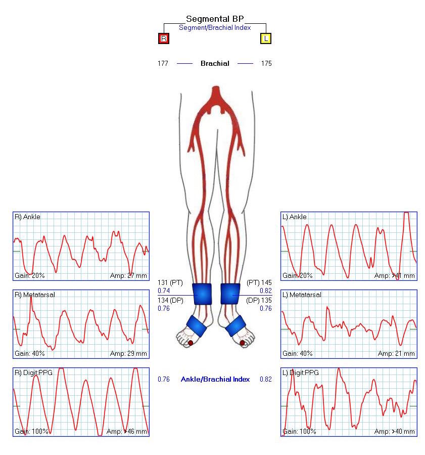
While there was no frank rupture on the CT, the outside report mentioned haziness of the posterior wall consistent with ongoing rupture. Examination was significant for hypotension, abdominal and back pain, and a large pulsatile mass in the abdomen.

centerline 3D

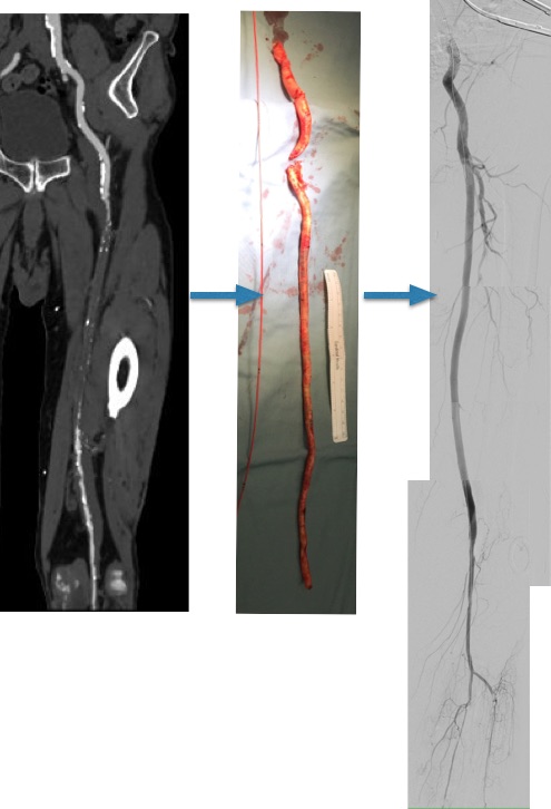
Despite the lack of contrast on this study, I was able to get a centerline reconstruction. The 3D virtual reality view then allows me to plan the operation virtually. The red and blue lines above bracket the beginning and end of the aortic aneurysm with the patient in a right lateral decubitus projection. A thoracoabdominal incision starting on the 8th rib was planned.

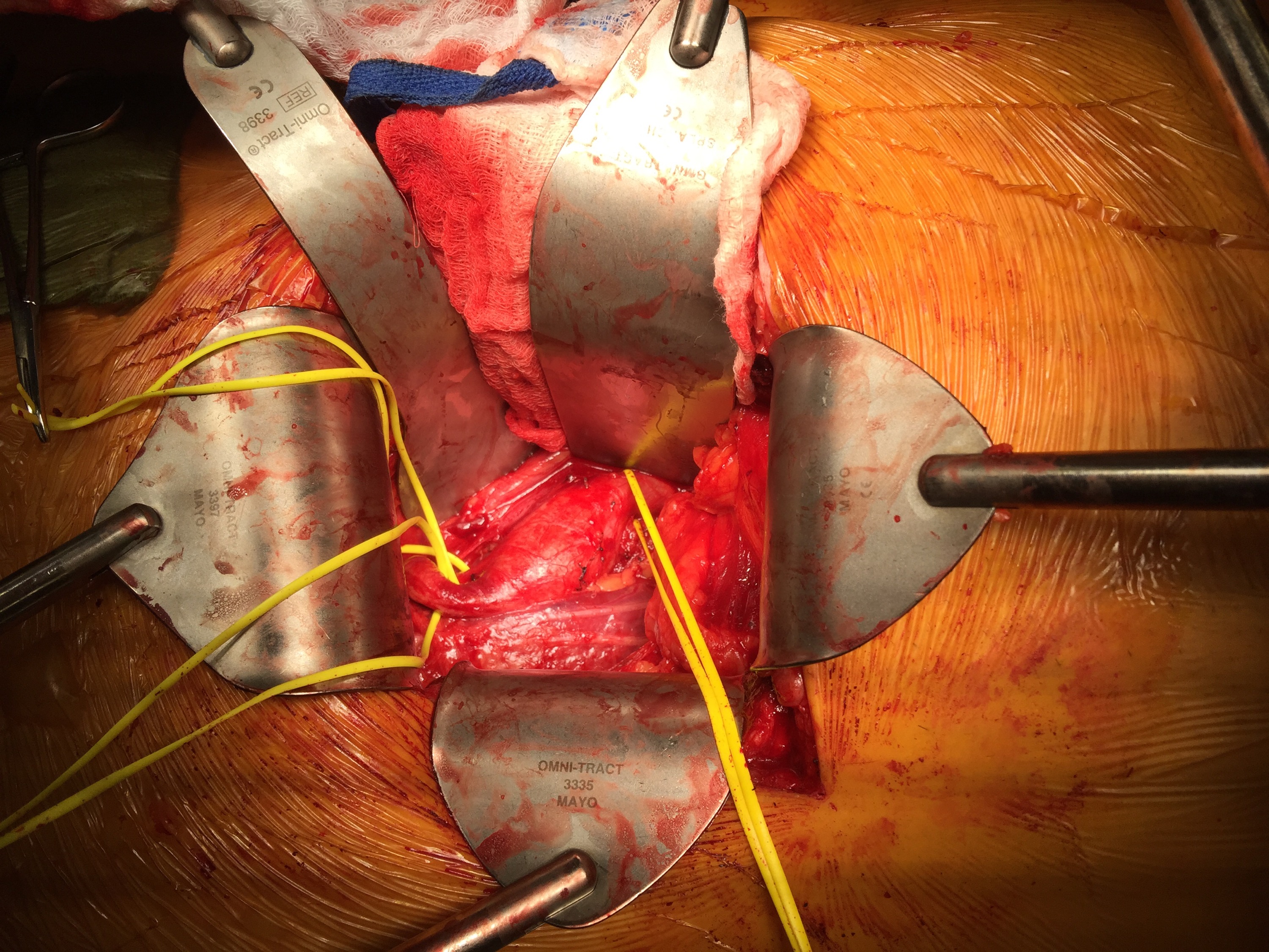
The patient remained stable through the intubation with a dual lumen endotracheal tube. The chest was entered and the left lung collapsed and the thoracic aorta in the chest was controlled for clamping. The retroperitoneum was dissected and the abdominal contents allowed to fall away exposing the remainder of the aneurysm. The diaphragm was taken down circumferentially. The aneurysm was leaking -not frankly but there was blood visible on the surface like a bruised, overripe plum of unusually large size.

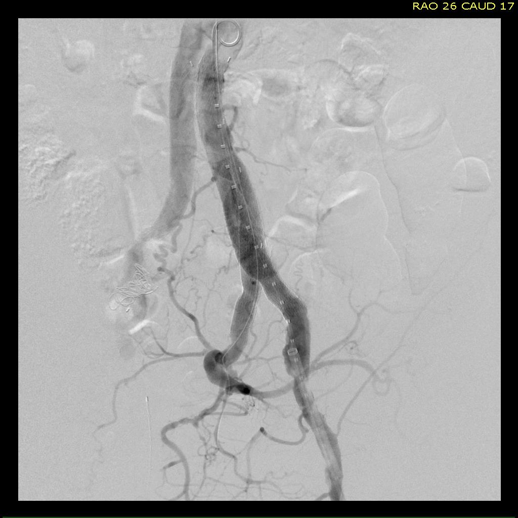
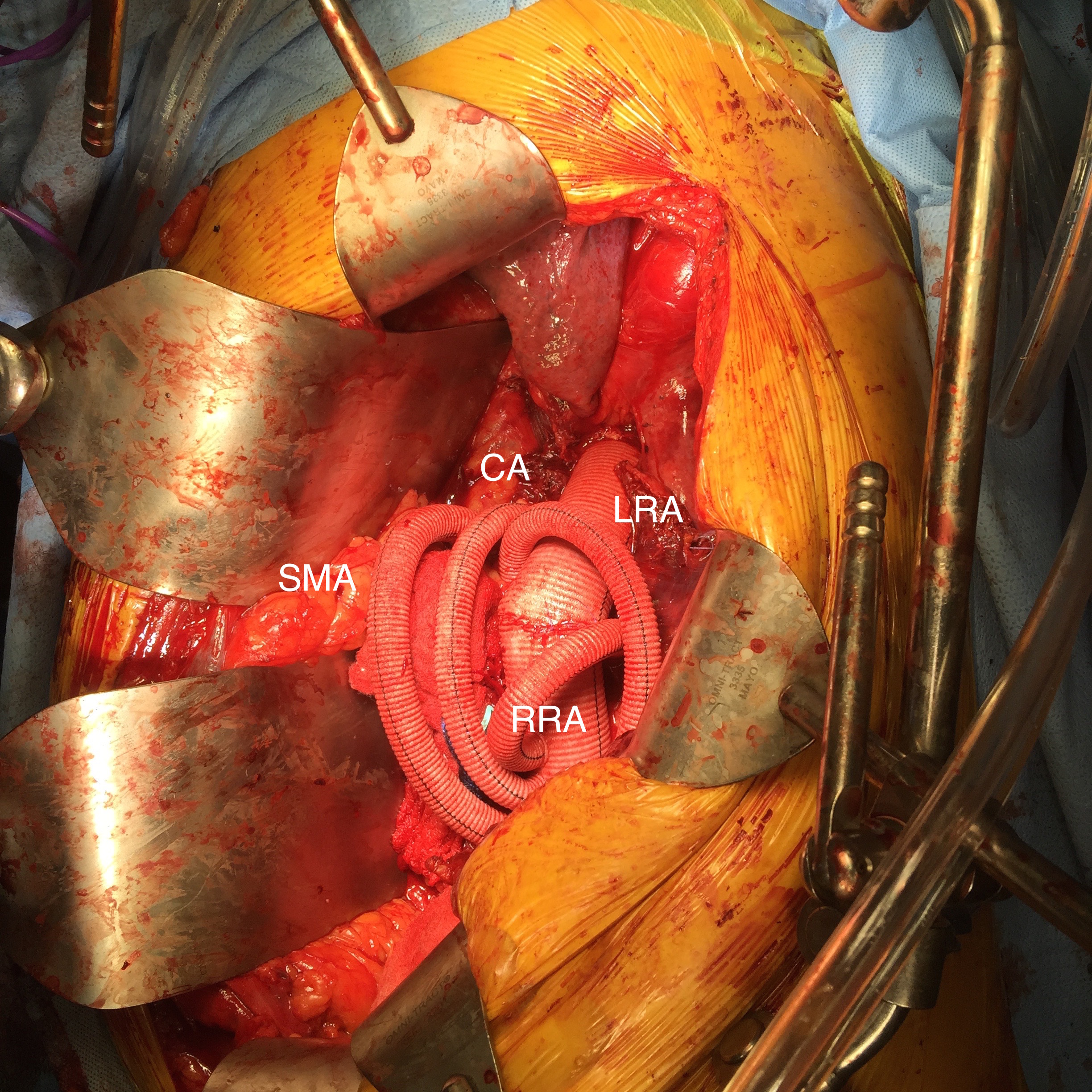
The aorta was clamped in the chest after giving the patient 5000 units of heparin -I often don’t if there is a lot of blood loss and I anticipate factor depletion. The transdiaphragmatic aorta was controlled and the celiac axis (CA), superior mesenteric artery (SMA), and left renal artery were controlled with vessel loops. The aortic bifurcation was controlled as well after I considered anastomosing to the narrow segment of aorta around the renal arteries. While saving the infrarenal aneurysm for later has an appeal, I feel that if you cut the graft and start sewing to the aorta and find that it is not of good quality, you have wasted time. The aortic clamp was moved down from the chest to the transdiagphragmatic aorta which was now mobilized. This avoided for me some spinal cord ischemia but can be a risky move because the aorta was not healthy even in the nonaneurysmal segments. A 32mm Dacron graft what had 4 branches was brought into the field and anastomosed proximally with 4-0 polypropylene suture.

I picked up using narrow gauge suture for aortic anastomoses from my cardiothoracic surgery confreres at the Clinic (Eric Roselli, MD). They will use 5-0 polypropylene with the idea that the smaller needles result in smaller needle holes. I used to use 2-0 suture with an MH needle and have seen my partners be successful at it, often buttressing the anastomosis with a gusset (Dan Clair), but this patient had the tensile strength to take suture well so I went with the smaller SH needle and smaller gauge suture. Other maneuvers include sewing to a strip of Teflon, or in the case of terrible aortic tissues, using interrupted sutures which give some added stability but at the cost of time (credit to Tom Bower).

Time is the killer. While cell salvage gives you some margin for blood loss, this is lost with coagulopathy and hypothermia. The grafts to the viscera were sent from distal to proximal -I feel this greatly eases wire access if needed from a femoral access. There can be a problem with twisting, and I avoid this two ways -by allowing for generous length with looping around the main graft to create forgiveness -closing the retroperitoneum inevitably twists the graft -this I credit to my former partner Pat O’Hara who retired last year. The right renal artery received the first graft while cold saline was given to the left renal artery which was revascularized last. Neither had ostial lesions which I have learned to stent with a bare metal stent directly with the artery open -this I credit to Jeanwan Kang, MD, one of my current partners. The CA graft resulted in great back bleeding from the SMA. The SMA graft and left renal artery grafts completed the visceral segment of the case.

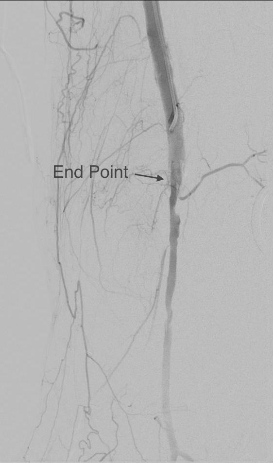
The distal anastomosis was challenging because the bifurcation was heavily calcified. I have to say, the distal often will give me fits when the proximal does not because of the calcium. I generally do perform an endarectomy, but this often results in very poor remnant adventitia. The advice here is be prepared to go distally, but consider that it may add time to the case.

Version 2

The hemostasis was obtained -the most important factor in hemostasis is early and successful repercussion. The wound was restored with repair of the diaphragm, closure of the chest over two chest tubes and closure of the abdomen.

The success of these patients only begins with the operation which I cannot do without the active participation of our cardiac anesthesia, nursing, and trainees -our fellow Eric Shang did his work competently. I am fortunate to have strong help in our vascular intensive care unit. There, my patient was actively resuscitated with blood product, stabilized, and weaned off the ventilator within 2 days. Fortunately, he was not paralyzed by this operation which can happen in up to 10% of patient. Also, his renal function stabilized and he never required dialysis. He was eventually discharged to rehab in under 2 weeks. He returned to my office about a month after the rupture, walked in, accompanied by his family. He was making progress with his rehab, and his wounds had healed well.

Various indices are formulated to predict outcome, which traditionally are viewed as poor for open repair on octogenarians. I am still old fashioned and rely on the “eyeball” test. Several risk stratifying schemes have been published. Most recently, the group from Harborview (link, another link) published a simple stratification scheme for infrarenal AAA rupture. Garland et al (in press) found that having combinations of the following factors predicted mortality well for ruptured AAA including:

  1. Age >76
  2. preop Cr>2.0mg/dL
  3. BP<70mmHg at any point
  4. arterial pH<7.2
Mortality risk based on number 1-4 of positive risk predictors
Mortality risk

If this was a ruptured infrarenal AAA, the patient had two of the risk factors -age>76 and BP<70mmHg, which conferred a risk of 80% mortality for open repair, which translates to a higher number for thoracoabdominal aortic aneurysm repair.

One of our recent aortic fellows, Muhammad Aftab, published the Baylor experience on open repair of TAAA when he was there and found that for open repair, rupture conferred an independent risk for death with a OR of 5.7.

rupture risk table from AFTAB paper

Despite the dismal statistics, several intangibles did favor survival in the patient. He was at 88 still a working professional. He exercised everyday and was fit. He did not drink to excess and never smoked. And he had complete understanding during our preoperative conversation and had a strong grip. And he survived waiting several hours at his hospital for workup and eventual transfer which is a stress test. This last factor accounts for the higher mortality rates for rupture that occurs in hospitals and in places like Seattle where the EMS transport is highly efficient, and better mortality rates at rural referral centers like Mayo where the filtering effect of time leaves a greater proportion of patients likely to survive an operation for rupture.

Reference

Aftab et al. J Thorac Cardiovasc Surg 2015;149:S34-S41

Categories
acute mesenteric ischemia aortic dissection tbad techniques TEVAR type b aortic dissection visceral malperfusion

Acute Aortic Syndrome Unit -TBAD with SMA Dissection and Thrombosis with Acute Mesenteric Ischemia

Figure SMA thrombus with dissection

The figure above shows the summarizes the problem that brought the patient to his local hospital and triggered his transfer to our acute aortic syndrome unit. The concept is that all chest pain of cardiovascular origin gets intake through a vast intensive care unit staffed by cardiovascular intensivists. Stabilization, workup, transfer to operating room or interventional suite all happens in an ICU that encompasses almost a city block.

The patient is an older middle aged man with sudden onset of back and abdominal pain. He was on coumadin for a prior SMV thrombosis and treatment for a ruptured appendicitis -antibiotics with plan for staged appendectomy. CT at his local hospital revealed a type B aortic dissection (TBAD) that extended into his superior mesenteric artery.

Bovine arch in 3DVR view on TeraRecon Aquarius reconstruction.
Bovine arch and TBAD in 3DVR view on TeraRecon Aquarius reconstruction.
The aortic dissection terminated in the infrarenal aorta. The celiac and SMA had true and false lumen perfusion, the right kidney was perfused through the false lumen, the left through the true. Both legs received true lumen flow.

Figure Centerline true lumen compression

The dissection started at the left subclavian artery origin. The false lumen compressed the true lumen up at the proximal descending thoracic aorta. This is an important finding because in this configuration with much of the filling of the dissection from the distal reentry sites, the false lumen acts like a pressurized, compressive lesion. With time, the adventitia around the false lumen may become aneurysmal if the false lumen fails to thrombose or obliterate. When the dissection is acute, the flap may cause a direct obstruction to flow or a dynamic one that depends on the pressure difference between true and false lumen.

Figure SMA thrombus with dissection

In this patient, thrombosis occured in the SMA beyond the origin due to dissection and decreased flow. This was consistent with the patient’s complaint of generalized abdominal pain and examination findings of pain out of proportion to the exam, indicating acute mesenteric ischemia.

His laboratory findings were within normal ranges, indicating this was early in the process. It is important to remember that no lab value correlates with acute mesenteric ischemia except very late in the process, and acute mesenteric ischemia remains a clinical diagnosis (reference 1) that is associated with a high mortality rate.

He was taken to the hybrid operating room. Right groin access was achieved and wire access to the arch was achieved. IVUS (Intravascular ultrasound, Volcano) was used to confirm the location of the wire -I believe this is an important adjunct as simply passing the wire doesn’t guarantee travel up the true lumen.

IVUS confirming true lumen access, and dissection flap compressing SMA origin
A conformable TAG endograft (CTAG, Gore) was delivered through a 24F sheath into position. Two devices were used to cover the thoracic aorta from the left subclavian artery to a position immediately above the celiac axis. The left subclavian was partially covered -the bare stents covering the rest.

Before deploying CTAG

Partial coverage of the left subclavian artery confirmed by persistence of strong left brachial artery pulse

Post Deployment of two 40mmx20cm CTAG (Gore) endografts
This excluded the proximal entry tear of the TBAD. IVUS (image below) showed improved lumenal diameter of the true lumen into the SMA.

After stent graft placement in the thoracic aorta, the true lumen into the SMA expands
Once this was done, wire access into the SMA was achieved. This was technically challenging from the groin, and the backup plan was access from the left brachial artery which had been prepped. With patience, 6French access into the SMA was achieved. The origin was stented with a balloon expandable 8mm x3cm stent -sizing was based on IVUS and CT. This creates an alarming arteriogram as the stent appears oversized on subsequent runs -it is important to remember that the false lumen still takes up space beyond. Arteriography located the thrombosed segment and the reconstituted SMA beyond.

SMA beyond occlusion

Wire access was achieved across the thrombus. At this point, I had a range of options for thrombectomy including simply aspirating which retracting a catheter. This was not optimal as I could lose subsequent wire access or reenter the false lumen. The other option was an open thrombectomy and patch angioplasty -the thighs were prepped in case we had to harvest vein. Again, in the setting of dissection and going into the mesentery, an open revascularization while feasible, is challenging.

Thrombectomy catheters like Angiojet were available, but I chose to try the Export aspiration catheter (Medtronic). It is simple to set up and goes over a 0.14 wire. It is designed for the coronaries which have a similar lumenal diameter as the SMA. It worked well in this setting in retrieving thrombus which had a pale element that may have indicated some chronicity.

IMG_6111

The completion arteriogram was satisfying.

Post thrombectomy SMA

The SMA completely filled as did the celiac axis and both renal arteries. I opted not to treat the right renal artery as we had given 250mL of contrast, and it was filling well without intervention. The patient was making excellent urine and his blood pressure had been maintained with mean arterial pressures above 70mmHg. At this point, IVUS confirmed good deployment of the stent.

IVUS after SMA stent

The sheath was removed and the access site repaired. The general surgeons explored the patient and found all the bowel to be well perfused with pulsatile flows seen in the mesenteric arcade. The appendix was removed.

On waking, the patient was noted to not move his legs. A spinal drain was expertly placed by our cardiac anesthesia staff and his blood pressure was raised to MAP’s above 80. He recovered motor function in his legs soon after. I usually don’t place preop CSF drains in this scenario in the presence of good pelvic circulation, no history of infrarenal aortic interventions, and patency of the left subclavian artery. That said, with TEVAR of TBAD, there is a small incidence of paraplegia (1-5%) which I emphasize in my preoperative discussion.

He was started on heparin anticoagulation postop because of his history of SMV and now SMA thrombosis, interrupting it briefly to remove the CSF drain. A CTA was obtained to confirm absence of bleeding showing obliteration of the dissection in the aorta and good patency through the true lumen of the SMA.

CT before and after

pre and pos t SMA

Most importantly, he had complete relief of his abdominal pain.

The treatment of acute mesenteric ischemia has greatly evolved since I presented my paper in 2002. While open revascularization remains a gold standard, it is becoming increasingly apparent that good to better results may be obtained with an endovascular approach. Dan Clair, our chair, has made the comment that early revascularization with endovascular technique is analogous to emergent PTCA in occlusions of the coronary system and that re-establishing flow is a critical first step.

Open exploration still is the mainstay of managing acute mesenteric ischemia, but laparoscopic exploration remains feasible. This patient underwent open conversion after an initial laparoscopic exploration to remove a ruptured retrocecal appendix that had been treated for over a month on antibiotics. Without bowel necrosis, a second look is usually unnecessary, but is critical when threatened bowel is left behind.

Reference

  1. Park WM et al. J Vasc Surg. 2002 Mar;35(3):445-52.
Categories
EVAR peripheral aneurysm techniques

Parallel Grafts in the iliac bifurcation -an option at least until branched grafts become commercially available

CTA_1

This patient had developed metachronous common iliac artery aneurysms after aorto-bi-iliac graft placement of a AAA a decade ago. This is not infrequent occurence in a significant number of patients with aneurysmal degeneration seen in the thoracic or visceral segment abdominal aorta, iliac arteries, and popliteal arteries, years after a primary AAA repair. The patients are often older than they were at the original repair, with concomitant risk factors, and so a minimally invasive option is preferred.

Right CIAA -vulnerable tissue

saccular r ciaa

The teaching during my fellowship was that aorto-iliac bypasses for aneurysmal disease were to be taken to the iliac bifurcation to go around vulnerable tissues. These tissues vulnerable to aneurysmal degeneration were infrarenal aorta up to the renal artery origins, common iliac and internal iliac arteries, and popliteal arteries. An anastomosis to the iliac bifurcation however normal appearing may degenerate given enough time. This patient developed a saccular aneurysm on the right iliac bifurcation and a small internal iliac artery aneurysm (1.5cm).

CTA_2

This was treated with coil embolization and stent graft from the right iliac limb to the external iliac artery.

RCIAA treatment

This is the standard endovascular therapy for common iliac artery aneurysms, and acceptable in the setting of unilateral disease, and in a staged fashion has been considered acceptable for bilateral disease, acknowledging there is a 10-40% incidence of buttock claudication and when the contralateral hypogastric is occluded or when the patient is diabetic, the risk of buttock or colorectal necrosis is not insignificant. The patient had transiently some buttock claudication and hip and thigh neuralgia with walking but this improved in the weeks leading up to treating his left common iliac artery aneurysm.

Left CIAA

The left common iliac artery bifurcation is sometimes challenging to access from a midline incision and exposure requiring a separate sigmoid mobilization. In men, the narrow pelvis can increase the challenge, so it is without fault that sometimes common iliac artery is left behind. This is what became aneurysmal, developing into a 3.0cm fusiform aneurysm beyond the left limb of the graft.

CTA_6

The internal iliac artery had a moderate 50-75% stenosis at its origin but was not aneurysmal, and I chose to revascularize this. The patient was sexually active and walked for exercise. My options included proceeding with left hypogastric embolization and stent grafting, mirroring the right but with a significant risk for buttock claudication, sexual dysfunction, and a small risk for colorectal ischemia. Other option is an external iliac or common femoral to internal iliac artery bypass which is an excellent option in good risk patients.

Endovascular options

Iliac branched stent grafts are undergoing trial. My center is participating in both available industrial FDA approval trials (disclosure, I am site PI for the Gore trial), but this patient’s presentation and anatomy exclude him from the trials. The final option is placing a parallel stent grafts -one to the internal iliac artery and the other to the external iliac artery from a large common iliac stent graft. While not ideal, until branched grafts become available, this remains a viable option. The principle is to size the grafts to minimize potential gutters between the grafts, and have long seal zones to minimize the impact of the gutters. Access from two points is required to get two grafts into position. With the acute angle of the aortobi-iliac graft, up and over access is generally not possible. The 10mm Viabahn graft that I chose to place in the hypogastric requires a 12Fr sheath, which cannot be placed from the brachial artery, so I prepped for an axillary cutdown. The left common femoral access was percutaneous.

Image-17

The left CFA access allowed placement of a 16mmx10cm Excluder iliac graft limb to cover the aneurysm down to the iliac bifurcation. The left axillary arterial cutdown access allowed placement of a 12Fr sheath (Flexor) to allow access of the left internal iliac artery and safe delivery of a 10mm Viabahn stent graft. The left external iliac artery was sealed with a 13mm Viabahn stent graft that was deployed simultaneously. Ballooning was performed to both.

Completion
Completion

No leak was seen. The axillary access was repaired directly and the CFA access was repaired with two Perclose S devices.

Discussion

Despite initial acceptance of bilateral hypogastric occlusion, even staged, it can be the cause of significant disability aside from buttock claudication, which sometimes does not remit with exercise. Ischemia of the pelvis can drive a plexopathy that can result in motor and sensory neuropathy and disability. Death can occur. Preserving one of the hypogastrics can go a long way to preventing these complications, and everyone eagerly awaits adding iliac branched grafts to the armamentarium.

Categories
PAD remote endarterectromy techniques

Arterial Restoration: more than just a pretty name

Progression
CTA on left shows occlusive plaque in SFA but contiguous plaque from external iliac origin into the popliteal artery. This was removed with EndoRE resulting in restoration of original artery patency -arteriogram on right. A single short stent was placed in the EIA origin and the above knee popliteal artery received a short stent as well.

This patient is a 90 plus year old man who developed ever worsening claudication to the point he was disabled and more worryingly, had developed pain over his left heel. His ABI’s were severely diminished.

preop ABI2

CTA showed that he had an occluded SFA with above knee reconstitute, but also had only single vessel runoff to the foot via a heavily diseased posterior tibial artery that had serial mild to moderate stenoses.

00016287326_20150730_1

00016287326_20150730_6

00016287326_20150730_7

An attempt at endovascular recanalization was performed at an outside institution, but the SFA lesion could not be crossed. Bypass was not a good option -the ipsilateral saphenous vein had been harvested for CABG, and a long operation was going to have a significant impact on this patient who also had mild dementia and drank 2-3 glasses of wine a day. It is not uncommon to have a successful operation, but have the patient lose 2-3 months in recovering from the physical effects of a long operation as well as from perioperative delirium.

I felt that removing the occlusive plaque from his arteries offered a minimally invasive solution. The plaque was easily accessible via an oblique, skin line incision in the groin, and clearance could be performed from the external iliac artery origin to the planned endpoint slightly beyond Hunter’s canal. While the outflow was not perfect, in my experience, aside from a single native vein bypass, long segment restoration of vessel elasticity results in very acceptable patency rates.

endoRE graphic

Remote endarterectomy is a bit of a lost art from the early days of vascular surgery. A ring dissector (Vollmer Ring Dissector, LeMaitre Vascular) is used to liberate the plaque from the remnant adventia. A cutting device (Moll Ring Cutter, LeMaitre Vascular) shown third from left below is used to divide the plaque.

LeMaitre

The common femoral artery plaque is usually contiguous with plaque in the external iliac artery and surgeons who perform a lot of CFA endarterectomy have various maneuvers to remove as much plaque as possible, up to stenting the end point of the plaque down to the endarterectomy patch. I have never been satisfied with this because the EIA behaves differenty than the CIA (am looking into this!) in my experience and placing stents even minimally across the inguinal ligament is not desirable. Sending the dissector up to the EIA origin frees the plaque to be removed completely with the CFA plaque. The clip below shows the Vollmer Ring dissecting plaque up to the EIA origin. I do this over a wire in the pelvis because in the rare instance of leak or rupture, rapid control is possible without having to open the abdomen.

Once freed, the cutter is used to transect the plaque and the end point is tacked down with a stent at the distal common iliac/EIA origin which is a better place for a stent than the inguinal ligament.

The PFA in this patient did not require endarterectomy and reconstruction, but if it did, I would have made the arteriotomy go onto the profunda from the CFA. The SFA plaque is then mobilized with the Vollmer ring. I don’t do this over a wire, but have a definite end point in mind based on what I see on CTA.

The CTA (images earlier) shows that the above knee popliteal artery has no significant calcified plaque. This doesn’t mean there isn’t fibrotic plaque. Cutting the plaque as in the clip below results in a coned in antegrade dissection which has to be crossed in the true lumen.

This is technically the most difficult part of the EndoRE procedure and it requires good imaging and wire skills. The trick here is that an ultrasound guided puncture of the popliteal or tibial vessel can give you distal true lumen access if needed. It was not necessary in this patient. The better maneuver is if the end point is surgically accessible is to cut down and tack down the plaque and patch the arteriotomy.

Angios -14

Angios -39

The patient regained multiphasic PT and DP signals at the end of the case, after the common femoral artery was patched and flow restored. The small groin incision was closed with a running absorbable monofilament after multilayer deep closure. The patient had a blood loss of 50mL. An ilioinguinal field block and local anesthesia provided excellent pain control. Postoperative ABI was improved to 0.82 from 0.34 and all pain was relieved. The patient felt good enough to go home on postoperative day 1.

postop PVR2

This illustrates what I feel to be a best application of both open and endovascular techniques. The above knee popliteal stent is short and in a position that is not going to result in fracture. The external iliac stent is in a protected position in the pelvis and quite large -10mm, which I expect will stay open for the life of the patient. The profunda femoral artery, the rescue artery, is widely patent, and numerous collaterals off the SFA have been restored to patency which I feel aid in maintaining the patency of this repair, along with the restored elasticity of the artery which mimics the biomechanics of autologous vein.

In most patients with compromised outflow, I start warfarin along with ASA at 81mg. Because of his age, I opted for Plavix+ASA. These fail with the development of random TASC A restenoses along the SFA which are amenable to balloon angioplasty. The role of drug eluting balloons in this situation is unknown but theoretically promising. Occlusion through thrombosis does not result in embolization and limb loss as in failure of prosthetic bypass grafts (another option in this patient), but rather leaves a situation where endovascular thrombectomy or lysis is technically feasible.

The great thing is that this is by far superior to stenting of a TASC D femoral arterial lesion.

Categories
imaging Lymphatic training

Mind the Lymphatics: managing a persistent postoperative seroma

figure 1

The patient was referred from an outside institution for the development of a large tender mass in her below knee incision after a femoropopliteal bypass done with PTFE for ischemia after a aorto-bifemoral bypass. The patient reported swelling that grew in the months after the operation to the point that she was unable to walk without pain. On examination, she had a Nerf football sized swelling in her previous below knee incision without erythema. It was quite tender. CTA showed a patent aorto-bifemoral bypass and a femoropopliteal bypass to the below knee popliteal artery. Incidentally noted was the absence or occlusion of the profunda femoral artery. There was bland lymphedema below the knee.

Differential diagnosis included:

  1. Graft Infection
  2. Seroma from PTFE ultrafiltration leak
  3. Seroma from lymph leak.

Graft infections can present like this, but also drive local and systemic inflammation and in the absence of fevers and white counts, was highly unlikely. Occasionally, indolent infections with S. epidermidis will present with fluid collections but typically this is a late presentation. Ultrafiltration leaks from PTFE are fairly rare in my practice but can occur randomly. Most PTFE grafts nowadays come with an external wrap that acts as a seal against microporosity, but on occasion, I have seen protein rich fluid accumulated around PTFE grafts. This typically is not high pressure and accumulates along significant or whole length of graft. I used to treat that with graft excision and replacement, but I have had success with relining the graft with PTFE based stent grafts and externally draining the seroma.

I suspected this to be a seroma from lymphatic leak. The lymphatics are an unusual system of vessels in that they are remnants of an earlier circulatory system that was designed to move and mix nutrients and primitive phagocytic immune cells throughout the external compartment of an organism. They are diaphanous vessels that have smooth muscles that periodically contract like cardiac muscle, propelling fluid and cellular components past valves. Typically, cautery, suturing, and the inflammation of wound healing are sufficient to close lymphatics, but when there is potential space and a large lymphatic trunk that has been divided within it, that space will be filled with fluid, particularly with edema fluid that accumulates post surgically with dependency.

This patient was treated with I&D, but the lymphatic was identified by injection with Isosulfan blue in the subcutaneous space of the foot (between the toes). The dye is avidly taken up by the lymphatics and it can be used to identify the leak, allowing for extirpation and closure.

Isosulfan blue is injected into the subcutaneous spaces between the toes.

The vital dye will be cleared by the kidney -the pee will be greenish blue for a day or two. This is contraindicated in patients with known sulfa allergies.

The dye is seen in the wound within minutes without any added measures -no pumping or massaging was required. The patient had begun spontaneously draining the night before her operation.

The dye concentrates in the lymphatics which are easily identified.

IMG_5298

The lymphatics were ablated and a VAC dressing was applied. Two weeks later, there has been significant healing with complete resolution of the seroma.

IMG_5858

Loss of lymphatics at this level does not cause permanent injury but clearance of edema is slowed. Clearly, the avoidance of lymph leaks is the first step in preventing seromata, but when they occur, it is simple enough to identify and treat them using this technique.

They are one way self circulating pipes and therefore treating the afferent termini is all that is necessary.
Categories
AAA EVAR techniques

The Last Vein

image

The deep femoral vein offers an important source of autologous conduit, particularly for aortic reconstruction or for limb salvage. It may be mobilized on one day and harvested another in a staged fashion to avoid a prolonged operation. While there is a period of leg edema postoperatively, most tolerate harvest of this vein which may be life saving.

Categories
peripheral aneurysm techniques

Open repair is preferred for younger patients

  
The patient had an isolated 3.0cm common iliac artery aneurysm. Patient is in his fifties and wants to avoid the need for annual CT scans, buttock claudication. He had also read about neurological complications with open aortic surgery like retrograde ejaculation. 

  
An older patient may be well served with hypogastric artery embolization and iliac stent grafting. In the absence of an aortic or contralateral common iliac artery aneurysm, it would be hard to justify placing a bifurcated aortic stent graft to then accessorize with snorkels. He was not a candidate for the branched iliac stent graft trial (disclosure: I am a site PI for the Gore iliac branched trial and the Cook iliac branched device is also available on trial) and he was not enthusiastic about the follow up -neither was I, when we discussed other endovascular options. 

When I broached open surgery, there was a pause because he had read about all the endovascular procedures that were possible, but truthfully, he had never had an honest discussion about open repair. 

In the current set up of care and training, there would be opinions favoring a purely endovascular approach. Ironically, in another time, the approach we chose would have been considered minimally invasive. The operation was planned with a left lower quadrant retroperitoneal pelvic exposure. The plan was to replace only the aneurysm and revascularize both the internal and external iliac arteries. The internal was revascularized with an end to side anastomosis to a 12mm graft and the common iliac to external iliac revascularization was end to end. 


The patient recovered and was discharged in two days. The good thing is that he won’t face buttock claudication and has a low risk of neurologic complications (primarily retrograde ejaculation). Future endovascular options were maintained in the way the graft was tailored -particularly in the creation of a generous landing zone for any future aortic endograft. The patient won’t need to come back for surveillance on the same rigorous schedule as an endograft. 

Categories
humor training

A Dozen Snippets of Advice to Graduating Trainees

IMG_2287.PNG

  1. Pass your boards and get your licenses. Board eligibility has the shelf life of a sack of dog food. After about two or three years, you better throw it out. While your apprenticeship with me and my partners has given you insight into the various styles and techniques of repairing arteries and veins, no hospital or insurance company will let you touch a patient without eligibility or certification. And while you are at it, maintain your certification  with CME’s. Apply for licenses early and diligently. You are like newly hatched baby sea turtles and the ocean is your board certification.
  2. Look the part. Stand up, look people in the eye, smile. Stay well groomed and wear clean clothes. Scrubs are acceptable only on days you are operating in the hospital, but no one should see you at the grocery store in them. Dress professionally, but don’t spend more money than your peers or partners. Clean fingernails a given.
  3. Remember, your first job is not like a first spouse and may not be forever. Exit strategies at a basic that can be negotiated from the start is coverage of a tail policy upon mutual separation. Triggers for retention salary (never bonus which is taxed differently) can be negotiated. For example, you take a rural job away from people you might want to marry –you may put in your contract that every year after a certain number you aren’t married, you get a raise. Same with partners who are said to be near retirement –people live longer and want to work longer, and you might find that promised increase in volume and salary does not come to fruition. Contracts can be structured for retention salary increases in those instances. Hard to recruit areas need to recognize that and be willing to increase your salary based on volume that would otherwise go to another partner if they could recruit them.
  4. While it can be viewed as a business transaction, you are setting out to take care of people in a community. Cultural competence is a huge advantage if you are not a native. Understanding the reluctance of an 80 year old Iowa farmer to get surgery in the fall because of the harvest may give you insights to head off argument –their fine sons or daughters may come home and help organize the harvest. Part of the process of getting to know the community is establishing some roots –I don’t mean marrying the mayor’s daughter or having three kids out of wedlock. It means joining clubs, churches, community organizations. It means attending the local fairs and buying from local stores even when Amazon would be a lot more convenient.
  5. Towns can be measured by metrics. How hard is it to get to New York from where you are. Is it in fact New York? How hard is it to get to your town from where your loved ones are? What is the swankiest brand of car sold in that town. Is sushi made by Japanese, is dim sum by Chinese, the pho by Vietnamese? Is there Korean food? Is there a Whole Foods? Is there a functioning public transportation system? Can you get fresh fish? How many pro sports teams are there? Is there a college nearby that you have heard of? How fast is the internet? Is there cell coverage? Do they drink the tap water? Is there a meth/heroin/oxycontin problem? Is the highest paid person in the state the football coach?
  6. Learn the limits of your hospital, your ICU, your floors, your consultants, your office staff, and yourself in equal measure of importance. Be patient and stick to simple straightforward low risk cases if possible and have partners co-scrub more challenging cases. Find and know the regional referral center if you are in a community hospital and don’t feel shy about referring patients beyond the capabilities of everything in the first sentence. Your results will be under a microscope, but the most important watcher is you.
  7. Take care of yourself. Exercise, eat right, and take up a leisure time activity that won’t result in lawsuits or court ordered DNA tests. Golf is great. Vacationing is okay, but spending every moment of time out of town sends the wrong message. Budget and start saving for retirement because you won’t be doing this forever. Pay down debts and don’t take on unnecessary debts. You don’t need a Porsche or a McMansion. If you have kids, stick to public schools and live modestly unless your spouse has a lot of money, then you’re a trophy spouse!
  8. Low hanging fruit of publicity –eating meals in the doctor’s lounge, chatting with staff in the OR lounge, attending staff functions, joining the local medical society. The ten minutes of conversation over a stale sandwich or rubbery, overcooked chicken works. Make sure to have business cards handy or your contact set up to share easily by text or email. Pro tip: having pens printed with your name and practice and number –the equipment and drug reps can’t give you swag but you can give them swag to give out. Give grand rounds or CME talks. Bring in your former faculty as guest speakers. Get an article in the local paper –it will end up on the web site, but mostly older people, ie your patients, will read actual papers. Social media and the internet –unless you are deeply committed to keeping a live presence there with frequent posts and comments, don’t bother. There are too many practice websites and doctors blogs that get refreshed every 3-5 years that they are a liability. You need to blog weekly or FB, Tweet, and Instagram post daily to get a following. That said, done right, you can control your image far better than the hive mind will. The people reading the internet won’t be your vascular patients, but it will be their kids who will search you out on the internet. The other tactic is to never, ever be on the internet.
  9. Humans, from the time of the Australopithecines and maybe before, are organized through direct personal relations in groups numbering up to ten or twenty. You will be in control of an OR or an office, and you have to learn how to do this well to be effective, and it will depend on forming good working relationships. This is not easy and mistakes will be made, but ultimately your success will depend on how well you orchestrate your team. Buying pizza for the team is a good way to get pizza for yourself, but will also earn the gratitude of your people.
  10. No amount of preparation on your part will make up for problems outside of your control. When managing these by “taking ownership,” usually by starting committees and study groups, takes up increasing part of your day and happen without compensation or acknowledgement, it is time to move.
  11. Surround yourself with smart competent people. No referral stream is worth the trouble of associating with stupid, incompetent people, because ultimately, you will become one of them. That said, graduating at PGY 5-7, maybe more, means that you are likely the most trained, most up to date individual in the medical community and to the degree you have to live and work there, you have to give something of yourself to take care of patients. If that means admitting a complex patient with an unrecognized exacerbation of a connective tissue disorder because they were referred to your clinic with foot pain, it may be simpler to simply admit the patient to your service and start the care and workup rather than trying to do an outpatient turf. Sending this patient to the emergency room or dismissing the patient with instructions to set up a specialist appointment washes your hands, but you are not taking care of this person are you?
  12. You are being paid to be smart and competent at vascular surgery like LeBron James was brought back to Cleveland to revive it economically and redeem its souls from perdition. Act accordingly.
Categories
Journal Club

May Journal Club

May Journal club is upon us. The winner of April’s was Dr. Hardy who gave an excellent review of Gwon et al’s paper on covered versus bare metal stents in malignant SVC occlusions. The presenters are (with link to PDF).

Dr. Wohlauer: EVLT vs. Stripping

Dr. Vargas: Compression stockings for PTS

Dr. Abbasi: Balneotherapy in advanced CVI

Categories
AIOD aortoiliac occlusive disease (AIOD) CTA EndoRE PAD remote endarterectromy techniques

EndoRE-ABF -an alternative to the EndoABF which is in turn an alternative to the ABF.

  
The patient is 70 year old woman with prior history of smoking who developed severe claudication and near rest pain. She was unable to walk more than 50 feet before having to stop due to severe leg pain. On exam, neither femoral artery pulses were palpable. PVR’s (pulse volume recordings) and ABI’s (ankle brachial index) are shown below.

PVR pre2

PVR’s demonstrate the presence of severe inflow (aortoiliac occlusive disease or AIOD). CTA was acquired and the findings were consistent with the PVR’s.

preop centerline CTA composite

There was diffuse bilateral iliac atherosclerotic plaque with occlusion of the right common femoral artery and left common and external iliac artery. The 3DVR (three dimensional virtual reality) reconstruction image below shows this as well as the abdominal and pelvic wall collaterals feeding the legs around the occluded iliofemoral system.

Pre CTA

Plans were made to perform a hybrid common femoral and profunda femoral endarterectomy, remote external iliac artery endarterectomy (EndoRE), and common iliac artery stenting. The specific challenges to this case was getting into and staying in the true lumen. Typically, this is easiest to achieve from a left arm access with wires being pushed antegrade, but in a smaller person, particularly woman, this increases the chances for access site complications. My plan was to expose both common femoral arteries and get control of the external iliac arteries at the inguinal ligament and the profunda femoral arteries at the point the proximal plaque dissipated -typically at the second branch point, and then get micropuncture access of the right iliac system by accessing from the common femoral plaque. This would give me true lumen access, and with a sheath and curved catheter (VCF in this case, but a similarly shaped OMNI Flush catheter would do as well), wire access up and across the occluded left iliac system could be achieved and the wire retrieved from the left common femoral artery. This up and over access with the wire allows for control of the aortic bifurcation and both iliac systems.

I perform EndoRE over this wire -this allows for quick access if the artery is ruptured. To minimize blood loss, I gain control of the common femoral artery in the following fashion -a 4cm segment of common femoral artery is left intact and looped above the inferior epigastrics -this loop is brought out in the lateral lower quadrant of the abdomen so that the loop doesn’t travel distally over the arteriotomy. The second loop adjacent to the arteriotomy is sent through periadventitial tissues behind the artery to keep the loop migrating over the arteriotomy. The arteriotomy is created from the distal CFA (common femoral artery) onto the profunda femoral artery (PFA) where the endarterectomy is started. A separate arteriotomy on the superficial femoral artery (SFA) allows me to divide the plaque and mobilize the proximal segment up to the SFA origin, freeing the CFA plaque in this manner. It also gives me the option to perform EndoRE of the SFA if warranted. The dissected plaque and system of loops which I call the blood lock is shown below:

  
The yellow loops are major control points (the blood lock loop is drawn in the picture above) and the red loops are around smaller branch arteries. At this point, micropuncture access through the plaque core was achieved into the true lumen of the yet patent EIA (external iliac artery, picture below).

 
The right EIA plaque was mobilized with a Vollmer ring dissector, and cut with a Moll ring cutter (LeMaitre).


This allowed for cutting and removal of the plaque. 

  
Up and over access and control of the wire from the contralateral (left) arteriotomy allowed for EndoRE on the other side. The occluded left common iliac plaque was ballooned and wire access into the aorta from the left was achieved. 


Kissing balloon angioplasty was performed with revascularization of the aortoiliac bifurcation and common iliac arteries. 


The stents were extended across the dissected end points of the external iliac artery origins. The arteriotomies were closed with bovine pericardial patches. Because the PFA were of small caliber, to avoid narrowing the distal end of the patch, the patches were sewn over Argyll shunts which also allowed perfusion of the legs during the suturing of the patches. The loops made this a straighforward maneuver. 

The completed CFA to PFA patch on the left is shown below:


Closure involved reapproximating the Scarpa’s type investing fascia of the femoral triangle and a running dermal layer of absorbable monofilament, dressed with a surgical glue. No drains were used, but if needed, they would be exited through the counter incisions created for the EIA loops. 

The patient recovered well. I always use cell salvage -sometimes, profundaplasties can be bloody, particularly if they are in reoperative fields. The ABI’s and PVR’s at the ankles improved significantly.

  The postoperative CTA shows good results as well. Below is the composite right and left centerline from aorta to the PFA’s. 

  
The 3DVR reconstruction images are shown below, with the comparison to preop shown in the first image of this blog entry:

  
The pre and postoperative images of the centerlines (composited) are shown below:

  
EndoABF is an established hybrid procedure involving an open endarterectomy of the common femoral and PFA/SFA with iliac balloon angioplasty and stenting, often taking the stents distally into the CFA and the patch to deal with complex distal EIA plaque. This procedure, which would be an EndoRE ABF, offers some advantages in eliminating the need for EIA stents which are often placed across the inguinal ligament and into the patch during EndoABF. In my experience, the EIA EndoRE performed as an extension of a CFA endarterectomy is safe, and made even safer by performing the EndoRE over a wire. Published results from Europe shows for TASC C and D disease, EIA EndoRE has excellent patency, and I would expect the same here. EndoRE and Endo ABF both offer advantages over traditional ABF, particularly in patients with medical comorbidities.