Categories
PAD remote endarterectromy techniques

Arterial Restoration: more than just a pretty name

Progression
CTA on left shows occlusive plaque in SFA but contiguous plaque from external iliac origin into the popliteal artery. This was removed with EndoRE resulting in restoration of original artery patency -arteriogram on right. A single short stent was placed in the EIA origin and the above knee popliteal artery received a short stent as well.

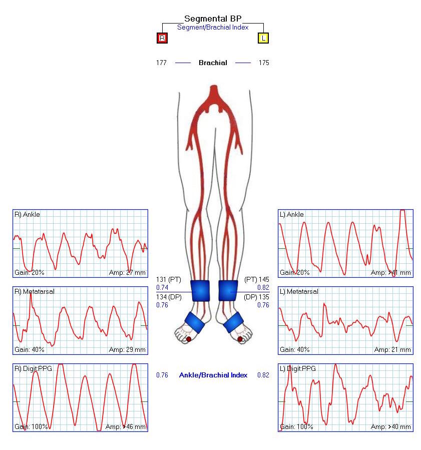
This patient is a 90 plus year old man who developed ever worsening claudication to the point he was disabled and more worryingly, had developed pain over his left heel. His ABI’s were severely diminished.

preop ABI2

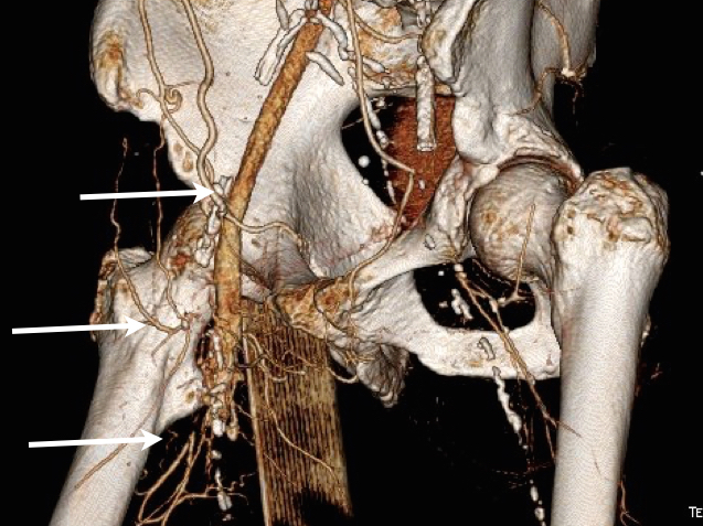
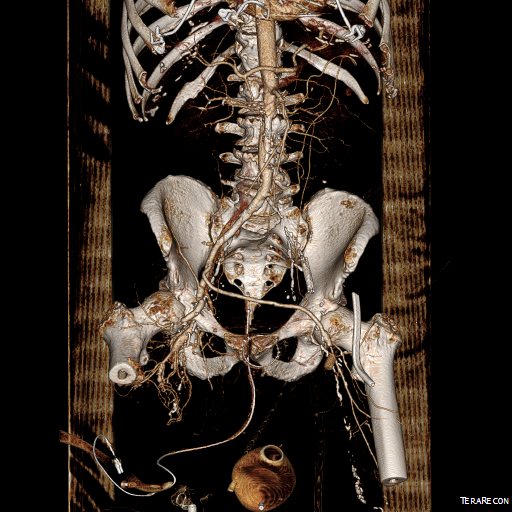
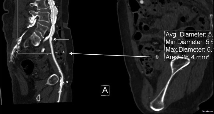
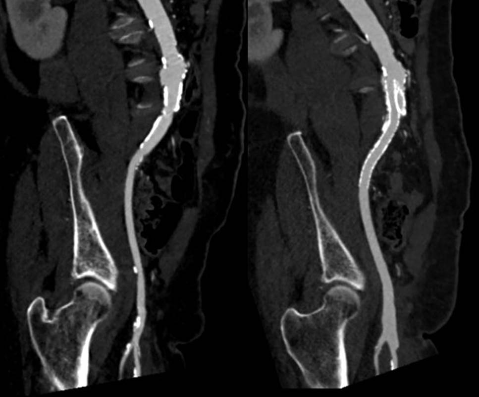
CTA showed that he had an occluded SFA with above knee reconstitute, but also had only single vessel runoff to the foot via a heavily diseased posterior tibial artery that had serial mild to moderate stenoses.

00016287326_20150730_1

00016287326_20150730_6

00016287326_20150730_7

An attempt at endovascular recanalization was performed at an outside institution, but the SFA lesion could not be crossed. Bypass was not a good option -the ipsilateral saphenous vein had been harvested for CABG, and a long operation was going to have a significant impact on this patient who also had mild dementia and drank 2-3 glasses of wine a day. It is not uncommon to have a successful operation, but have the patient lose 2-3 months in recovering from the physical effects of a long operation as well as from perioperative delirium.

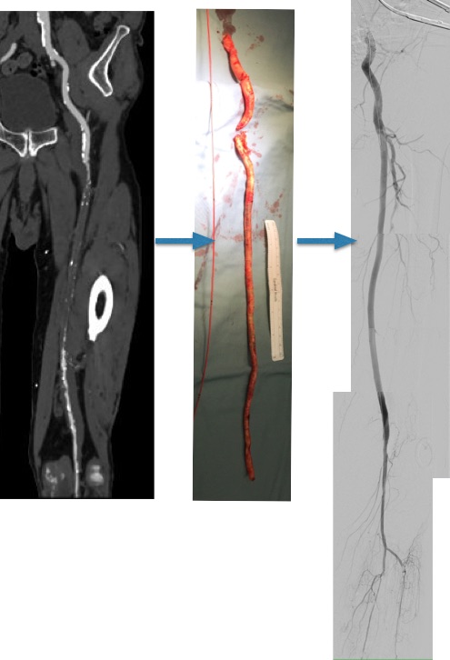
I felt that removing the occlusive plaque from his arteries offered a minimally invasive solution. The plaque was easily accessible via an oblique, skin line incision in the groin, and clearance could be performed from the external iliac artery origin to the planned endpoint slightly beyond Hunter’s canal. While the outflow was not perfect, in my experience, aside from a single native vein bypass, long segment restoration of vessel elasticity results in very acceptable patency rates.

endoRE graphic

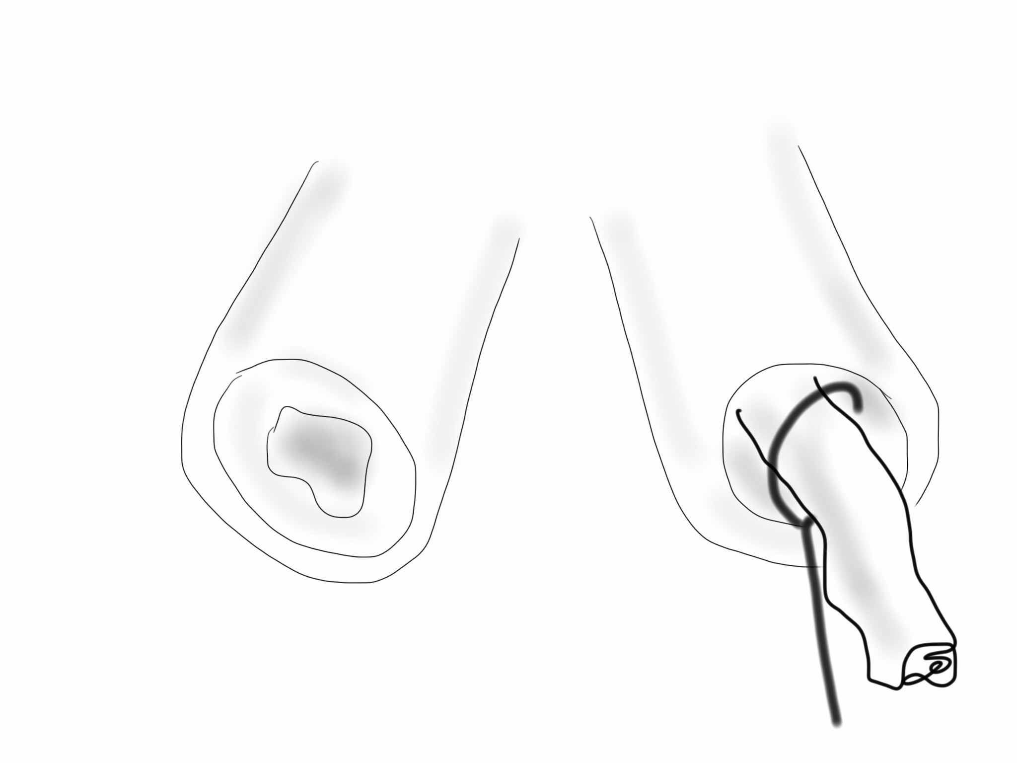
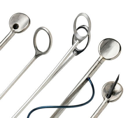
Remote endarterectomy is a bit of a lost art from the early days of vascular surgery. A ring dissector (Vollmer Ring Dissector, LeMaitre Vascular) is used to liberate the plaque from the remnant adventia. A cutting device (Moll Ring Cutter, LeMaitre Vascular) shown third from left below is used to divide the plaque.

LeMaitre

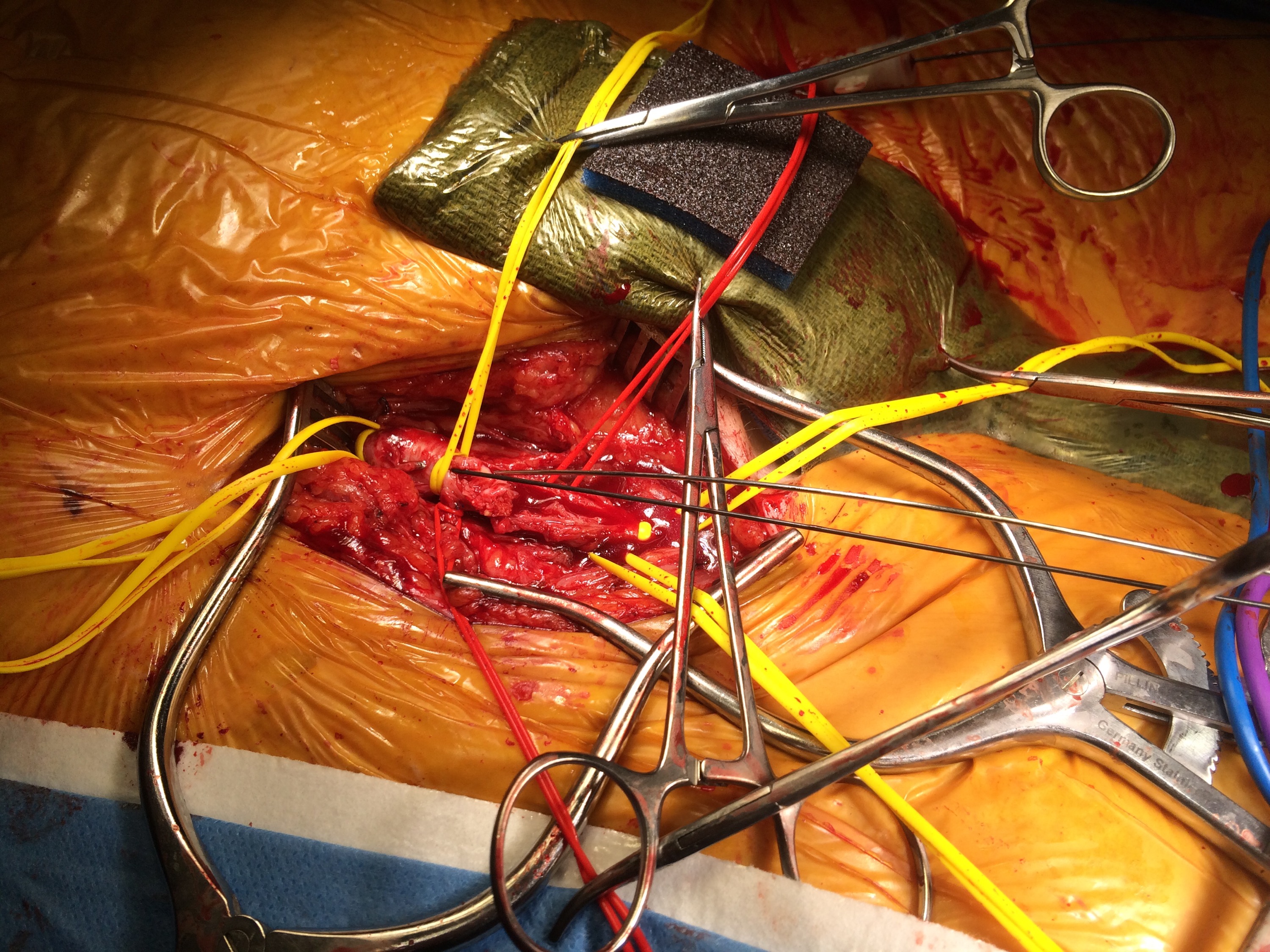
The common femoral artery plaque is usually contiguous with plaque in the external iliac artery and surgeons who perform a lot of CFA endarterectomy have various maneuvers to remove as much plaque as possible, up to stenting the end point of the plaque down to the endarterectomy patch. I have never been satisfied with this because the EIA behaves differenty than the CIA (am looking into this!) in my experience and placing stents even minimally across the inguinal ligament is not desirable. Sending the dissector up to the EIA origin frees the plaque to be removed completely with the CFA plaque. The clip below shows the Vollmer Ring dissecting plaque up to the EIA origin. I do this over a wire in the pelvis because in the rare instance of leak or rupture, rapid control is possible without having to open the abdomen.

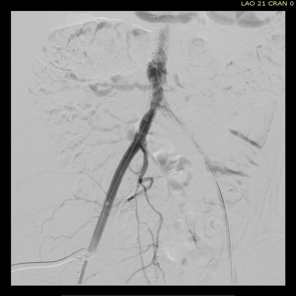
Once freed, the cutter is used to transect the plaque and the end point is tacked down with a stent at the distal common iliac/EIA origin which is a better place for a stent than the inguinal ligament.

The PFA in this patient did not require endarterectomy and reconstruction, but if it did, I would have made the arteriotomy go onto the profunda from the CFA. The SFA plaque is then mobilized with the Vollmer ring. I don’t do this over a wire, but have a definite end point in mind based on what I see on CTA.

The CTA (images earlier) shows that the above knee popliteal artery has no significant calcified plaque. This doesn’t mean there isn’t fibrotic plaque. Cutting the plaque as in the clip below results in a coned in antegrade dissection which has to be crossed in the true lumen.

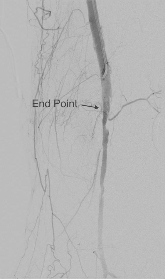
This is technically the most difficult part of the EndoRE procedure and it requires good imaging and wire skills. The trick here is that an ultrasound guided puncture of the popliteal or tibial vessel can give you distal true lumen access if needed. It was not necessary in this patient. The better maneuver is if the end point is surgically accessible is to cut down and tack down the plaque and patch the arteriotomy.

Angios -14

Angios -39

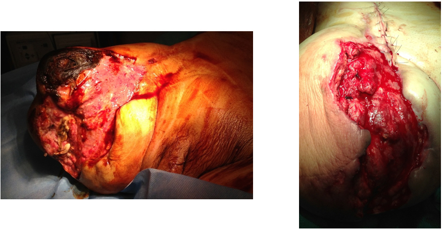
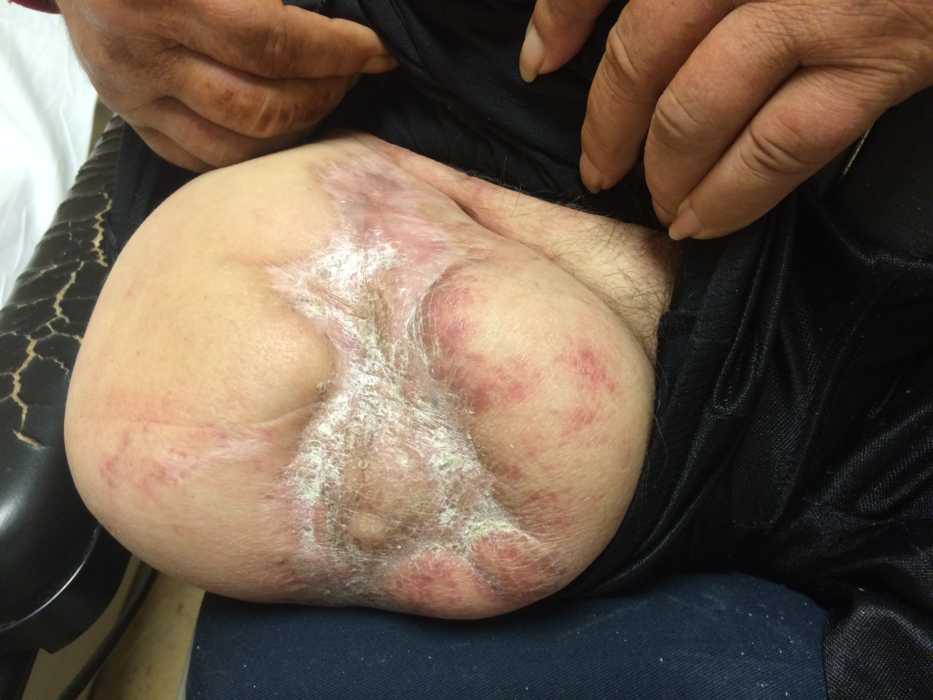
The patient regained multiphasic PT and DP signals at the end of the case, after the common femoral artery was patched and flow restored. The small groin incision was closed with a running absorbable monofilament after multilayer deep closure. The patient had a blood loss of 50mL. An ilioinguinal field block and local anesthesia provided excellent pain control. Postoperative ABI was improved to 0.82 from 0.34 and all pain was relieved. The patient felt good enough to go home on postoperative day 1.

postop PVR2

This illustrates what I feel to be a best application of both open and endovascular techniques. The above knee popliteal stent is short and in a position that is not going to result in fracture. The external iliac stent is in a protected position in the pelvis and quite large -10mm, which I expect will stay open for the life of the patient. The profunda femoral artery, the rescue artery, is widely patent, and numerous collaterals off the SFA have been restored to patency which I feel aid in maintaining the patency of this repair, along with the restored elasticity of the artery which mimics the biomechanics of autologous vein.

In most patients with compromised outflow, I start warfarin along with ASA at 81mg. Because of his age, I opted for Plavix+ASA. These fail with the development of random TASC A restenoses along the SFA which are amenable to balloon angioplasty. The role of drug eluting balloons in this situation is unknown but theoretically promising. Occlusion through thrombosis does not result in embolization and limb loss as in failure of prosthetic bypass grafts (another option in this patient), but rather leaves a situation where endovascular thrombectomy or lysis is technically feasible.

The great thing is that this is by far superior to stenting of a TASC D femoral arterial lesion.

Categories
bypass PAD techniques Wounds

Deep rescue from a hospice: saving a patient from hip disarticulation with advanced hybrid inflow procedure and vein bypasses

PREOP.001

The patient is an elderly man who had bilateral above knee amputations after failure of aortobifemoral bypass grafts at an outside institution. Unfortunately, he had no femoral pulses and his amputation on the right broke down (image above). His left stump had erosion of his femur to the skin with rest pain as well, but was at least covered by skin for now. He was declared too sick for hip disarticulations and was sent to a hospice where he failed to pass away. After a year there, he was sent to us for an evaluation.

He was suffering from rest pain and had complete breakdown of the skin over his amputation stump. More worrisome was the development of gangrenous scrotal and decubitus ulcers which were small but persistent and also foci of pain. CTA showed the following:

PREOP CTA.001

The aorta was occluded below his renal arteries. An AV fistula near his common femoral vein lit up his right iliac vein on the CT above. He had had a prior aortobifemoral bypass but this was occluded. Gratifyingly, it was anastomosed proximally end to side, giving us options. As with any revascularization, we had an inflow source -his aorta, and several potential outflow sources (CTA below, contrast filling iliac vein from AVF’s).

OUTFLOW.001

In particular, his distal profunda femoral artery showed promise. Vein mapping revealed a short segment of basilic vein in his arm to use as bypass, but we needed inflow from the aorta.

I have come to appreciate two things about aortoiliac recanalization. First is that passing the wire antegrade is far likelier to stay in the true lumen at least in the aortic inflow segment -retrograde wire passage inevitably dissects the occlusive aortic plaque and reentry into the true lumen of the diseased aorta is just as challenging as in the leg. The second is vein bypasses have excellent patency in challenging conditions -you just need excellent inflow and an arterial bed to perfuse.

My plan was to cross the aortoiliac occlusion with a wire from the left arm. Once the right iliac system was entered, it didn’t matter if I was in a subintimal plane. The wire could be seated in the common femoral artery to access with a surgical exposure. Once this was done, my intention was to perform remote endarterectomy of the external iliac artery and stent from the aorta to the common iliac artery. The endarterectomized external iliac artery would be the inflow source of a later staged ilio-cross femoral bypass to revascularize his left AKA stump. The common femoral artery at its origin would provide inflow to a short vein bypass to his profound femoral artery.

The wire passed readily into the right iliofemoral system and a groin exposure and common femoral arteriotomy allowed me to retrieve the wire which had been passed from the left arm. A remote endarterectomy was performed over the wire which I do to ensure access in case the artery ruptures (specimen below).

OR IMAGES.001

This allowed me to place a sheath into the right iliac system in the now reopened external iliac artery. Balloon angioplasty of the aortoiliac segment created working space for placement of balloon expandable stents from the infrarenal aorta to the common iliac artery, restoring an excellent pulse in the right groin.

The profunda femoral artery was encased in scar tissue, but following the occluded PFA from the CFA, I was able to expose an open segment and cut it open in the scar tissue. There was back bleeding, and I controlled the artery by placing a small Argyll shunt into the artery and reperfusing it from the recanalized right iliac system.

OR IMAGES.002

The Doppler flow in the shunt was excellent, suggesting great outflow potential. The bypass was performed over the shunt with reversed basilic vein. Completion arteriography showed excellent flow.

PLANNING SLIDE.001

The amputation stump was debrided of dead bone and muscle and the graft was covered with a sartorius muscle flap.

OR IMAGES.003

Before and after images are shown. The remaining open wound granulated well, and ultimately accepted a split thickness skin graft. His scrotal and decubitus ulcers healed as well (below at 6 months post op).

IMG_2380

His left AK stump subsequently degraded while he recovered so three months after this operation, he underwent a right external iliac to left profunda femoral artery bypass with cadaveric vein.

00018205637_20131006_1

00018205637_20131006_6

I don’t like using cadaveric vein, but we really had no options. The right external iliac artery was approached through a right lower quadrant (transplant) incision and a punch biopsy of the artery revealed only normal adventitia on pathology. The EIA was soft and sewed well -essentially a normal artery brought back from the dead. The left profunda femoral artery was large after endarterctomizing its origin and accepted the bypass flow well.

The mortality from hip disarticulation in the setting of gangrene and infection is very high, and I feel that standard approaches to this problem -prosthetic axillo femoral bypasses, thoracobi-femoral bypasses, in the setting of advanced infection and gangrene were unlikely to succeed. In over 1.5 years of followup, everything has remained patent, and the patient lives independently.

Categories
PAD techniques TEVAR

External iliac remote endarterectomy in lieu of a conduit for TEVAR

IMG_1281

The patient had diffuse atherosclerosis with small luminal area even in areas without calcified plaque. It predicted inaccessibility for the 22 French sheath required to deliver the 32mm C-TAG device to be placed for a symptomatic type B thoracic aortic dissection associated with a small but expanding proximal aneurysm.

IMG_1277

My options included direct aortic puncture, an aortofemoral conduit, or an endoconduit. The aorta was heavily calcified and the bifurcation was narrowed by circumferential plaque down to 6-7mm at its narrowest and the left iliac had a severe narrowing due to this plaque. The common femoral artery was severely diseased with a lumen diameter of 4mm due to heavily calcified plaque.

I have come to favor direct aortic puncture over conduits, but the heavily calcified aorta and the absence of safe areas to clamp made me think about other options. My experience with endoconduits has been limited to revising problems of endoconduits from elsewhere, but others report it as a feasible option.

The problem with a long artery narrowed with irregular plaque and even intimal thickening is that it will readily expand to accommodate a large sheath but removing it involves the frictional resistance of the whole artery and typically the “iliac on a stick” avulsion involves the whole length of external iliac artery, likely because the common iliac is anchored by the aortoiliac plaque, the smaller diameter of the EIA, and the longer more tortuous path offering greater resistance in the EIA compared to the aorto-common iliac segment.

IMG_1272

Remote endarterectomy, a technique involving endarterectomizing an artery through a single arteriotomy, offers the possibility of increasing the lumen of even a mildly diseased artery and reducing the frictional coefficient, assuming the remnant smooth adventitia is less resistant than rough irregular intimal plaque.

IMG_1275

The plan was to expose the right common femoral artery and endarterectomize it and gain wire access from the R. CFA. A wire would be placed on the left iliofemoral system to protect it for later kissing iliac stents. A right EIA remote endarterectomy would be performed, and then the right aorto-common iliac segment would be balloon dilated to 8mm.

IMG_1278

The operation went as planned. The external iliac plaque was removed in a single piece from the EIA origin.

IMG_1279

Arteriography showed the right EIA to be free of intimal disease, and dilators and ultimately the 22F sheath went in easily.

IMG_1276

The TEVAR also went uneventully -the left subclavian which had a prior common carotid to subclavian bypass, was covered and the aneurysm and flap were excluded from the left CCA to the celiac axis.

IMG_1280

The most difficult part of the operation was removing the sheath, as is usually the case with a tight iliac, but the friction point was largely at the common iliac and not the external iliac. No artery could be seen extruding with the sheath at the groin while steady tension was applied to the sheath under fluoro. The aortic bifurcation was repaired with kissing iliac stent. The patient recovered well and her chest pain resolved.

I have done this for EVAR, including reopening occluded external iliac arteries, and even for a 26F access for TAVR, avoiding the need for placement of a conduit in selected patients.

Addendum: in followup, I had the chance to check up on the repair -the EIA remained large and patent.

before after