Categories
bypass EndoRE PAD tibial revascularization

The Ilio-femoral-popliteal remote endarterectomy -The Concept Behind Extended Remote Endarterectomy is Moving Inflow from the Groin to the Knee

IMG_6805

Why perform such an extensive endarterectomy when just a few stents will do? This is a valid question, given the relative safety of interventions and the durability of bypasses. There are three reasons why ilio-femoral-popliteal endarterectomy works well in my practice.

  1. Minimally invasive
  2. Restore elasticity and collaterals
  3. Move the inflow point from the groin to the knee

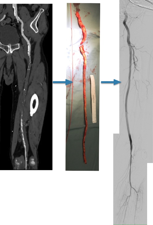
The procedure is minimally invasive. Take for example this patient whose plaque is shown above. He had a common femoral occlusion for which a common femoral endarterectomy was aborted when the prior surgeon ran into excessive bleeding. Workup for coagulopathy was negative and the patient came to me with rest pain. Pedal level pulses were not palpable, and the signals were barely there.

pvr2

CTA showed that he had a CFA occlusion as well as SFA occlusion.

no annotation_9

Because the common femoral plaque is contiguous with the external iliac plaque, it is often simpler to complete a remote endarterectomy over wire up to the external iliac origin than to try to get a satisfactory end point at the inguinal ligament -I do not like stenting across the ligament into the patch which is the usual bailout if the end point causes a stenosis. It is far simpler to apply a stent at the external iliac origin.

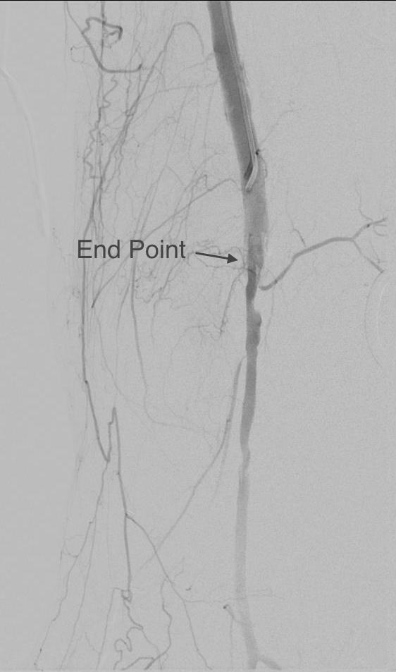
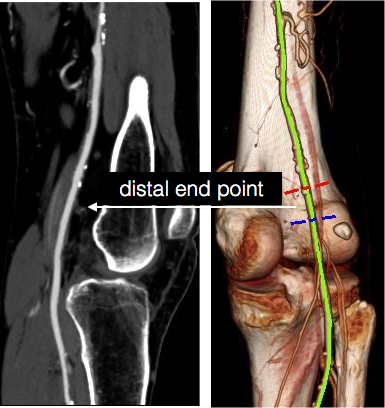
The popliteal end point was chosen where the visible plaque was no longer apparent in the patent artery. The goal is to cut across thin intima, and frequently no distal stent is required because a secure end point is achieved much like the “feathered endpoint” seen in carotid endarterectomies.

Termini

distal end point

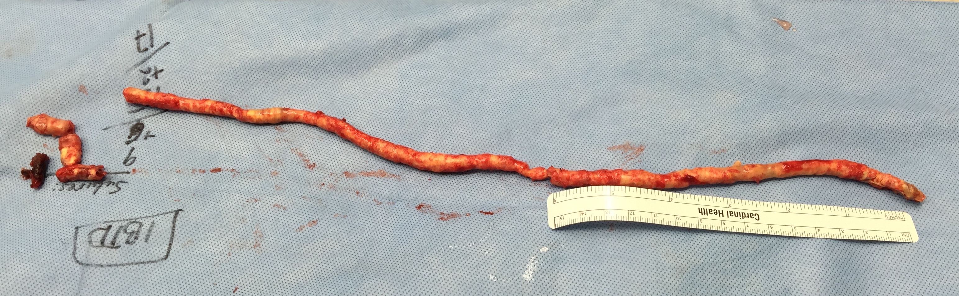
My intention was to endarterectomize the atherosclerotic plaque from the external iliac origin to popliteal artery via the groin incision marked in orange.

3DVR allows for planning the operation in great detail

The video shows the setup and motion in dissecting the plaque.

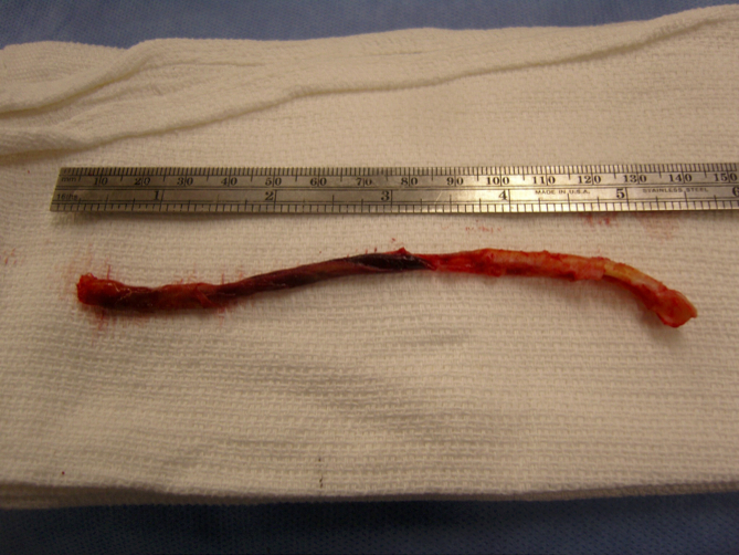
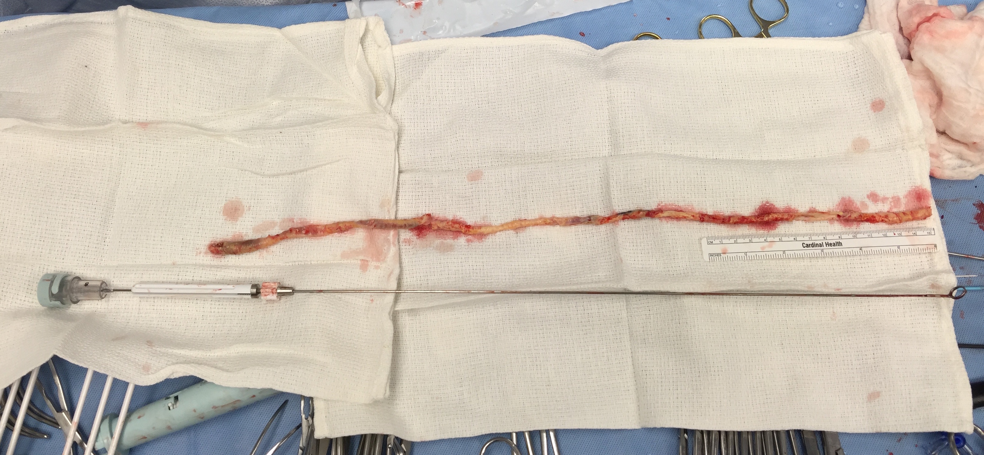
The plaque came out easily (first image, top).The proximal and distal end points required stents.

Before and after
Before and after

The patient regained palpable dorsalis pedis and posterior tibial artery pulses. Total OR time was less than 2 hours. An ilioinguinal field block allowed for good pain control and the patient was discharged the next morning, having to heal only a 10cm wound. There is no good endovascular option for common femoral disease, and while stenting the whole SFA can be done, on more than a few occasions I have had to treat occluded “full metal jacket” SFA stents, usually by removing them. EndoRE has been shown to be superior to PTFE and almost as good as vein in the REVAS Trial when compared to fem-AK POP bypass. Going home the next day after such an extensive revascularization is not a stunt -it’s the direct result of limiting the incision and blood loss and OR time.

2. Restore Elasticity and Collaterals -Arterial Restoration

One of the components of arterial flow that is lost with atherosclerotic disease is arterial elasticity. That is the stretchiness of the artery in response to pressure. Elastic distension and recoil account for significant portions of forward flow during diastole which is lost with atherosclerotic plaque. As plaque builds up, and the artery becomes stiffer. The artery that goes through remote endarterectomy regains this elasticity. Ultrastructure from a recanalized external iliac artery sampled from a punch arteriotomy for a cross ilio-femoral bypass showed that three months after endarterectomy, the external iliac artery was ultrastructurally normal per pathology report.

Also, collaterals that were previously occluded are seen to be restored to patency. This has an important impact on patency and any future failures. The endarterectomized arteries fail due to the presence of isolated, random fragments of medial smooth muscle which cause focal TASC A restenoses. These are easily amenable to balloon angioplasty. If the revascularization fails, there is no catastrophic thromboembolism that is typical of PTFE thromboses -rather the collaterals keep segments open and it is straightforward to thrombectomize or lyse the artery and intervene as necessary.

3. Moving the inflow point from groin to the knee.

This is an important concept. One of the principles of inflow restoration is delivering large flow and pressure directly from the aortic source to the leg. Recanalizing from the external iliac to the below knee popliteal artery creates this situation below the knee, allowing for very short bypasses to be performed from the popltieal artery to tibial targets -a very useful circumstance when vein is limited. This next patient is a presented with gangrene of his fifth toe after esophagectomy for cancer, and had severe diabetes.

prepvr2

He had useful saphenous vein in his thigh only, some of it having been harvested in the proximal thigh for a common femoral endarterectomy. CTA showed a dilated common femoral and profunda femoral artery, severely calcified SFA and popliteal artery which were occluded, and only a patent peroneal artery as runoff.

no annotation -_3

The plan was to harvest the short segment of vein then through the same incisions, below the knee and in the mid thigh, expose the below knee popliteal artery and tibial origins, and the mid SFA. I intended to avoid the groin. The plaque was removed from the tibioperoneal trunk to the SFA origin, and the origin was stented.

IMG_6637

This reestablished a normal inflow at the level of the below knee popliteal artery. I also did an eversion endarterectomy of the anterior tibial artery which resulted in significant back bleeding -a good sign. A short bypass was performed from the below knee popliteal artery to the peroneal artery.

IMG_6639

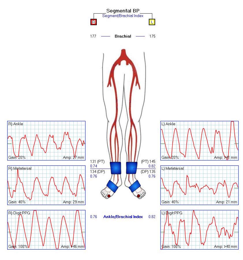
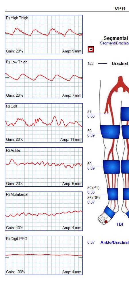
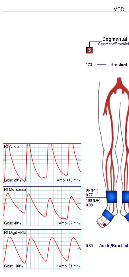
This resulted in a palpable dorsalis pedis artery pulse and excellent peroneal and posterior tibial artery signal.

completion

The ABI improved and the waveforms predicted healing for his 5th ray amp.

post abi2

This last case illustrates the point that once the conceptual inflow point is moved to the below knee popliteal artery, bypasses can become short, and durable tibial revascularizations become feasible. By avoiding a redo groin, avoiding multisegment arm vein bypass, and keeping the procedure time under 5 hours, the operation remains less invasive.

Categories
PAD remote endarterectromy techniques

Arterial Restoration: more than just a pretty name

Progression
CTA on left shows occlusive plaque in SFA but contiguous plaque from external iliac origin into the popliteal artery. This was removed with EndoRE resulting in restoration of original artery patency -arteriogram on right. A single short stent was placed in the EIA origin and the above knee popliteal artery received a short stent as well.

This patient is a 90 plus year old man who developed ever worsening claudication to the point he was disabled and more worryingly, had developed pain over his left heel. His ABI’s were severely diminished.

preop ABI2

CTA showed that he had an occluded SFA with above knee reconstitute, but also had only single vessel runoff to the foot via a heavily diseased posterior tibial artery that had serial mild to moderate stenoses.

00016287326_20150730_1

00016287326_20150730_6

00016287326_20150730_7

An attempt at endovascular recanalization was performed at an outside institution, but the SFA lesion could not be crossed. Bypass was not a good option -the ipsilateral saphenous vein had been harvested for CABG, and a long operation was going to have a significant impact on this patient who also had mild dementia and drank 2-3 glasses of wine a day. It is not uncommon to have a successful operation, but have the patient lose 2-3 months in recovering from the physical effects of a long operation as well as from perioperative delirium.

I felt that removing the occlusive plaque from his arteries offered a minimally invasive solution. The plaque was easily accessible via an oblique, skin line incision in the groin, and clearance could be performed from the external iliac artery origin to the planned endpoint slightly beyond Hunter’s canal. While the outflow was not perfect, in my experience, aside from a single native vein bypass, long segment restoration of vessel elasticity results in very acceptable patency rates.

endoRE graphic

Remote endarterectomy is a bit of a lost art from the early days of vascular surgery. A ring dissector (Vollmer Ring Dissector, LeMaitre Vascular) is used to liberate the plaque from the remnant adventia. A cutting device (Moll Ring Cutter, LeMaitre Vascular) shown third from left below is used to divide the plaque.

LeMaitre

The common femoral artery plaque is usually contiguous with plaque in the external iliac artery and surgeons who perform a lot of CFA endarterectomy have various maneuvers to remove as much plaque as possible, up to stenting the end point of the plaque down to the endarterectomy patch. I have never been satisfied with this because the EIA behaves differenty than the CIA (am looking into this!) in my experience and placing stents even minimally across the inguinal ligament is not desirable. Sending the dissector up to the EIA origin frees the plaque to be removed completely with the CFA plaque. The clip below shows the Vollmer Ring dissecting plaque up to the EIA origin. I do this over a wire in the pelvis because in the rare instance of leak or rupture, rapid control is possible without having to open the abdomen.

Once freed, the cutter is used to transect the plaque and the end point is tacked down with a stent at the distal common iliac/EIA origin which is a better place for a stent than the inguinal ligament.

The PFA in this patient did not require endarterectomy and reconstruction, but if it did, I would have made the arteriotomy go onto the profunda from the CFA. The SFA plaque is then mobilized with the Vollmer ring. I don’t do this over a wire, but have a definite end point in mind based on what I see on CTA.

The CTA (images earlier) shows that the above knee popliteal artery has no significant calcified plaque. This doesn’t mean there isn’t fibrotic plaque. Cutting the plaque as in the clip below results in a coned in antegrade dissection which has to be crossed in the true lumen.

This is technically the most difficult part of the EndoRE procedure and it requires good imaging and wire skills. The trick here is that an ultrasound guided puncture of the popliteal or tibial vessel can give you distal true lumen access if needed. It was not necessary in this patient. The better maneuver is if the end point is surgically accessible is to cut down and tack down the plaque and patch the arteriotomy.

Angios -14

Angios -39

The patient regained multiphasic PT and DP signals at the end of the case, after the common femoral artery was patched and flow restored. The small groin incision was closed with a running absorbable monofilament after multilayer deep closure. The patient had a blood loss of 50mL. An ilioinguinal field block and local anesthesia provided excellent pain control. Postoperative ABI was improved to 0.82 from 0.34 and all pain was relieved. The patient felt good enough to go home on postoperative day 1.

postop PVR2

This illustrates what I feel to be a best application of both open and endovascular techniques. The above knee popliteal stent is short and in a position that is not going to result in fracture. The external iliac stent is in a protected position in the pelvis and quite large -10mm, which I expect will stay open for the life of the patient. The profunda femoral artery, the rescue artery, is widely patent, and numerous collaterals off the SFA have been restored to patency which I feel aid in maintaining the patency of this repair, along with the restored elasticity of the artery which mimics the biomechanics of autologous vein.

In most patients with compromised outflow, I start warfarin along with ASA at 81mg. Because of his age, I opted for Plavix+ASA. These fail with the development of random TASC A restenoses along the SFA which are amenable to balloon angioplasty. The role of drug eluting balloons in this situation is unknown but theoretically promising. Occlusion through thrombosis does not result in embolization and limb loss as in failure of prosthetic bypass grafts (another option in this patient), but rather leaves a situation where endovascular thrombectomy or lysis is technically feasible.

The great thing is that this is by far superior to stenting of a TASC D femoral arterial lesion.

Categories
PAD techniques

The EIA is more like SFA than CIA with regard to stent patency

CTA

From my case files, this was a case which I performed in 2010 and published in a prior blog.

CCx: Patient is a 56 year old man with complaints of pain in right leg with walking short distances and discomfort in the foot at night.

HPI: The patient has had cramps in his right calf with walking about a block for over a year, but over the past three months, he has developed pain with walking less than half a block which is incapacitating. He has developed pain at night which wakes him and he has taken to sleeping with his right foot dangling off the edge of the bed. This has resulted in some swelling of that leg which makes it doubly uncomfortable to wear shoes. He works as a manager at a local big box store and walks constantly. He used to smoke but quit last week. He feels this has worsened the pain.

Past Medical History: Hypertension, dyslipidemia, acid reflux

Past Surgical History: Ruptured appendix at 22

Medications: Zantac, Hydrochlorothiazide, Lipitor, Aspirin

Allergies: Penicillin (rash in 1972)

Social History: Employed, 30 pack year smoking history, quit last week

Examination: T 98 BP 142/88 HR 88 RR 12 Ht 68inch Weight 192lbs

HEENT: PERL, EOMI

Chest: Bilaterally CTA

Cor: RR

ABd: Soft, scar right lower quadrant

Ext: Cool right foot with dependent rubor, elevation pallor. Warm left foot

Neuro: Motor and sensory examination normal

Skin: Loss of hair over toes of right foot, and distal right leg pretibium

Pulses: No palpable pulses right leg. Left leg femoral, popliteal, and dorsalis pedis artery pulses are easily palpable

Labs: WBC 9.8 Hb 13.2 HCT: 40 PLT 332 Cr 0.8

Testing: Segmental pressures R/L: Brachial 144/138 High Thigh 88/150 Low Thigh 77/140 Calf 72/132 Ankle 71/140 Metatarsal 68/122

Pulse volume recordings notable for moderately diminished signals right high thigh cuff.

CTA: Moderate atherosclerosis of infrarenal abdominal aorta and its bifurcation with severe plaque of the right common iliac artery and occlusion of the external iliac artery. There was reconstitution of the common femoral artery on the right via collaterals. The left common iliac artery was affected by a moderate (50-75%) stenosis due to low density plaque.

Impression: PVD with rest pain of right leg due to severe aortoiliac occlusive disease and occlusion of right external iliac artery.

Plan: After discussing treatment options, we decided to try a right external iliac artery remote endarterectomy with angioplasty and stenting of his common iliac disease. This was chosen over aorto-bifemoral bypass because he had limited time off from work and work did require that he lift more than 20 pounds.

angio
Up and Over Wire during remote endarterectomy ensures wire access if rupture occurs.

Operation:

Remote endarterectomy of right external iliac artery with aortography, bilateral common iliac artery angioplasty and stenting.

This operation was done via a single right groin exposure and percutaneous access of the left groin. The common femoral artery had severe posterior plaque which was the starting point of the endarterectomy. Up and over access of the right external iliac artery was achieved and a wire was passed across the occluded external iliac artery and into the right femoral system. With clamping of the common femoral artery, the wire was brought out and controlled with a Fogarty clamp -this allowed for excellent stabilization and control and possible emergent balloon occlusion in the case of a perforation.

A Vollmer ring dissector was sent over wire and plaque up the external iliac artery under fluoroscopy and dissection was stopped at the iliac bifurcation which was heavily plaqued. A Moll Ring cutting device (LeMaitre) was used to transect the plaque which was removed.

plaque

The right and left common iliac arteries were stented with self expanding nitinol covered stents and post-dilated. I chose this as I have had occlusions occur in the setting of diffuse TASC C disease with low density plaque -I suspect that thrombus propogates across open cells like weeds growing through chicken wire. The stents on the right were extended across the iliac bifurcation.

A completion angiogram is here to the right. The common femoral artery was repaired with a patch angioplasty (bovine pericardial patch, LeMaitre).

The groin was closed and the patient recovered and was discharged in a few days with excellent palpable pulses on the right and improved pulses on the left. He was without symptoms of claudication or rest pain in the right leg.

Discussion:

Remote endarterectomy allows for removal of plaque via a single groin incision, obviating the need for an abdominal exposure required in an aorto-bifemoral bypass. This minimally invasive technique is associated with a low complication rate and earlier return to full work status because the abdominal incision is avoided.

Smeets et al [reference] reviewed with 7 year experience with 48 patients and had a technical success rate of 88%. One patient died due to a myocardial infarction within 30 days of the operation. The complication rate was low. 6 patients required coversion (retroperitoneal flank exposure) for additional arteriotomy (3 patients) and bypass (3 patients). The primary and assisted patencies shown to the right were acceptable with a secondary patency of 94% at 3 years.

graph

These cases require more surveillance than an aortobifemoral bypass. Intimal hyperplasia does occur in random loci in the SFA remote endarterectomy and this should apply to the external iliac artery. I chose the title because the external iliac artery biologically behaves like the superficial femoral artery in relation to endovascular patencies and not like the common iliac artery or aorta -probably because it shares a common embryology with the SFA, not the CIA. It is a troublesome artery that is often overlooked by vascular surgeons when femorofemoral bypass is performed for occlusive disease -the supplying external iliac artery though patent is usually diseased and has a small lumen. With a fem-fem bypass, both legs are supplied often through an artery with the caliber of a child’s drink straw. I have seen the donor leg become symptomatic through what is termed steal, but in fact reflects the hemodynamic inadequacies of a diseased external iliac artery.

I feel that 5mm is the minimal lumen caliber for an external iliac artery, and a 4mm lumen in an adult will clearly show a hemodynamic effect particularly after exercise or application of vasodilators in the endo suite. Stenting an occluded external iliac artery though technically feasible even in this case is not a durable solution in my experience. This operation allowed the patient to return to work without an extended convalescence.

I think removing the plaque offers advantages over stenting to the inguinal ligament. The common iliac stents have superior potency to external iliac artery stents and moving the stent point to the CIA and not stenting the EIA in my experience has better long term potency.

Reference

Smeets L, et al. J Vasc Surg 2003;38:1297-1304.