Categories
bypass complications graft infection mycotic aneurysm open aneurysm surgery peripheral aneurysm pseudoaneurysm skunk works techniques

EIA pulldown transposition: another way to treat infected common femoral pseudoaneurysm

Patient with infected femoral pseudoaneurysm, skin necrosis, about to blow

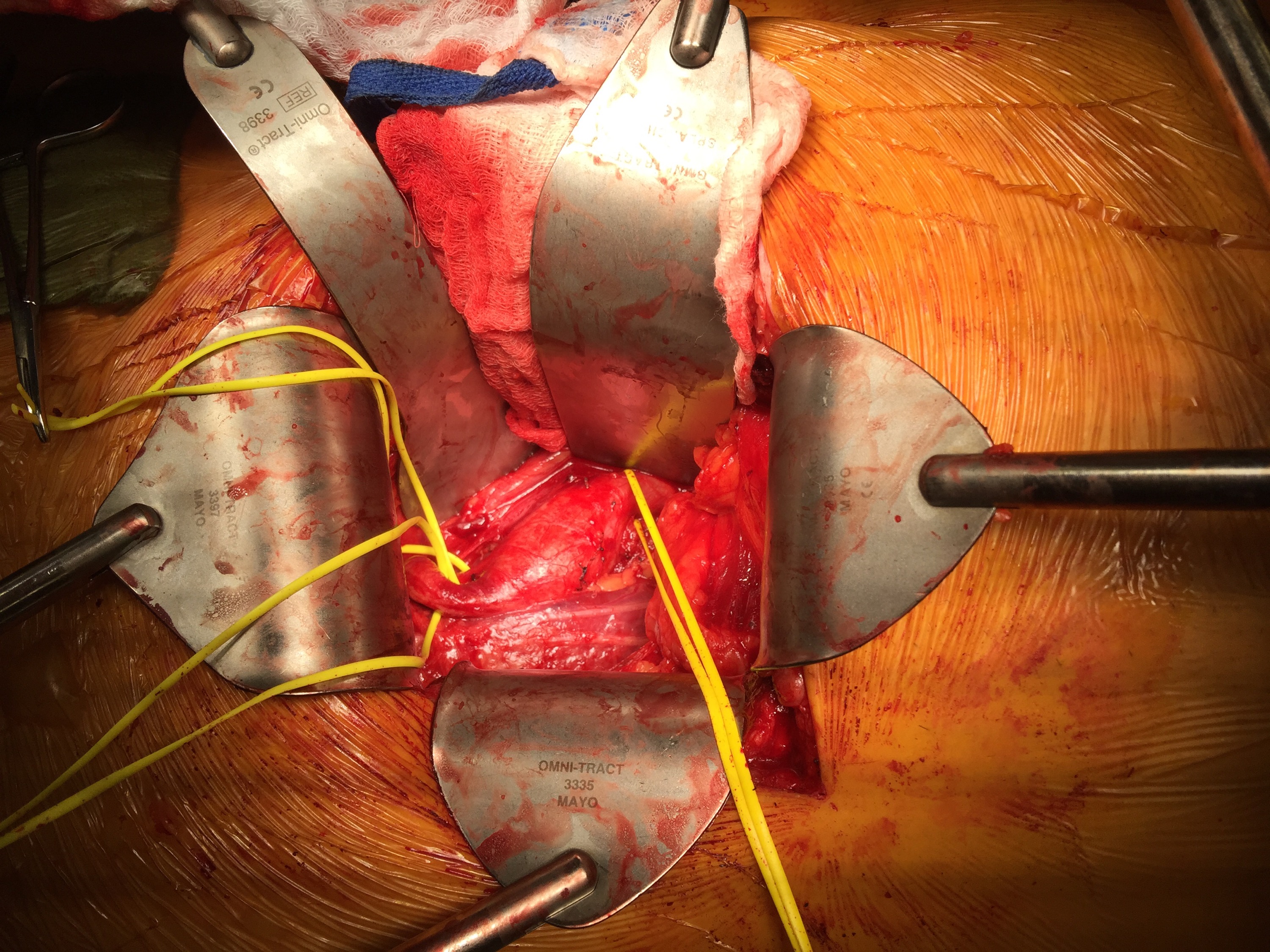
A surgeon from Nepal posted a case of a ruptured common femoral pseudoaneurysm infected from IV drug abuse on LinkedIn. The comments centered around typical textbook responses which were:

  1. Ligate, debride, obturator bypass
  2. In situ bypass with femoral vein +/- sartorius flap
  3. Rifampin soaked graft or crypreserved allograft
  4. Ligate only

My preferred treatment is #2, in-situ bypass with harvest of adjacent deep femoral vein. I never liked that procedure because in general in these patients, everything bleeds. Then I had a thought -how about if you mobilize the external iliac artery in the pelvis over its entire length and pull it out from under the inguinal ligament to sew to the femoral bifurcation or SFA? That is, when you enter the pelvic retroperitoneum to gain proximal control:

The external iliac artery is usually redundant and elastic in young people

You mobilize the external iliac artery from the iliac bifurcation to the inguinal ligament, detaching the inferior epigastrics as a last step. And then you pull it out from under the inguinal ligament, and anastomose it to the femoral bifurcation or the SFA.

The mobilized external iliac artery is pulled down to reach normal femoral artery. The distance x is the length of CFA that needs replacing

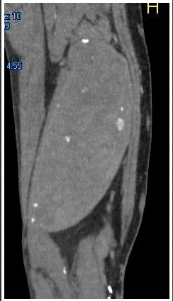
This makes sense because in young people and those with AAA and minimal atherosclerosis, the external iliac artery is both redundant and elastic, making it suitable for a pull down transposition. But then, how do you know as you mobilize the artery in the pelvis that you have enough to pull down?

Pythagoras figured that out two an a half millenia ago. If you measure the straight line distance from iliac bifurcation to the takeoff of the inferior epigastric arteries, you get the straight line external iliac artery distance. The length of the common femoral artery which is the excess EIA length needed, is assigned the value x. Then the height of the stretched artery off the line between the iliac bifurcation and the inguinal ligament will determine how much extra artery you have.

Taking these values, I did some maths.

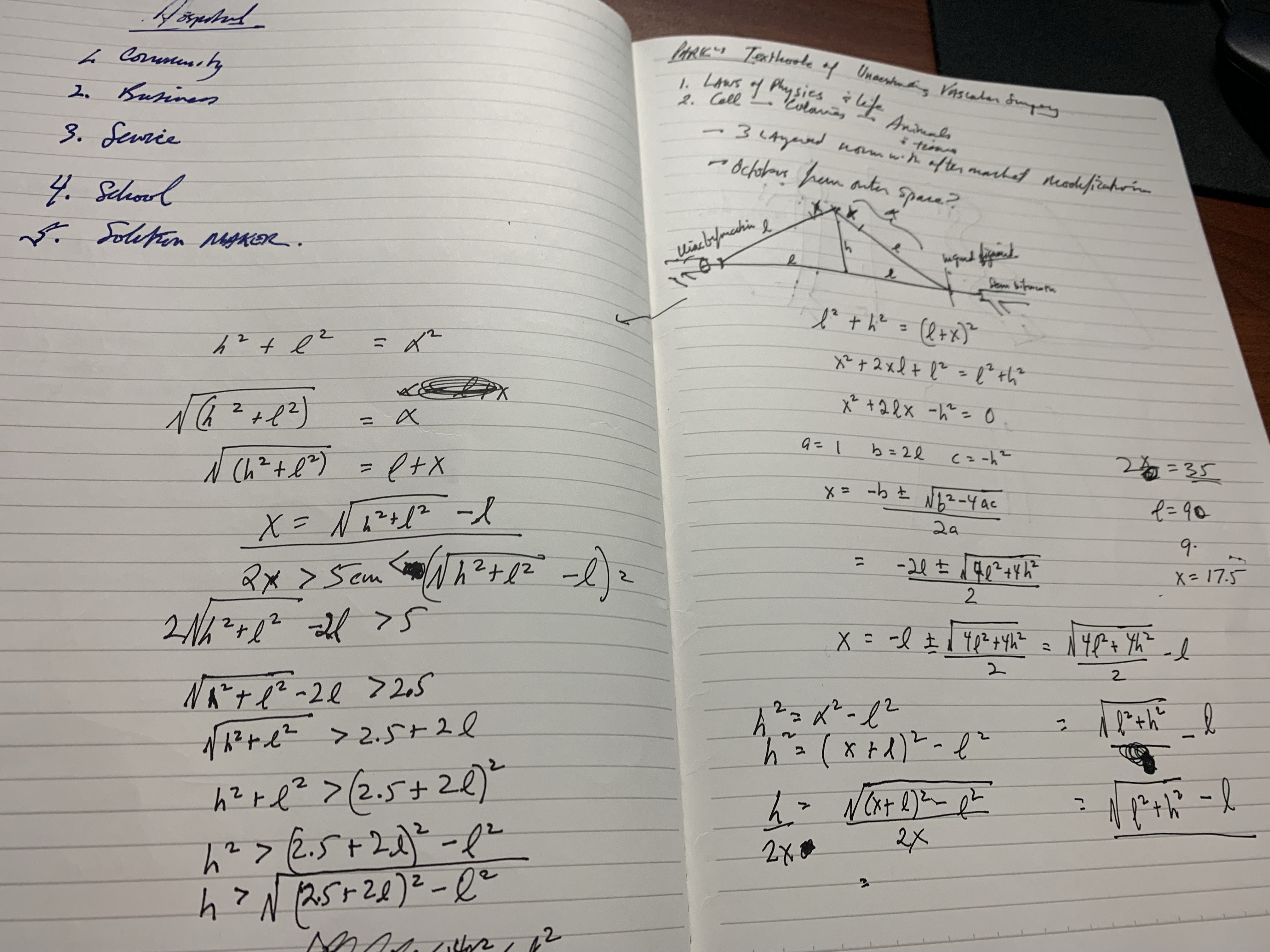
The solution for h, the height, is highlighted in yellow below. (note, the variable x in my notes is half the length of CFA, l is half the length of EIA, ie. 2x is CFA length).

Creating a spreadsheet for CFA lengths from 2 to 6cm and EIA straight distances of 5-10cm, the ratio of height H to CFA length varies from a minimum of 0.7 to maximum of 1.7 with an average of 1.1. That means the majority of the time, if you get 1.5x the length of CFA height off the pelvis, you should reach.

If you are short, you can detach the profunda and mobilize the SFA, pulling upwards, then reattach the PFA. Though this is entirely a thought experiment, there is no reason why it should not work. As with most things, I predict that it already has been done!

The advantages are using autologous tissues and leveraging the natural anatomy. There is a cost benefit in that OR time is shorter with less time for venous harvest and avoiding grafts, patches, and devices. The patient would avoid ischemia as would happen in the staged repair. The disadvantage is when you are short, but if you mobilize the appropriate amount (height off pelvis at least 1.5x the CFA length) you should be okay. The more curvature and tortuosity seen on 3DVR recontstruction and absence of significant atherosclerosis would predict feasibility.

Categories
median arcuate ligament syndrome opinion peripheral aneurysm techniques

Nonoperative management of median arcuate ligament syndrome (MALS)

The patient is a young woman who presented with classic symptoms and findings of median arcuate ligament syndrome (MALS). She avoided food because eating triggered severe pain in her upper abdomen. Over a year, this resulted in 15 pounds of weight loss. As a result, she no longer had the energy to work or exercise. She had an extensive gastrointestinal workup including blood work (LFT’s, amylase, cholesterol panel), abdominal CT scan, and upper endoscopy which were normal except for the finding of narrowing of the celiac axis due to compression by the median arcuate ligament. Examination was notable for upper abdominal pain exacerbated by pressure and seated, hunched-over posture. Unfortunately, due to her health insurance, surgery was not covered and she did not want any. So I recommended she try the following. 

  1. Eat standing up with good posture, shoulder back, back arched, taking deep breaths and holding once food passes
  2. Practice upward facing dog yoga pose (figure) 5 reps daily with deep inhalation breath holds. If this is difficult, do this standing up. 
  3. Improve the posture during seated work, never hunch over and pressed forward with “shrimp back,” periodically take a deep breath and hold with excellent posture. 
Upward facing dog yoga pose -shoulders square, outward collar bone tension, with deep inhalation breath holds

Over the past several years, I’ve noted that most patients respond to this, even in acute MALS pain situations (yes, there is acute MALS like slipped discs, for another post). That patient came back a few weeks later reporting that she was able to eat more food, more frequently. She also acknowledged compliance with the exercise and postural adjustments at work. A month later she reported regaining her lost weight and only mild pain with sitting in a car for a long time. She was still eating standing up, and she was grateful for having been treated without surgery.

From reference below, a mechanism for celiac plexus compression, injury, fibrosis, and development of neuropathy

We have postulated that MALS is a nerve compression syndrome of the celiac plexus by the median arcuate ligament (reference). There are two consequences to MALS, neither of which is mesenteric ischemia. The first is this compression of the celiac plexus with injury resulting in inflammation and fibrosis, resulting in further compression and a neuropathy of the celiac plexus. This neuropathy triggers aberrant pain sensations in response to eating. The other consequence is remodeling and injury due to arterial compression. The celiac axis can develop post-stenotic dilatation, growing large enough to be considered aneurysmal. The compression can damage the intima resulting in dissection. The artery can be injured and a pseudoaneurysm can develop. Finally, the aneurysmal segments may develop thrombosis and be the source of thromboembolism, usually to the spleen. Even when the celiac axis clots off, unless there has been resectional surgery such as a Whipple or splenectomy, the stomach gets enough collateral flow that ischemia is rare when celiac axis occlusion occurs. So similarly to  thoracic outlet syndrome (TOS), there is a neurogenic MALS and an arterial MALS. 

The first line of therapy in neurogenic TOS is physical therapy. With symptomatic MALS, I wondered if there could be physical therapy as well. This young woman and others I have managed nonoperatively suggests good response in some,  and partial response in most to exercises and maneuvers designed to address the compression.

Currently, in lieu of celiac plexus block, I have patients treat their MALS nonoperatively using the above protocol  for 2-4 weeks, typically while they undergo further workup to rule out more common gastrointestinal etiologies of their abdominal pain, and many have been able to improve their circumstances with these measures alone. This patient chose not to have operation as she was able to live symptom free and regain lost weight with these recommendations alone. 

Addendum

I received a comment from Ms. Suzanne Peek, president of the National MALS Foundation, who correctly pointed out not everyone presents in this way. I agree each patient undergoes a unique journey that is often marked by diagnostic delays because this is a rare condition. As I stated, not every patient responds to this regimen and after work up typically will undergo surgery to which in our published results 85% have a positive response to when diagnosed with MALS. Is certainly an area of further study to see if more people can respond to this non-operative approach. Our previous protocol which we instituted after the publication of our report was to use celiac plexus blockade as a diagnostic study and occasionally permanent blockade as therapy for those with prohibitive operative risk. A positive response to this regimen may signal a positive response to surgical celiac plexus ablation.

Reference

Weber JM, Boules M, Fong K, Abraham B, Bena J, El-Hayek K, Kroh M, Park WM. Median Arcuate Ligament Syndrome Is Not a Vascular Disease. Ann Vasc Surg. 2016 Jan;30:22-7. doi: 10.1016/j.avsg.2015.07.013. Epub 2015 Sep 10. PMID: 26365109.

Categories
EVAR peripheral aneurysm techniques

Parallel Grafts in the iliac bifurcation -an option at least until branched grafts become commercially available

CTA_1

This patient had developed metachronous common iliac artery aneurysms after aorto-bi-iliac graft placement of a AAA a decade ago. This is not infrequent occurence in a significant number of patients with aneurysmal degeneration seen in the thoracic or visceral segment abdominal aorta, iliac arteries, and popliteal arteries, years after a primary AAA repair. The patients are often older than they were at the original repair, with concomitant risk factors, and so a minimally invasive option is preferred.

Right CIAA -vulnerable tissue

saccular r ciaa

The teaching during my fellowship was that aorto-iliac bypasses for aneurysmal disease were to be taken to the iliac bifurcation to go around vulnerable tissues. These tissues vulnerable to aneurysmal degeneration were infrarenal aorta up to the renal artery origins, common iliac and internal iliac arteries, and popliteal arteries. An anastomosis to the iliac bifurcation however normal appearing may degenerate given enough time. This patient developed a saccular aneurysm on the right iliac bifurcation and a small internal iliac artery aneurysm (1.5cm).

CTA_2

This was treated with coil embolization and stent graft from the right iliac limb to the external iliac artery.

RCIAA treatment

This is the standard endovascular therapy for common iliac artery aneurysms, and acceptable in the setting of unilateral disease, and in a staged fashion has been considered acceptable for bilateral disease, acknowledging there is a 10-40% incidence of buttock claudication and when the contralateral hypogastric is occluded or when the patient is diabetic, the risk of buttock or colorectal necrosis is not insignificant. The patient had transiently some buttock claudication and hip and thigh neuralgia with walking but this improved in the weeks leading up to treating his left common iliac artery aneurysm.

Left CIAA

The left common iliac artery bifurcation is sometimes challenging to access from a midline incision and exposure requiring a separate sigmoid mobilization. In men, the narrow pelvis can increase the challenge, so it is without fault that sometimes common iliac artery is left behind. This is what became aneurysmal, developing into a 3.0cm fusiform aneurysm beyond the left limb of the graft.

CTA_6

The internal iliac artery had a moderate 50-75% stenosis at its origin but was not aneurysmal, and I chose to revascularize this. The patient was sexually active and walked for exercise. My options included proceeding with left hypogastric embolization and stent grafting, mirroring the right but with a significant risk for buttock claudication, sexual dysfunction, and a small risk for colorectal ischemia. Other option is an external iliac or common femoral to internal iliac artery bypass which is an excellent option in good risk patients.

Endovascular options

Iliac branched stent grafts are undergoing trial. My center is participating in both available industrial FDA approval trials (disclosure, I am site PI for the Gore trial), but this patient’s presentation and anatomy exclude him from the trials. The final option is placing a parallel stent grafts -one to the internal iliac artery and the other to the external iliac artery from a large common iliac stent graft. While not ideal, until branched grafts become available, this remains a viable option. The principle is to size the grafts to minimize potential gutters between the grafts, and have long seal zones to minimize the impact of the gutters. Access from two points is required to get two grafts into position. With the acute angle of the aortobi-iliac graft, up and over access is generally not possible. The 10mm Viabahn graft that I chose to place in the hypogastric requires a 12Fr sheath, which cannot be placed from the brachial artery, so I prepped for an axillary cutdown. The left common femoral access was percutaneous.

Image-17

The left CFA access allowed placement of a 16mmx10cm Excluder iliac graft limb to cover the aneurysm down to the iliac bifurcation. The left axillary arterial cutdown access allowed placement of a 12Fr sheath (Flexor) to allow access of the left internal iliac artery and safe delivery of a 10mm Viabahn stent graft. The left external iliac artery was sealed with a 13mm Viabahn stent graft that was deployed simultaneously. Ballooning was performed to both.

Completion
Completion

No leak was seen. The axillary access was repaired directly and the CFA access was repaired with two Perclose S devices.

Discussion

Despite initial acceptance of bilateral hypogastric occlusion, even staged, it can be the cause of significant disability aside from buttock claudication, which sometimes does not remit with exercise. Ischemia of the pelvis can drive a plexopathy that can result in motor and sensory neuropathy and disability. Death can occur. Preserving one of the hypogastrics can go a long way to preventing these complications, and everyone eagerly awaits adding iliac branched grafts to the armamentarium.

Categories
peripheral aneurysm techniques

Open repair is preferred for younger patients

  
The patient had an isolated 3.0cm common iliac artery aneurysm. Patient is in his fifties and wants to avoid the need for annual CT scans, buttock claudication. He had also read about neurological complications with open aortic surgery like retrograde ejaculation. 

  
An older patient may be well served with hypogastric artery embolization and iliac stent grafting. In the absence of an aortic or contralateral common iliac artery aneurysm, it would be hard to justify placing a bifurcated aortic stent graft to then accessorize with snorkels. He was not a candidate for the branched iliac stent graft trial (disclosure: I am a site PI for the Gore iliac branched trial and the Cook iliac branched device is also available on trial) and he was not enthusiastic about the follow up -neither was I, when we discussed other endovascular options. 

When I broached open surgery, there was a pause because he had read about all the endovascular procedures that were possible, but truthfully, he had never had an honest discussion about open repair. 

In the current set up of care and training, there would be opinions favoring a purely endovascular approach. Ironically, in another time, the approach we chose would have been considered minimally invasive. The operation was planned with a left lower quadrant retroperitoneal pelvic exposure. The plan was to replace only the aneurysm and revascularize both the internal and external iliac arteries. The internal was revascularized with an end to side anastomosis to a 12mm graft and the common iliac to external iliac revascularization was end to end. 


The patient recovered and was discharged in two days. The good thing is that he won’t face buttock claudication and has a low risk of neurologic complications (primarily retrograde ejaculation). Future endovascular options were maintained in the way the graft was tailored -particularly in the creation of a generous landing zone for any future aortic endograft. The patient won’t need to come back for surveillance on the same rigorous schedule as an endograft. 

Categories
AAA peripheral aneurysm

Consult QD post regarding iliac branched grafts

IMG_2914.JPG

Link to Clinic blog post regarding iliac branched grafts (link).

Categories
peripheral aneurysm

Popliteal Artery Aneurysm Sac Growth From Endoleak After Bypass

PAA figure 1

AUGUST 5, 2008 9:59 PM

The femoro-popliteal aneurysm originally ruptured four years prior to surgery and I had bypassed it with a PTFE graft. For the trainees, I have to emphasize that these aneurysms rarely rupture, and are rarely found at these sizes. The patient had internal iliac aneurysms as well, and while no connective tissue disorder was ultimately diagnosed, he had suffered from prior inguinal hernias and was over 6 feet tall.
In the intervening time, the aneurysm had steady growth from an endoleak. At the time of rupture, the size was indeterminate, but short followup recorded a size of 4cm. I had stapled off the proximal and distal femoropopliteal artery at the original surgery around the aneurysm. Over a 4 year period, during which the patient was briefly lost to follow up, the aneurysm regrew to over 6cm.

figure 4

At time of resection, there were several geniculate collaterals that were actively feeding the aneurysm. These were ligated.These would be the sources of the endoleak.

figure 2

The specimen is below.

figure 3

In general, for larger aneurysms, simply ligating them prior to bypass may leave you susceptible to growth, and even after a rupture, an aneurysm like this can regrow to prodigious size from endoleak. Resection, partial or complete, at time of first repair addresses this potential problem, but was not possible at original operation due to blood loss and shock.