Categories
AAA complications Endoleak EVAR imaging open aneurysm surgery opinion ruptured AAA Uncategorized

Off the guidelines: type II endoleak can derail the perfect EVAR

Every once in a while, I will make an exception to the SVS guidelines on AAA repair with regard to size at time of repair (link). I have a bunch of excuses. When I trained in 2000-2002 with several giants of vascular surgery, there was some controversy when the first guidelines came out in 2003 (link). The board answer became 5.5cm that year, but where I trained, it was a minority opinion held by Dr. Jeb Hallett. The majority was in the belief that as long as operative mortality was low, even high risk AAA repair could be undertaken (link). The published risk for Mayo was low, and that came from both technical excellence and high volume (more on that later). The criteria during my fellowship was 4.5cm in good risk patients for open repair based on data generated in the 1980’s and 90’s during Dr. Hollier’s tenure.

Then as now, the debate centered around the balance of risk. At specialty centers that achieved less than 1% mortality rate for elective open AAA repair, 4.5cm in good risk patients would seem perfectly reasonable. But given the 5-10% mortality seen in the Medicare database at that time for community practice, the 5.5 cm criteria was not only good science, it was prudent. The first set of guidelines held off the contentious volume recommendations that was the nidus of conflict within other surgical societies.

The advent of endovascular was a game changer -the mortality rate in the Medicare databases was 1-2% for EVAR in the community setting, meaning more surgeons in most hospitals could achieve tertiary center levels of mortality with this new technology. The issue was never really settled in my mind through the 2000’s, even with the PIVOTAL Study. I enrolled patients into the PIVOTAL Study (link) at that 4.5cm threshold during my time in Iowa. Eventually I lost equipoise and I stopped enrolling after a handful of patients. It had to do with graft durability.

Around that time, I took two patients in a row to the operating room for sac expansion without identifiable endoleak. They were Dacron and stent-based endografts placed about 5-7 years before by another surgeon and aortography failed to show type I or III endoleak. Sac growth was over a centimeter in 6 months and the aneurysm size was over 6cm in both. I chose to marsupialize the sac and oversew any leaks, with the plan to replace the graft if there was a significant leak. On opening the sac, no significant lumbar or IMA leaks were encountered but in these patients a stream of blood could be seen coming from the sutures securing the stents. It was the same graft that was in the trial, the AneuRx, and that was when I realized that these grafts have the potential to fail in the same way that patio umbrellas leak after years of use -cloth sewn to rigid metal with movement wears open the cloth wherever there is stitching. This did not happen with open repair. I lost enthusiasm for the trial as I lost faith in this graft which was retired from the market. I placed pledgetted sutures to close the leaks on both patients, and closed the aneurysm sac tightly around the graft in one patient who was higher risk, and replaced the stent graft in the other.

There are some exceptions to justify repair of 4.5-5.5cm AAA. During my time in practice, there were patients who lived far away from major medical centers who would not survive a ruptured AAA even if the rupture rate was low and who confessed they only came into town every five years or so. There were patients who suffered from clinical anxiety whose AAA was documented by a psychiatrist to amplify their anxiety. There were patients with vague abdominal pain for whom thorough workup have ruled out gastrointestinal causes and every visit to the ER triggered a CT scan to rule out AAA rupture. And there seemed to be some patients who seemed to have such perfect anatomy for EVAR, whose risks were low, and whose growth rates were so consistent that their repairs could be timed on the calendar. Some combination of these factors and lobbying on the part of the patient got them their repair in the 5cm range. And they still do.

The patient is a man in his sixties with hypertension who presented with a 4.7cm AAA which in various reports he came with described 5.2×4.7cm. After review of his images, it was clear it was 4.7cm. If measured on a typical axial cut CT scan or a horizontally oriented ultrasound probe, a cylindrical aortic aneurysm will be seen as an ellipse in cross section. A radiology report will typically report an aneurysms length and the anteroposterior and lateral dimensions. If you cut a sausage at an angle, the ovals you cut can be quite wide but the smaller length of the oval reflects the diameter of the sausage.

Looking back at his records, for three years he had multiple CT scans for abdominal  pain showing the AAA and a well documented record of growth of about 2-3mm annually -the normal growth rate. He asked me to prognosticate and so I relayed that 4.7cm in 2017 with a 3mm growth rate, we would be operating in 2020. The anatomy was favorable with a long infrarenal neck and good iliac arteries for distal seal and access. He was quite anxious as whenever he had abdominal pain, his local doctors would discuss the AAA and its risks or order a CT. After a long discussion and considerable lobbying by the patient and family, I agreed to repair his 4.7cm AAA.

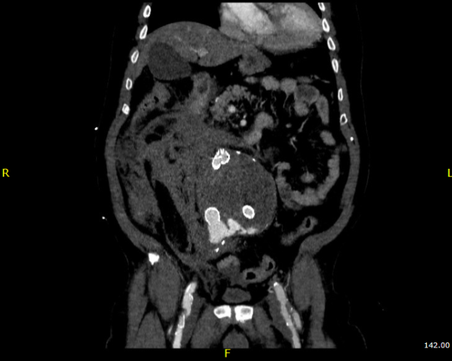
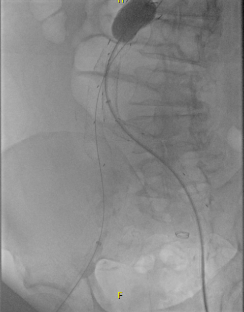
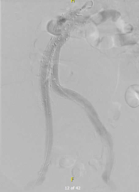
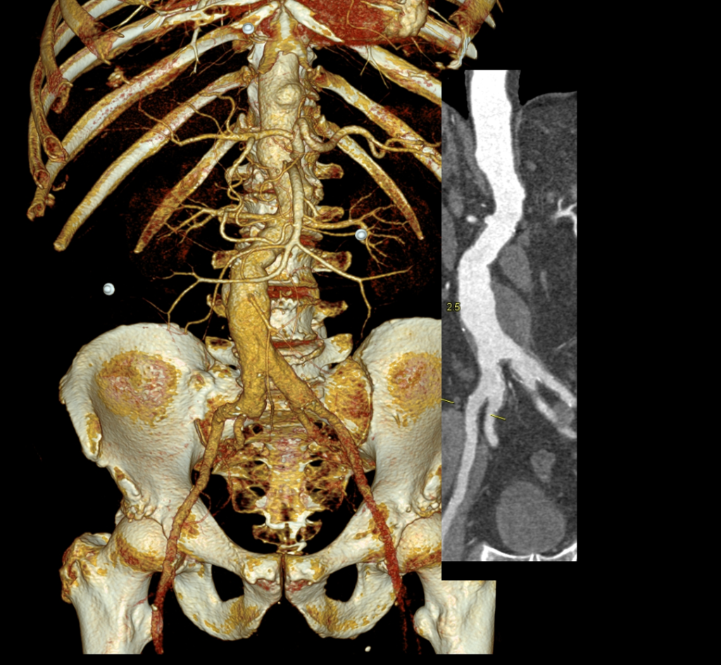
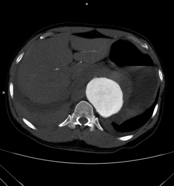
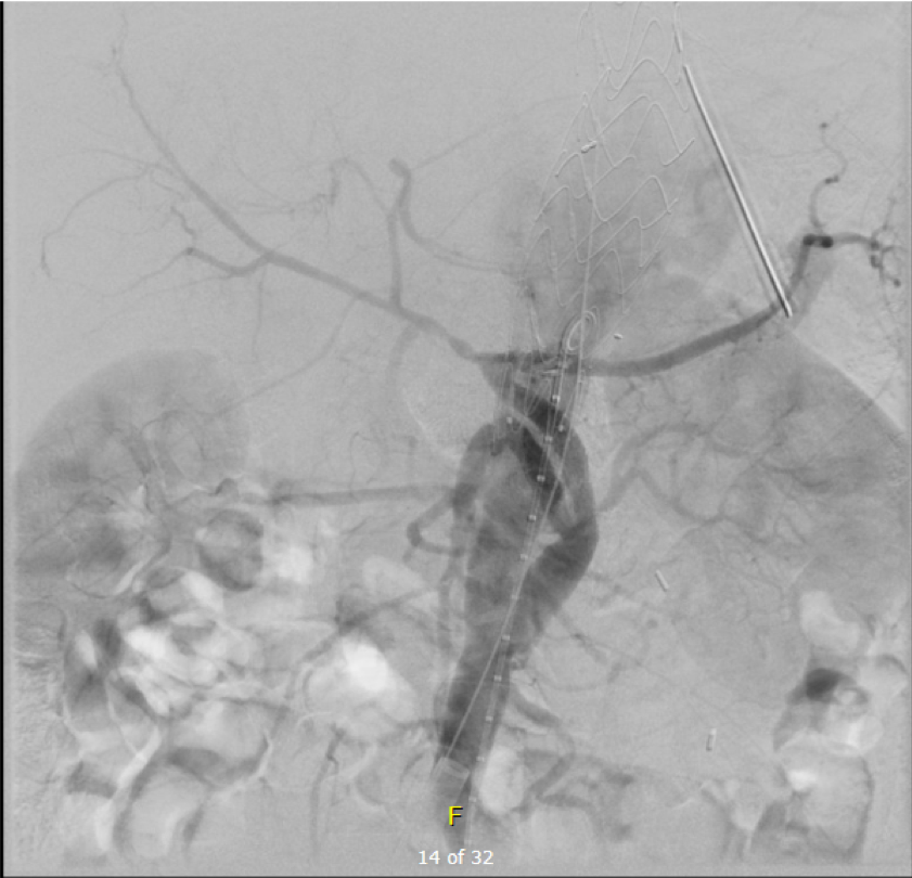
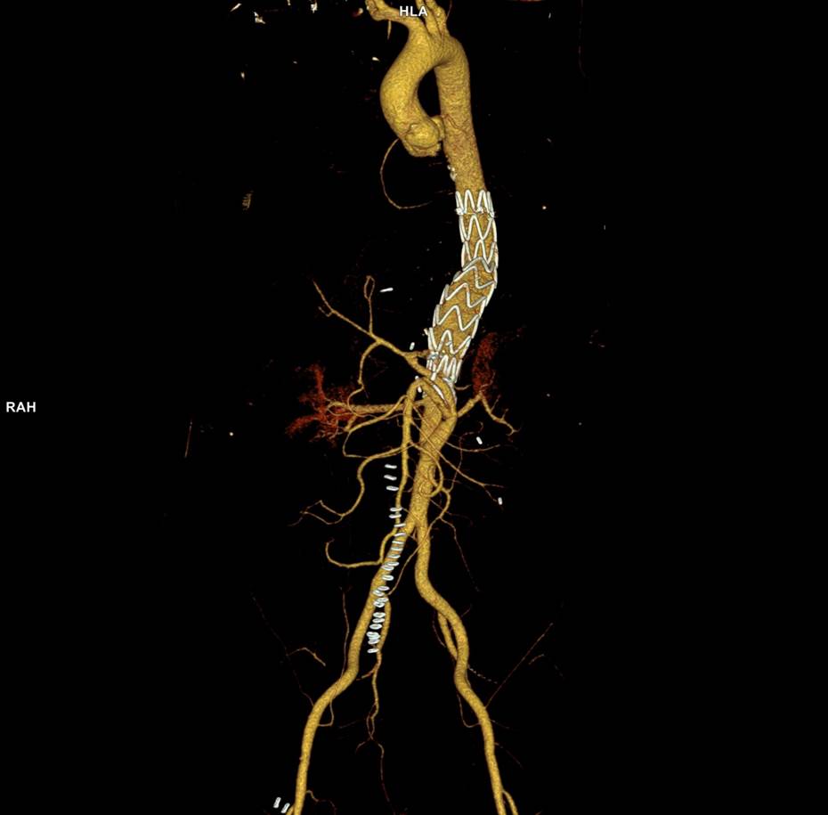
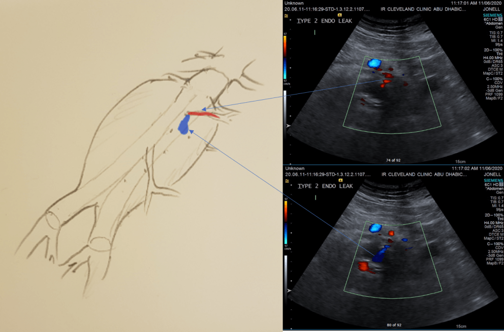
The EVAR was performed percutaneously. No endoleak was detected by completion arteriography (figure). He was soon discharged and was grateful. In followup, CT scan showed excellent coverage of the proximal and distal zones and absence of type III endoleaks. There was increased density to suggest a type II leak, but his inferior mesenteric artery was not the source of it. over a three year period, his aneurysm sac continued its 2-3mm of annual growth despite the presence of the the stent graft.

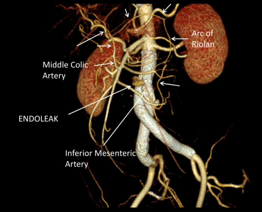
While CT failed to locate this endoleak, abdominal duplex ultrasound did showing flow from a small surface vessel (duplex below, figure at beginning of post). It was not the inferior mesenteric artery which can be treated endovascularly (link) or laparoscopically (link). CT scan suggested that it was one of those anterior branch vessels that one would encounter in exposing the aorta. Usually these were higher up as accessory phrenic arteries, but these fragile vessels, larger than vasovasorum, but smaller than named aortic branches, are seen feeding the tissues of the retroperitoneum.

Ultrasound revealed the type II endoleak from an anterior retroperitoneal branch artery.

Type II endoleaks are not benign. The flow of blood into the aneurysm sac after stent graft repair is almost never benign. It is a contained hemorrhage. There are three components to the pressure signal  seen by the aortic aneurysmal wall that could trigger breakdown, remodeling, and aneurysm growth. They include pressure, heart rate, and the rate of change of pressure. The presence of fresh thrombus may play an inflammatory role. Some endoleaks clearly have a circuit and others are sacs at the terminus of their feeding vessels, never shutting down because the AAA sac can both accept and eject the blood flow. Changes in AAA sac morphology due to sac growth can cause problems with marginal seals, component separation, and component wear. Sac growth can cause pain. Ruptures, while rare, can cause death. Mostly, type II endoleaks generate more procedures because it is hard to ignore continued growth.

Review of aortogram from device implantation showed a small anterior artery arising from the proximal aortic sac (arrow)

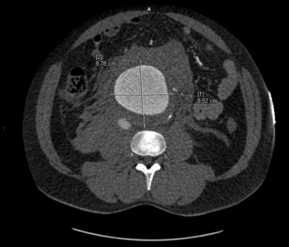
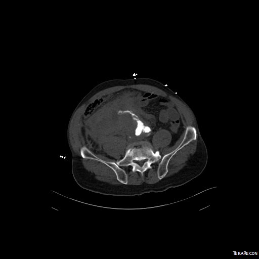
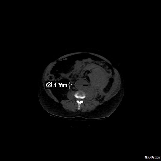
Three years of followup showed growth of the AAA sac to 5.5cm, which ironically threshold for repair. Again, no type I or III endoleak could be seen. He reached his calculated repair date, and I discussed our options in detail.

1. Do nothing, keep following

2. Endovascular attempt

3. Open surgery, marsupialization

4. Laparoscopic ligation of target vessel

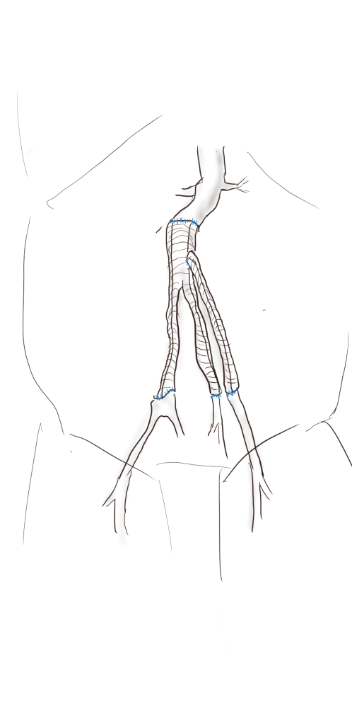
Doing nothing hasn’t worked for 3 years. What would more time buy? Endovascular -to where. The IMA is the usual target for an endovascular attempt, although iliolumbar access is possible (link), we really needed to fix this with one attempt. Open surgery is a great option -a short supraumbilical incision is all that would be needed to open the AAA sac and oversew the collaterals. The patient did not want a laparotomy. There are reports of laparoscopic guided endovascular access with endovascular coiling of the remnant sac with fluoroscopy. This adheres to the letter of the claim of minimal access, but really?

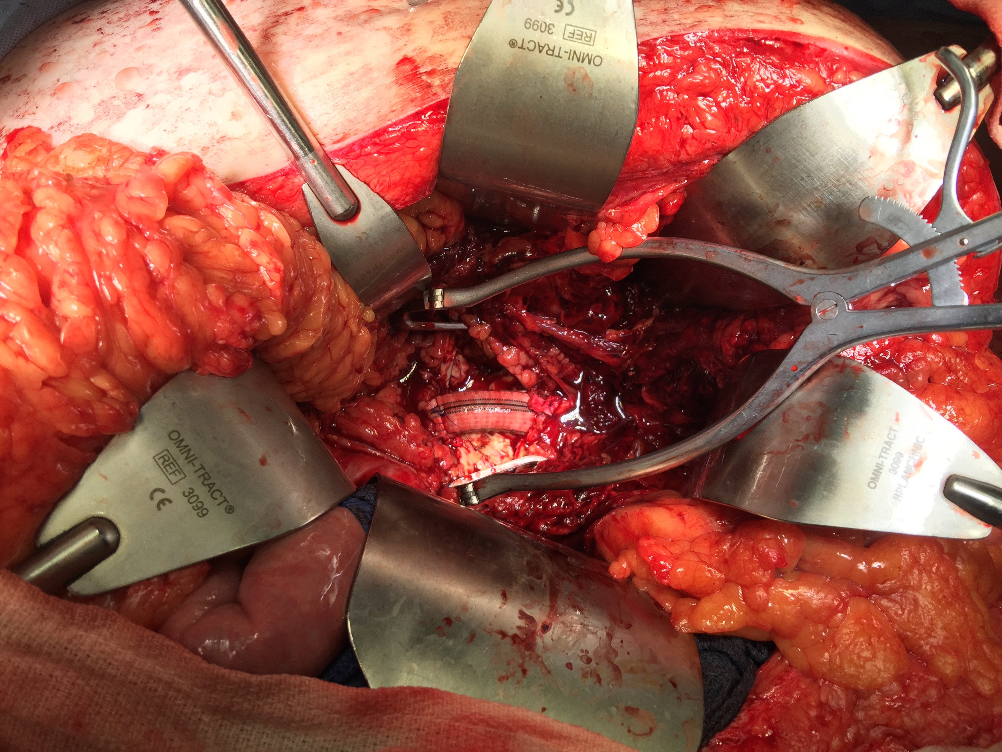
I compromised with the patient and offered laparoscopy. I have ligated the IMA a handful of times laparoscopically -these are relatively fast and straightforward cases. As I had the location of the endoleak, I felt it should be straighforward to dissect out the anterior sac much as in open repair and clip this vessel.

Use of ultrasound allowed localization of the leak and identification of the artery for clipping.

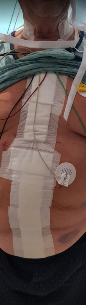
Of course, what should have been a 30 minute procedure through a minilaparotomy became a two hour enterprise getting through scar tissue (not the first time encountering this after EVAR) while pushing away retroperitoneum. I recruited the help of general surgery to get extra hands, but the patient was well aware that there was a good chance of conversion. Patience won out as the artery was ultimately clipped and endoleak no longer seen on ultrasound.

I waited a year before putting this together as I wanted CT followup. The sac stopped growing and has shrunk a bit back to 5cm or so. There will be those who argue that nothing needed to have been done about this leak as it would have stopped growing eventually, but I would counter that an aneurysm sac that kept growing like the stent graft never went in is one demanding attention. The key role of duplex ultrasound cannot be minimized. We have an excellent team of vascular scientists (their title in Europe), and postop duplex confirmed closure of the leak.

Not seeing the leak anymore is a positive, but the stent graft remains.

The patient is quite satisfied having avoided laparotomy. His hospital stay was but a few days. During my conversations with our general surgeons who are amazing laparoscopists, that this would have been a nice case with the robot. That’s a post for another day.

The definition of success in this case and many EVAR’s plagued by type II leaks leaves me wondering. Excellent marketing of the word “minimally invasive” has subtly defined laparotomy as failure, and not just in vascular surgery. When costs and efficacy are reviewed as we come out of this pandemic, I suspect that open surgery will selectively have its day in the sun. A ten blade, a retractor, a 3-0 silk is so much more cost effective than five ports and disposable instruments. And a stent graft system?

Maybe I am just a dinosaur.

Categories
AAA Commentary common iliac artery aneurysm complications CTA EVAR innovation ruptured AAA techniques training

Lifelong surveillance after EVAR -is it worth it?

About ten years ago, I had a patient who came to see me for moderate carotid disease. While his carotid disease was asymptomatic, he also had metastatic colon cancer. With colectomy, cryoablation of liver mets, and chemotherapy, he was in remission. Every 6 to twelve months he had some kind of CT scan with contrast. His renal function was poor and this was blamed on his chemotherapy. While it had nothing to do with this patient, I thought to myself, “Having an aortic stent graft was a lot like having metastatic cancer in remission.” After a stent graft, the patient is forever tied to the health care system. Without surveillance, there may be an endoleak, sac expansion, rupture, and even death. Patients and vascular surgeons can make choices that lengthen life, improve its quality, and avoid the complications of disease. But what if a treatment becomes a condition and a burden on healthcare resources and the patient’s finances?

Fool me once…

Type Ib Endoleak Causing re-Rupture of a previous r-AAA after no surveillance

Take this patient who had previously ruptured his AAA and undergone EVAR. Several years out from his rupture, he ruptured again from a type Ib endoleak due to aneurysmal degeneration of his right common iliac artery. Per his family, he never followed up. Perhaps he assumed he was cured of his disease? Repairing this was tricky, primarily because I hopped up and down, thinking, “I could cure this!” An open revision with a bifurcated graft would eliminate the need for EVAR surveillance, avoid abdominal compartment syndrome, and the physiologic consequences of a large retroperitoneal hematoma. But who wants a laparotomy? Not this patient, who was hypovolemic shock, and whose family chose the minimally invasive option that everyone assumes is better.

Not a clamp

I took him to the hybrid operating room, balloon occluding to stabilize his blood pressure, embolizing the normal internal iliac artery and extending the stent graft into the external iliac artery.

Completion -there is an Amplatzer plug in the right internal iliac artery

This patient stabilized and had abdominal tightness due to his large hematoma which did not need evacuation. After a stay lengthened by concern for abdominal compartment syndrome, moderate pain, fevers, and bilirubinemia (due to the hematoma), he was discharged and never showed up for followup. None of the phone numbers work. Without followup, EVAR is a menace. We will keep trying.

Regrets, I’ve Had a Few…

The great feature of EVAR is that the complications up-front at the time of surgery are wonderfully low. This patient pictured above here presented in middle age with a rupture into the retroperitoneum. He was unconscious and had hemorrhagic shock.

The decision to perform EVAR was made late in the transfer because I did not have the images from the transferring hospital (another subject for another blog post) so I set up for both open repair and EVAR. En route to the OR, I scanned, slowly, through the CT images sent via CD-ROM, and my internal discussion went something like this.

He’s a 50-something smoker in shock with a contained rupture of a 8cm infrarenal AAA with a good neck. Let’s take care of this in 30 minutes with a percutaneous endovascular aneurysm repair (p-EVAR).”

He’s a 50-something smoker in shock with a contained rupture with a good neck -let’s take care of this in 90 minutes with a tube graft, open aortic repair (OAR).”

With p-EVAR, he’s going to have just two groin punctures and much lower complication rate, shorter length of stay, similar to lower mortality. Look -his blood pressure is 75mmHg systolic!

That hypotension is permissive to minimize bleeding. With OAR, he’ll avoid abdominal hypertension and complications of a giant hematoma. Because he’s young, he’ll avoid lifelong surveillance. If anyone can clamp this AAA, it’s me...”

Pride cometh before the fall. Get this man off the table and figure out the logistics later. p-EVAR. You open him up, he’ll exsanguinate and expire before you get the clamp on.

I sighed, looked up at the gathered team, and announced, “p-EVAR.” The percutaneous EVAR is something I’ve been doing since 2004, long before it was a big deal, and we were done under an hour. His blood pressure stabilized, but general surgery was consulted for his abdominal compartment syndrome. With sedation, fluids and time, his urine out put recovered but his belly remained distended and his bladder pressures which were never seriously elevated, trended down.

It was made known to me that the patient had very limited insurance making followup surveillance challenging. Due to his coverage, he had to have his imaging done at designated hospitals, so I wrote a detailed note -basically the timings of his followup CT scan, and asked that the reports should be sent to me. I ordered a CTA prior to discharge which showed a type II endoleak adjacent to the graft and connected to both lumbar and inferior mesenteric arteries (first image above). After some thinking, I took the patient for an aortogram, accessed the IMA via the SMA and coiled into his AAA sac and the IMA.

It was only a few weeks ago one of my colleagues across town contacted me that the patient had been admitted with abdominal pain, a CT showing a type II endoleak from his lumbars, but a smaller AAA sac than his pre-repair size of 8.5cm. The patient is seeking to repatriate, and I doubt he would be able to get adequate followup in his home country without paying in cash. Happy that the patient survived his rupture, I still have persistent regrets at not getting him through an open repair, which I am sure he would have done fine with… Or maybe not.

Je Ne Regret Rien…

Recently I admitted a patient, in his 70’s, with a symptomatic 6.5cm infrarenal AAA with bilateral common iliac artery aneurysms, the right being 25mm, the left over 30mm. Because of the pandemic, he was stuck here, seeking to repatriate. Over ten years before, he had a segment of descending thoracic aorta repaired for a traumatic tear -probably one of the last before the wide adoption of thoracic stent grafts which work great by the way.

Cardiac risk evaluation revealed an ejection fraction of 35% with reversible ischemia on nuclear stress test. CTA of the coronaries revealed triple vessel coronary artery disease corroborated by catheterization. Off-pump CABG was planned which would eliminate the effects of cardiopulmonary bypass.

Preparations were made for EVAR with IBE of the left iliac aneurysm as a contingency, but there was no question that if the patient recovered well from his CABG, he would undergo open repair. This was because bell-bottoming or IBE must have regular coordinated surveillance which was not going to be easy with the patient leaving for another country in the middle of a pandemic. It is difficult to get followup to happen in normal circumstances (see above cases). I expressed my opinion to the patient and family and we agreed to see how the patient responded to off-pump CABG.

He underwent off-pump CABG with three vessels revascularized. He was extubated POD#1 and mobilized. By POD #4, he was on a regular patient floor, being co-managed by hospital medicine who takes care of all of our patients. The patient expressed readiness for the next operation. His kidney function remained normal. He was transfused 2 units of PRBC to bring his hematocrit to 30%. He was taken off Plavix, but kept on aspirin. On POD#6, he was taken back to the operating room for open aortic bypass. This would not have been possible without close coordination of cardiology, cardiac surgery, and vascular surgery. Choosing off-pump CABG was a critical element in being able to proceed with open aortic surgery.

Right branch taken to iliac bifurcation, separate bypasses sent to left internal and external iliac bypasses.

I do several things to decrease the physiologic impact of the operation. First is keeping all the viscera retracted under the skin. This simple move has the effect of decreasing the rate of intestinal paralysis and amount of fluid shifts that occur postop, akin to going retroperitoneal. This decreases the space you potentially have if you use standard clamps, but I use the Cherry Supraceliac clamp (image), DeBakey Sidewinder (transverse), or just a Satinsky clamp oriented transversely. This minimizes the occupation of volume over the anastomosis which always happens with standard aortic cross clamps. The anastomosis is easier without the clamp taking up valuable space.

Cherry Supraceliac Aortic Clamp

The iliacs are always clamped with Wylie Hypogastric clamps, again, with the principle of eliminating clamp overhang. Suturing is done with 4-0 Prolene on SH needles -this is plenty (link). The proximal anastomosis wants to bleed, and sewing to a fully cut ring of aorta ensures good posterior bites but also allows for sliding a band of graft down over the anastomosis (Dan Clair calls this a gusset) which works well at creating a hemostatic proximal anastomosis rapidly-trust me, getting this done well is the key step of the operation. Before closing, I infiltrate the rectus sheath and preperitoneum bilaterally with local anesthesia -lidocaine 1% with epinephrine 1:1 with bupivicaine 0.5%. The skin is closed with absorbable dermal sutures because staples create as many problems as they solve. The patient had cell salvage through the case and no extra units of transfused blood.

The patient was extubated that night and started on clear liquid diet. The next day his lines were removed and he was moved to the floor and started on regular diet when he expressed hunger. On POD#2, he was pacing floor, asking when he could be discharged.

Sternotomy and Laparotomy POD#2, walking the floor

As he was eating, walking, talking, breathing, evacuating bowel and urine, and pain free (well controlled), I saw no reason to keep him beyond POD #3 AAA/#9 CABG. I have kept in touch with him and his family and he is doing well and has given permission for this posting.

Satisfaction

This final case has confirmed several of my beliefs. First, calling something high risk can drive one to make bad choices and in fact endanger patients. This last patient would qualify as high risk on any international criteria, and you would not be wrong in quoting upwards of 30% major morbidity and mortality for cardiac revascularization and AAA repair, but you would also be tying your hands from offering the best solution for this man who fortunately was able to undergo two prodigious operations. He will not require much in the way of followup. Coronary revascularization with arterial conduit and open aortic grafting frees him from the need for close followup and reassures us that his repair is durable.

Second, calling something advanced and minimally invasive gives one cachet in the marketplace but forgoes careful discussion and consideration of what is being abandoned. The first two patients survived their ruptures but now face the consequences of having stent grafts. It is a shame when podium speakers at international symposia declare surgery to represent failure because this affects training by encouraging abandonment of hard to acquire skills. It seeps into patient perceptions and expectations. I hope that a balanced approach prevails. You have to be capable of both open and endovascular approaches to be able to offer the best treatment for a particular patient.

Finally, these old operations are cost efficient and there is a lot of room to improve these procedures with new perspectives, techniques, and data. I don’t operate the same way I trained, and it is only through continued application of operations that improvements can come about. As budgets tighten and economies are stressed, cost efficacy will rein in much of the interest and demand in new stuff unless it adds value. That said, I am grateful to our stent graft representatives who have worked to get us bell bottoms and IBE’s for when they will be needed. These grafts will be used when the time is right.

Categories
AAA EVAR hybrid technique mycotic aneurysm ruptured AAA skunk works techniques training

The Hands of a Surgeon

My partner, Lee Kirksey, Vice-Chair of Vascular Surgery, just got a paper on-line (link) about the curiously increasing volume of open surgical repairs we were experiencing from 2010-2014 at the Cleveland campus of the Cleveland Clinic. When I joined in 2012, my impressions at that time were mostly the paucity of straightforward EVAR cases that I had seen in private practice, and the high prevalence of stent graft explantation, infected aortic grafts, and open aortic aneurysm repairs (OAR) for juxtarenal and thoracoabdominal aortic aneurysms for nominally high risk patients. My TAAA muscles had atrophied during my years out of fellowship and I eagerly took the opportunity to recruit the help of my partners and scrub in on these cases with Pat O’Hara, Jean Kang, Dan Clair, Ezequiel Parodi, and Lee Kirksey. It is without any shame that I sought out not just extra expert hands, but interrogated these experts for different ideas and approaches, and absorbed feedback. At the time, I was ten years removed from graduation, a full-fledged vascular surgeon who thought he could do any operation put before him. I cannot imagine the thoughts churning through the head of a recent graduate faced with the choice of taking on an open aortic operation with only 5 cases under their belt, referring the case on to the regional tertiary center, or trying to McGyver an endovascular solution. I contributed probably about 35-50 cases to this paper, but the outcomes were a collective effort. Even today, I will run cases by Sean Lyden, Christopher Smolock, or Lee, if only for the company and gossip.

“We explain this distribution of cases as a function of several factors: a unique, broad regional quaternary referral practice whereby patients with complex aneurysmal disease are referred to our institution; an institutional practice evolution resulting from a critical analysis and understanding of EVAR failure modes that lead to explantation, thus generating a different perspective in the EVAR vs open decision-making process; a parallel high-risk IDE fenestrated graft study; a historical willingness to accept all physician and self-directed patient referrals (ie, a willingness to manage more complex cases); and a published expertise in the area of EVAR device explantation with an annually growing volume of commercial device removals” -from El-Arousy et al.

Reading through that paper, I have come to the conclusion that the increasing open aortic volumes at the Cleveland campus has as much to do with the ongoing retirement of experienced surgeons regionally as it does with the ability to attract these cases. Loss of these surgeons has a cascade effect like losing a species in an ecosystem. The operating rooms forget where the OMNI retractor is because nobody asks for it anymore. The ICU’s are no longer familiar with the ebb and flow of the postoperative open aortic operation. The floors lose institutional memory of the care of these vascular patients as the stent grafts and interventions go home within 48 hours, sometimes the same day.

If you were a vascular surgeon born before 1970, your approach to the scenario of the ruptured abdominal aortic aneurysm may differ substantially from that of surgeons born after the Carter administration. Most of my cohort, Gen-X and older, feel comfortable applying some betadine, opening the belly and placing a clamp. Those younger than us have told me they feel more comfortable putting up a large balloon and deploying a stent graft. In this generation, it is normal to call a general surgeon to decompress the abdominal compartment syndrome and manage the abdominal vacuum dressing. For our generation, the giant industrial robot arms and 80 inch monitors creates a barrier to the problem at hand, and gathering all the extra staff after hours and on weekends requires the logistical skills of a wedding planner.

We prefer an operating table, a willing anesthesiologist, a cooler full of O-neg blood, Prolene and a graft, strong suction, and an extra set of hands. The data suggests either method is equivalent in outcome, but I would argue that depending on the circumstance, there is an optimal method for that patient and you have to have the ability to do either open or endovascular or some hybrid combination. Unfortunately, it is clear that open vascular surgery is year over year diminishing, and and it might not be so great when we start rupturing our aneurysms.

The open approach is preferred because we got good at it by doing a lot of these cases. Your hands -it becomes natural to change the course of the disease and the fate of the patient with your hands. One of the things you lose with a wire based approach is the tactile feedback from the organ that you are treating. Yes, there is a subtle feedback from the flexible tip of a Glidewire, but that’s missing the point. The thing that is rarely considered with open surgery is the tactile aspects of operating.

Your fingers are your point of care ultrasound. As an intern, one of my earliest tasks was reaching in through a 2cm incision with my index finger, feeling for what I would describe as a rotten shrimp, and delivering it out by hooking my distal phalanx around its base. Adhesions were rubbed like money between finger and thumb to judge if you could bovie through it. If you felt a sliding sensation, it was mucosa to mucosa and you looked for another spot to cut. Into my fellowship which could be called the triple-redo, no-one else wants to do-, difficult vascular operations fellowship, the pulse or the plaque felt under the finger would guide me to carve away scar tissue from blood vessel, visualizing the feedback from the fingers. In a rupture, with the belly under a dark mire of blood, there is no seeing, only feeling. Your hands reach into the lesser sac or transverse mesocolon and strangle the aorta -your fingers while clamping, feel and avoid the caudate lobe, the NG tube in the esophagus, and split the crura of the diaphragm like a pick pocket. Once the pulse returns as anesthesia refills the tank, you scratch free the aorta with your thumb and forefinger, then slide the jaws of the aortic cross clamp over your fingers and against the spine and clamp. This takes about 60 to 90 seconds (link).

When a patient is bleeding out, this is the way to control the bleeding. In practice, no amount of rehearsing for getting a patient into a endovascular suite, getting airway and access, swinging in the industrial robot arm, and getting everyone into lead aprons, sending up wire, placing a 12F sheath and an aortic occlusion balloon, will be satisfactorily smoothly and efficiently as a STEMI or STROKE alert. The rAAA is for most hospitals, unless you are in Seattle, a once in a while occurence. Many more people can find a scalpel and an aortic clamp than they can find a 32 inch aortic balloon, a stiff exchange length wire, and a 12F sheath.

When a patient presents with a slowly bleeding, contained rupture, there is time to assemble the teams required for an endovascular repair, and for opening and decompressing the abdomen, for anesthesia to get IV’s, central lines, arterial lines, and order crossmatched blood. One has the time to get and review CT scans and choose grafts. One can even do things backwards, debranching after securing the leak (link) with a stent graft. The luxury of time should signal to you that the endovascular option is the preferable route, as all the advantages of minimally invasive repair are possible. Rural hospitals sending patients two hours by ambulance or arranging for a helicopter -this is the great filter through which those likely to survive make it into the endovascular suite. These patients do great with EVAR, because everything moves more or less like a routine elective EVAR.

The setting up the operating room for tackling rAAA is quite simple. Keep everything nearby. Nothing should ever be stored out of sight, retrievable only by arcane codes whispered in the ears of people down in the basement or across the street. Amazon gives itself a day to get that gadget to you, but the rupturing patient does not have the time to have a 28mm stent graft ordered by looking up a Lawson number, finding the materials person in the faraway room to find it, running it over a city block. The stuff has to be next to the OR. Every scenario is unique, and the best planning is assuming no one person knows where everything is but everything is close at hand -major vascular sets, retractors, C-arm, cell saver, stent grafts, open grafts, balloons, cardiopulmonary bypass -every gewgaw is few steps away. The inventory is what you see, because if you can’t grab it, it does not help the hemorrhaging patient.

If you are a vascular surgeon born after 1980, it is likely that you may have trained in a 0-5 residency and all the old people harumphing about the old ways seem biased. Rather than being rational about their awful upbringing, the old people seem to suffer from Stockholm Syndrome, turning from victims of a harsh and brutal system inherited from the original, Halsted, a cocaine addict, to willing collaborators now mooning about the good old days of every other day call and 120 hour work weeks.

There might be a growing suspicion about advocating for open surgery when fewer people can perform it. One of the truisms of surgery is if only one surgeon claims to be able to do a rare operation with great results when everyone else abandons it, like venous valve surgery or robot assisted mastectomies, it can mean that surgeon might be uniquely talented or shamelessly selling something. It is a shame that open vascular surgery is devolving into that category of arcana, like the Jedi. I have no doubt that the last open vascular surgeon will be a reclusive, bitter, wild eyed hermit like Luke Skywalker was in episode VIII, if we let it get that far.

Bald eagles were saved from extinction. The methods of species reclamation may be what is needed to save open vascular surgery. Financial metabolism drives behavior, and there must be recognition of the time and dedication required to perform good open vascular surgery in the form of increased RVUs and reimbursement. The surgeons retiring in their mid 60’s with straight backs and steady hands need to be incentivized to stay around and coach the next generation in the ways of the Jedi. Call it the master surgeon designation. Every 0-5 graduate needs to focus on getting 100 leg bypasses, 50 carotid emdarterectomy, and 25 open aortas within the first five years of practice with a master surgeon if they did not get this experience during training. Like dead Jedi, it would help even if they were just virtually present, shimmering on Facetime in their (bath)robes to go over planning and approaches, but being physically present and reimbursed for it would make the most sense.

There is always self service in any human activity. One mildly prominent vascular surgeon that I have come across is famous for saying he did not have a vascular fellowship because he did not want to train his competition. It is easy for the fifty somethings to sit and proffer their open skills and profit from its scarcity but it goes against decency to not pass on this collective body of hard won knowledge and skills. There must be stewardship of this great thing we do, this honorable and treasured endowment.

Of the concrete ways we are trying is creating a network of advanced open surgery capable surgeons regionally organized by Martin Maresch, capitalizing on social media and electronic communications. Here at CCAD we are in the organizing phase of a vascular residency, and I very fortunate to have Houssam Younes join us as he shares my interest in surgical education and open vascular surgery. We are contemplating a non-accredited fellowship. We have general surgery residents coming through our service as well as medical students.

One of my mentors told me, “I can train a monkey to do cardiac surgery,” as he was training me to do cardiac surgery. And he was right. The final comment I have is you have to demystify surgery, take away the Instagram perfection, the romance, and list in practical terms the toolkit of maneuvers that form the component parts of all operations and propagate it. Let us not kid ourselves. The technical skills of surgery can be taught to anyone. The Mayo brothers were performing surgery as teenagers before medical school. The knowledge and experience and judgement -that varies as much as people vary and we have a curriculum for that, but the physical acts of surgery need to be taught starting at the medical school level. Standardized drills and exercises need to be created so that proficiency can be metered.

“The individual per trainee OAR volume did not decrease during this period. In the training program, the use of “component separation” (separation of each
operation into discrete, instructionable steps that facilitates trainee mastery) is integral to instruction of open aortic aneurysm repair techniques and permits the
trainee to master all of the technical exposure and repair skills necessary to address and to manage both straightforward and complex aneurysm anatomy. Component
separation is essential to maximize trainee experience across all levels” –from reference 1

Here is my list of things a trainee must accomplish by the time they graduate from a vascular residency or fellowship.
1. Tying knots with gloves on with 6-0 Prolene inside a pickle jar without lifting or moving a 12 ounce lead fishing weight to which the suture is being tied, fast, one handed, two handed, left and right handed.
2. Holding forceps, needle holders, and clamps
3. Correct operation of the OMNI retractor, Weitlander retractor, Balfour retractor, Thompson retractor
4. Incise skin through dermis through correct depth and length with both #15 and #10 blade
5. Open the abdomen through midline and flank incisions and close these incisions
6. Harvest saphenous vein
7. Vascular anastomosis on a table, inside a pickle jar, inside a short Pringle’s can
8. Dissection of adhesions and scar tissue around blood vessels and organs
9. Dissect and expose the common femoral artery via vertical and oblique incisions and close these incisions
10. Dissect and expose the carotid bifurcation, left and right side, and close these incisions
11. Dissect and expose the tibial vessels in various parts of the leg and foot
12. Dissect out the brachial artery at the elbow
13. Dissect out the axillary artery and vein below the clavicle
14. Dissect out the axillary artery and vein from the axilla
15. Dissect out the subclavian artery, vein, and brachial plexus above the clavicle
16. Dissect out the arm veins
17. Dissect out the iliac artery via a lower quadrant pelvic retroperitoneal exposure
18. Dissect out the abdominal aorta via midline laparotomy
19. Dissect out the abdominal aorta via retroperitoneal approach
20. Dissect out the thoracoabdominal aorta via a thoracoabdominal exposure
21. Dissect out the popliteal artery via suprageniculate, infrageniculate incisions and prone position
22. Dissect out the inferior vena cava
23. Dissect out the iliac veins
24. Harvest deep femoral vein
25. Temporal artery biopsy
26. Endarterectomy of carotid, femoral artery, any artery with patch angioplasty
27. Exposure and control of supraceliac aorta, suprarenal aorta for clamping
28. Exposure and control of thoracic aorta
29. Exposure and control of the great vessels via sternotomy and supraclavicular incisions
30. Exposure and control of the vertebral artery
31. Safe removal of vascularized tumors
32. Amputations of digits, legs and arms up to pelvis and shoulder
33. Exposure and control of radial and ulnar arteries
34. Hand surgical techniques of exposing arteries, tendons, and nerves in forearm and hand
35. Plastic surgical techniques of skin grafting and basic rotational flaps
36. Fasciotomy of arms and legs, hands and feet.
37. Exposure and control of celiac axis
38. Exposure and control of superior mesenteric artery
39. Exposure and control of left renal vein
40. Exposure and control of hepatic veins, portal vein
41. Exposure and control of renal arteries
42. Exposure and control of profunda femoral arteries
43. Safe removal of spleen
44. Transabdominal retroperitoneal exposures of the abdominal aorta and inferior vena cava
45. All of the above in a reoperative field
46. All of the above with limited visualization and by sense of feel only
47. Laparoscopic and thoracoscopic techniques
48. Orthopaedic surgical techniques of myodesis, bone grafting, precision osteotomies, infection control, external fixation, spinal exposure
49. Safe resection and anastomosis of bowel
50. Drainage of infection
51. Intensive care of SIRS, MOFS, CHF, Septic shock, postoperative fluid shifts
52. Nonsurgical and surgical management of lymphedema, seromas, and edema
53. First rib resection
54. Spinal exposure
55. Organ harvest and transplantation
56. Planning of complex open, hybrid, and endovascular procedures

Every year, it is apparent that endovascular options suffer from some flaw when outcomes are studied beyond 2 years, but progress will march on in that sphere. It has to. The loss of open capable surgeons to early retirement is accompanied by overapplication of endovascular techniques at least partly due to the lack of knowledge of these open surgical options and achievable good results and partly due to financial incentives. The solution lies in redistribution of reimbursement to open procedures and creation of open surgical fellowships and identifying and empowering mentors who still walk among us.

Categories
acute mesenteric ischemia aortic dissection bypass chronic mesenteric ischemia complications ruptured AAA taaa tbad techniques TEVAR thoracabdominal aortic aneurysm training type b aortic dissection visceral malperfusion

Life imitates social media: a ruptured type V thoracoabdominal aortic aneurysm or IT saves a life

ARTERIAL (2) (2)
Type V TAAA Rupturing into Right Pleural Cavity

It was only last month when I came across a post of an aortic aneurysm in a difficult spot (link) and I couldn’t help chiming in some comments. Reading it now, I sound insufferable, because I wrote,

“Depends on etiology and patient risk stratification. Also assuming aneurysm goes to level of SMA and right renal artery origins and involves side opposite celiac. Options depending on resources of your institute: 1.Open repair with cannulation for left heart bypass and/or circulatory arrest 2.Open debranching of common hepatic, SMA, R. Renal from infrarenal inflow and then TEVAR 3.FEVAR from custom graft from manufacturer on protocol 4.Parallel grafting to CA, SMA, R. RA with TEVAR 5.Surgeon modified FEVAR 6.Open Repair We would have a multidisciplinary huddle around this patient -Vascular, Cardiac Surgery, Cardiology, Anesthesia, and ID (if needed) to help choose. Be ready to refer to a center with more resources -including prepping patient for transfer and imaging -including uploading images to the cloud for transfer with patient’s permission. More info please”

More Info Please, Indeed

The post I commented on was of a saccular aneurysm in the transdiagphragmatic segment of aorta. Ironically, only a few weeks later, I got called from hospital transfer center about a patient with a leaking aortic aneurysm, a type V thoracoabdominal aortic aneurysm as it turned out, from an outside hospital, needing urgent attention, and we accepted in transfer based on the conversation I had with the tranferring physician. And that was the problem -usually in taking these inter-hospital transfers, you have to pray that the precious CT scans come along with the patient burned correctly onto a CD-ROM. You can buy and watch a movie in 4K resolution over the internet -about 4 gigabytes, but a patient’s CT scan which is about 200 megabytes -because of various self imposed limits, overly restrictive interpretations of laws, and lack of computer skills, these life saving images get transferred on CD in 2019. That last time I purchased a CD for anything was over 15 years ago.

An Interested Party

The technical solution –to use the internet to transfer critical life saving information between hospitals – came about when our IT folks took an interest in my quarterly complaint email about using the newfangled internet for sharing files. After mulling various solutions ranging from setting up a server to using commercial cloud solutoins, we came upon the compromise of using our internal cloud with an invitation sent to the outside hospital. I would send this invitation to upload the DICOM folder of the CD-ROM to an outisde email address. It was simple and as yet untried until this night. “Would the patient agree to have his CT scan information transmitted to us electronically?” I asked the other physician. He assured me that the patient was in agreement.

It Takes Two to Tango

Of course, being able to transfer these pictures requires a willing partner on the other side, and the referring physician made it clear he did not have the technical expertise to do so. It took a bit of social engineering to think about who would have that ability. Basically, aside from myself, who spend all their time in dark rooms in the hospital in front of giant computer monitors? The radiologists! I got through to the radiologist who had interpreted the report and explained the simple thing I needed. Gratefully, he agreed, and I sent him a link to our cloud server. I explained to him, “When you receive this, clicking the link opens a browser window and then you open the CD-ROM and find the DICOM folder and drag and drop it on the browser window.” The 200-500 megabytes of data then get sent in electronic form, as it was meant to in 2019.

The Internet Saves a Life

The brutal truth is that in locking down a computer system, many hospitals make it impossible to even load an outside CD-ROM, creating many self imposed barriers to care. Thankfully, at CCAD, we were able to work together to find a secure solution. With the CTA on our servers, I was able to review the study within 15 minutes of accepting the patient, and arrange for the right team to be assembled, and confirm that we had the right material (stent grafts) for treating the patient. When the patient arrived, OR was ready to go, saving hours of time that normally would have been required if the CT scan had to be reviewed from the CD-ROM that came with the patient. Sometimes, the CD-ROM does not come, and in a critical situation, the CTA has to be repeated with some risk to the patient for complications of the contrast and radiation.

What to Do

The patient had a 8cm sphere shaped aneurysm arising in the transdiagphragmatic aorta, leaking into the right pleural cavity.

Screen Shot 2019-06-19 at 9.29.15 PM

The patient was otherwise a healthy middle aged man with risk factors of smoking and hypertension. The centerline reconstructions showed the thoracic aorta above the aneurysm to be around 20mm in diameter and same below, with the celiac axis and superior mesenteric artery in the potential seal zone of a stent graft. The only plaque seen was around the level of the renal arteries and was focal and calcified. Looking at the list I had made as a comment to the Linked-In post, I realized that I really only had one viable option.

ARTERIAL (10).jpeg

Open repair, usually the most expeditious option, was made challenging by the right hemothorax, making a left thoracotomy hazardous if the lung had to be deflated. Cardiopulmonary bypass would have to be arranged for, and that adds a metabolic hit that greatly raises the stakes. Of the endovascular solutions, the only viable option was TEVAR to exclude the rupture and debranching of the celiac axis and superior mesenteric artery. To those who would advocate for parallel grafts, there was no room in the normal 20mm diameter aorta. And branch systems for rupture are some time in the future. Also, the patient was becoming hypotensive. So the planned operation was first TEVAR to stop the bleeding, and then open surgical debranching. A hybrid repair.

plan sketch.PNG

The smallest stent graft we have is a 21mm graft, but it would not be suitable for this aorta. In practice, the normal aorta is quite elastic and will dilate much more than what is captured on a CTA. The next size we have is 28mm graft and I chose this to exclude the rupture, which was done percutaneously.

TEVAR angio.png

As seen below, the graft excluded the celiac and SMA. Late in the phase of the final aortogram (second panel) there was an endoleak that persisteed despite multiple ballooning. The timing suggested the intercostals and phrenic vessels contributed to a type II endoleak, but it was concerning.

postangio.png
Find the Endoleak

The bypasses were sent from the infrarenal aorta to the common hepatic artery and the SMA close to its origin, and the origins of the celiac axis and SMA were clipped. The bypasses were then done with a 10x8mm bifurcate Dacron graft originally for axillofemoral bypassing. It had spiral rings which I removed at the anastomosis and this resulted in a kink at the closer bypass. Usually, I loop this for iliomesenteric bypass but there was not enough distance from the infrarenal aorta. I have to add a little trick I modified from my pediatric surgery experience as a resident -a Heinecke-Mikulwicz graftoplasty:

IMG_2646.JPG

This worked to relieve the kink as evidenced on the aortogram above. After closing the laparotomy, I placed a chest tube in the right chest. The patient had a course prolonged by a classic systemic inflammatory response syndrome, with fevers, chills, and leukocytosis. He bled for a while but stopped with correction of his coagulopathy. All blood cultures were negative, but a CT scan was performed out of concern for the endoleak, and the possibility of continued bleeding.

postCTA.jpg

No endoleak was detected as the sac was fully thrombosed. There was a consolidation of the blood in the right chest, but it resolved with fibrinolytic therapy.

Discussion

This case illustrates several points I have been making on this blog.

  1. Hybrid repairs are not some kind of compromise but the full realization of a complete skill set. When students ask me how to judge a training program, one of the best metrics is how frequently are hybrid operations performed. It means either the endovascularist and open surgical operator are in complete synchrony or there are individual surgeons competent in both open and endovascular surgery. Hybrid operations, rather than being a compromise, are an optimization.
  2. Time -Laying the stent graft across the celiac and SMA origins starts a warm ischemia clock. The liver and intestines, in my reckoning, should be able to tolerate the 2 hours of work to get the bypasses working. Cardiopulmonary bypass may give you less ichemia but at the metabolic cost of the pump time. These negative factors add up, but were surmounted by the fact that early control of hemorrhage was achieved. Stopping the bleeding and restoring flow are the core functions of vascular surgery.
  3. Planning and preparation. The ability to see the CT images and prepare the teams and materiel before the patient transferred was lifesaving. This is where our IT gets credit for responding to a critical need and formulating a solution that meets internal policies, external regulations, and saves a life. It illustrates so many opportunities particularly with electronic medical records and their processes which focus more on documentation for billing. A discharge summary should be multimedia like this blog post and it should be normal and easy to generate. And finally, as clinicians, we should mind technology with as much attention as we give to the latest medical devices and techniques.
Categories
AAA common iliac artery aneurysm iliac artery aneurysm ruptured AAA training

If the odds are against the patient, who is for the patient?

IMG_1484

The first patient, a man in his late 70’s, ruptured in the emergency room at around four in the afternoon on a weekday, which was fortuitous, as the hospital was fully staffed, fully armed. The patient had arrived only a bit earlier with the complaint of severe abdominal pain, and soon after getting his CT, arrested. CPR commenced as I arrived by Dr. Timothy Ryan, our chief resident at that time. 

Ruptured CIAA with CPR 1 -_1
a rupture
The patient was wheeled upstairs with ongoing chest compressions. The anesthesia and operating room staff started a bucket brigade of blood -there was enough staff to start a symposium. Within 5 minutes of hitting the operating, I poured betadine on the chest and belly and took a blade and cut open the abdomen. Blood poured out onto our scrubs and to the floor and our shoes. I pushed my hand into the retroperitoneum, gently sweeping aside the torn tissues and blood clot to feel the hill of the aneurysm. I walked over the slope of the aneurysm and tweedled my fingers around the aorta above the aneurysm. The cross clamp rode my fingers into position around the aorta. The patient, so very dead minutes before in the ED, came back as I began to feel a pulse above the clamp. The patient lived through the operation and the night where grim data -pH of 6.8, lactates in the double digits, four figure LFT’s, kidney failure all predicted a bad outcome. And yet he survived, and a few days later, a second operation to washout and close his belly which had been left opened and packed occurred, and he recovered. We still talk about that day now three years out, and while he thanks me, I thank the whole hospital because I don’t remember speaking very much -the right things just happened around me as we worked, the whole hospital and me.

More recently, while I was finishing up two urgent cases, I got a call that the patient with the leaking aneurysm had arrived from across town and was becoming hypotensive.

ruptured AAA -_1
another rupture
Gratefully, one of my partners, Dr. Christopher Smolock, was rounding that Saturday and stepped in to finish up the last of the two cases while I ran down to the patient, a man in his late sixties, who had arrived in our acute aortic syndrome unit.

IMG_7195 (1)
Dr. Christopher Smolock
We conversed, the patient and I, and he understood what laid ahead. We rolled him up to the OR, and while we were prepping and draping, my fellow, Dr. Francisco Vargas, looked to me gravely and said with certitude, “I think he’s dead.

IMG_7197
Dr. Francisco Vargas
Chest compressions commenced and again, knife in hand, I cut him open from sterum to pubis and got the clamp on. It took 15 minutes of CPR to get a pulse back. I was very pessimistic as during the case, ridiculously bad lab data came back like a pH of 6.9, lactate above 10, no urine.

IMG_7234
Graft Repair of Rupture
The blood bank sent down coolers like the kind you take to tailgates, only filled with blood and plasma. The aneurysm had grown like a rotten apple on a stick and the graft we needed to repair it was surprisingly short. He too made it to the ICU, and after a long recovery which included dialysis, a tracheostomy, and a reboot of the brain -the brain takes a while to recover from the anoxia, but his went “bonnnnng” like a waking Mac after days of spinning beach balls, and he started to follow commands. The morning before he transferred to rehab, we talked about what he could have done to prevent the rupture. Not knowing about it, not much, I replied. People traditionally lived to about 20-30 years of age, I said, before dying of disease, violence, or predation. Longevity has meant wear and tear on irreplaceable parts. We agreed it was good to be alive.

Ruptured aortic aneurysms are the sine qua non of vascular surgical practice. As a junior resident back in the antedeluvian 90’s, I remember one of my chiefs, Dr. Eric Toschlog, now a trauma surgeon out East, running a patient upstairs from the ER with a rupture, and before the attending arrived by taxi, had the graft in. When it became my turn, as a fellow working on a patient who had been helicoptered in from the frozen wastes of Minnesota, I remember playing a trick with my mind -that the patient was proportionally the same size as the rabbits I was working with in the research lab, that I was really big and the patient’s aneurysm very small. This works to calm the heart, steady the hand. Nowadays, my mind is blank, and my hands working reflexively.

There has been a series of papers that create scores that allow prediction of odds for survival, and both of these patients, particularly with their prolonged CPR, have greater than 90% predicted mortality on these measures. In this month’s JVS, Broos et al, in the aptly named paper, “A ruptured abdominal aortic aneurysm that requires preoperative cardiopulmonary resuscitation is not necessarily lethal” describe a 38.5% survival rate among their series of patients with rupture who had CPR (ref 1).

Practically speaking, no one I know would use these scores to decide to not operate. While many series show better survival for emergency EVAR compared to open repair, several randomized control trial failed to show better results when these methods were directly compared. A retroperitoneal approach is preferred by some in our group, but having tried both closed chest CPR with the patient in right lateral decubitus position and open cardiac massage -(both died), I prefer supine.

There is no survival if there is no attempt.

Reference

  1. J Vasc Surg 2016;63:49-54.