In George Orwell’s Animal Farm, under the dictatorship of the alpha pig, Napoleon, the pigs who represented the nomenklatura of the farm chant the slogan “four legs good, two legs better,” after initially declaring “four legs good, two legs bad” during their revolution. They clearly understood the luxury afforded by a bipedal lifestyle, because in rising on two legs, you get arms and hands which can do many things like caress a baby or wield a cudgel. What the pigs in the parable weren’t realizing were the consequences of a bipedal lifestyle.
When Lucy, the Australopithecine, bipedal hominid ran about on two legs, she did have the use of two arms and hands. Possibly an adaptation to living in tall grasslands with few trees, the ability to stand tall like meerkats, allowed the biped to see far into the horizon for big cats who probably loved the big brained hominid for the high calorie meal inside the hard skull -many fossils from this time show puncture marks from the incisors of medium to large cats.
The walking and running put heat stress on the brain, and the tool use which happened incredibly early and is observed in the chimpanzee, likely drove the selection for a larger brain (more neurons will allow for one to lose some neurons to heat stress but stay in the game), but it created likely the first problem for our ancestors -discharging a cantaloupe sized head through a pelvis that was small to begin with but now also reshaped for bipedalism. We still suffer from a childbirth process that no other mammal faces -birthing a less than fully cooked baby -a tradeoff for that giant head.
Standing also meant the load bearing was shifted 90 degrees with long term consequences. For our ancestors who only lived about 20-40 years if the chimps are correct, this wasn’t a big deal as arthritis and tendinitis didn’t preclude eating and breeding and didn’t affect them until they were old. But with modern sanitation and social structures, we are reaching 100 years and the majority of the problems of the integument -the bones and ligament, the low back pain, the sore knees, the ratchety hips, can all be explained by our bipedal lifestyle. Your arm is 30-50 pounds of meat and bone and supported only by muscles off your spine, and your blood vessels and nerves traverse a narrow passage through these muscles and your first rib. Your diaphragm with 5-10 pounds of heart, lungs, and blood sits on first branch artery off of your aorta. Your veins, designed to drain blood from your organs, have to do so with over a meter of static water pressure and your sump pumps only work when you are walking. Muscles and their tendons are stretched tight in the odd way that upright walking and running demands, compressing blood vessels and nerves. All of this weight is put on your feet which have to deal with up to a ton of pressure with running…
I’ve talked about this concept many times before but never had a chance to put it together like this talk. I may write an article. Looking back, I did this blog post (Link).
I am grateful to Ms. Mei Nortley and Mr. John Raphael for the invitation to give this talk.
Narrow shoulders means the arm hangs off the first rib tethered by the brachial plexus
When Australopithecus began to walk upright, there were many consequences. It freed the hands from weight bearing, but it also burdened the shoulder girdle with the weight of the extremity. Boticelli’s Venus shows the Renaissance ideal of the narrow shouldered maiden, but that bulk of shoulder and arm meat and bone, roughly equal to the weight of a jamón ibérico (vacation pic below), hangs off the neck with pathologic consequences. Also, the australopithecine usually died by disease or trauma by the second decade if chimpanzees are to be believed, but we live for nearly a century wearing down tissues designed for 10-20 years.
jamón ibérico
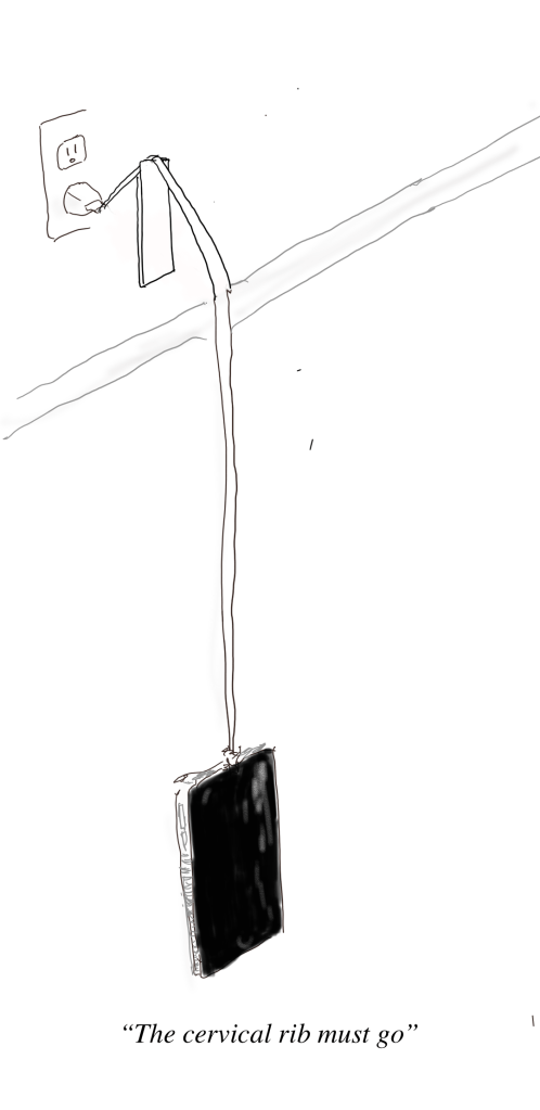
Mechanically speaking, there is a weight and a rope. The brachial plexus is the rope and it is draped over the first rib. The muscles of the shoulder girdle should support the weight of the arm but with bad posture, another consequence of bipedalism, the muscles may not be up to the task.
When there is a cervical rib, the situation is worse. The thoracic outlet is narrowed, and the weight of the arm, the jamón, is borne on the cervical rib. The brachial plexus becomes stretched over this anomalous bone and the trauma results in symptoms of pain, weakness, and strange sensations. I am of the opinion that cervical ribs in general should just go when found as nerve (and arterial) injury when found late may be irreversible.
CASE
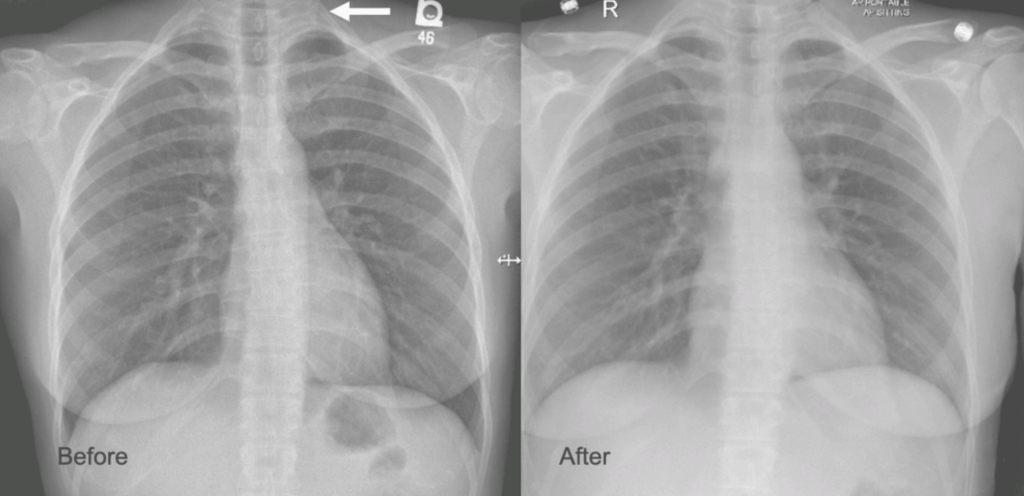
The patient is a younger woman who was referred from cardiology after workup of chest pain and left arm pain was negative for cardiac disease. She also had occasional right arm pain. The only finding of note during an extensive cardiac workup were cervical ribs found bilaterally on chest x-ray (below).
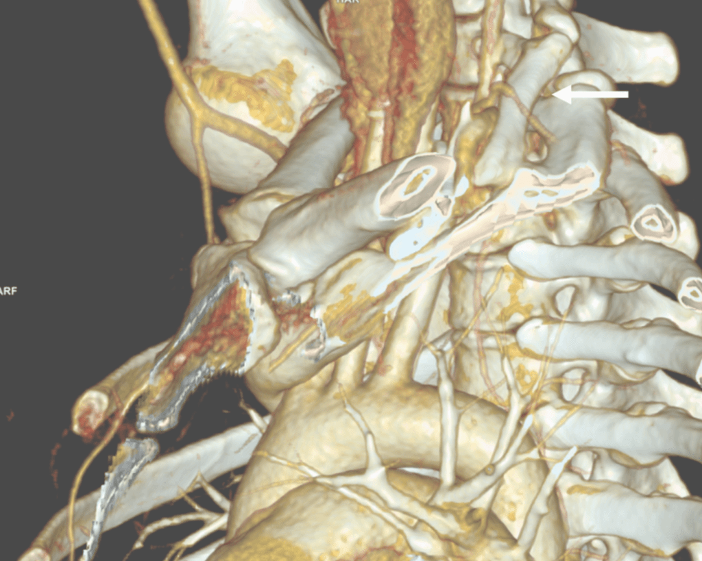
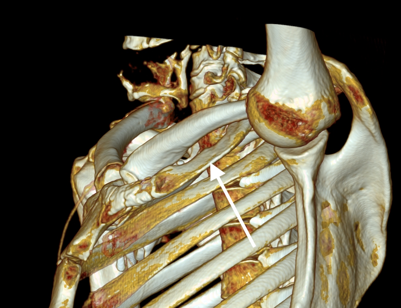
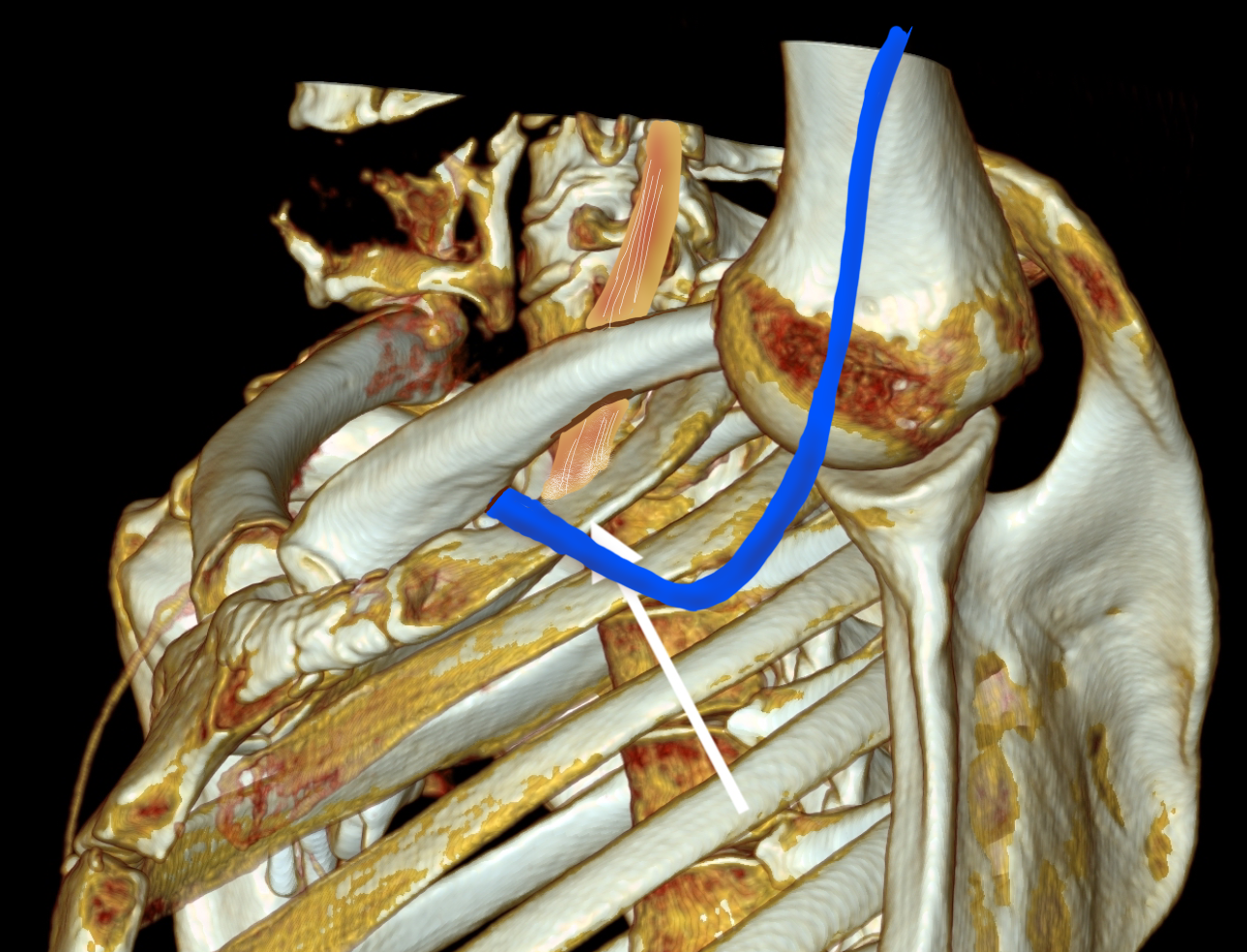
On examination, she was petite and had narrow shoulders. Stress maneuvers extinguished the arterial pulse in both extremities. EAST test was vaguely positive -weakness and numbness in the fingertips. Tapping on the cervical ribs which were prominent bumps in the supraclavicular fossae triggered shooting discomfort in both arms. I ordered a CT scan with contrast -this helps with operative planning as the 3D reconstructions allow you to view the operation before you perform it.
White arrow points to left cervical rib. The artery and plexus which is not seen are draped over it.
I recommended cervical rib resection, bilateral, staged. The left side was chosen first as it was the more symptomatic side. I recruited the assistance of Dr. Nader Habela, our spine surgeon.
Cleveland Clinic Abu Dhabi, has its roots in Cleveland Clinic,and was founded over a century ago in the vasty fields of wartime France by the four founders in an US Army tent. The observation at that time formed the root of the culture, the meme being that without barriers, the distinctions of competitive, siloed specialities made less sense than collective knowledge. It is encapsulated in our words, “To Act As a Unit.” CCAD is close to those roots. Lacking trainees in vascular (working on that), we totally depend on each other for extra pairs of hands in the OR. While it drives my nurses crazy, working with experts in other specialties exposes me to different techniques, instruments, and gadgets, which I load my trays with. Working with Dr. Habela, I saw that for cutting bone in tight spaces, an osteotome and mallet (hammer and chisel) worked with diamond cutter precision is faster and more precise than chewing your way through with a Kerrison. I do wish I had a surgical light saber.
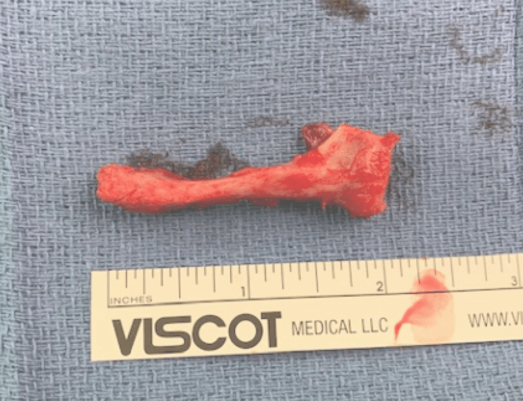
Exposure was via supraclavicular approach. The cervical rib had a joint on the first rib and it was removed. The picture below shows its dimensions.
The artery and brachial plexus were tented up and there was inflammation around these structures. The anterior scalene was released for added measure, but first rib resection, I felt, was unnecessary.
The patient underwent contralateral cervical rib resection a few months later and had significant relief of symptoms but not total relief which I believe has to do with the slower relief time course with neuropathy. The chest pain never recurred. While I do know that diaphragmatic issues refer to the shoulder because of the emryologic origins of the diaphragm and shared roots of the phrenic nerve with brachial plexus, I do not know of a brachial plexus issue referring symptoms to the chest outside of autoimmune neuropathies which the patient did not have. The chest x-ray, which I always get after these procedures to check on diaphragm innervation and for pneumothorax showed the removal of the left cervical rib only, but no insight in why she had chest pain along with the extremity symptoms.
Cervical Ribs Must Go
I have never comes across a benign cervical rib. Because they are easy to remove, they should come out. While no arterial injury had occurred in this patient, we see many instances where compression and aneurysmal degeneration beyond the compression with embolization results in tissue loss, frequently misdiagnosed as rheumatologic arteriopathies. The cervical rib is a special case of neurologic thoracic outlet syndrome (nTOS) where the pathoanatomic mechanism is magnified by the extra bone. While physical therapy has a role in standard nTOS, no amount of PT will address the cervical rib. The inclusion of the Boticelli Venus has to do with the fact that stature and posture plays a significant role in nTOS. Even after first rib resection, there are some people who need a second rib resection to clear the space.
The critical need to treat this is that nerve injury is sometimes irreversible if left untreated. The worst outcome is a causalgia -the feeling that the upper extremity is being electrocuted, put on fire, eaten by flesh eating ants, that is perfectly and completely disabling because our function is defined by our ability to use out upper extremities. Once this sets in, surgical neurolysis or any reoperation has very little chance of working.
So many problems from a bipedal lifestyle
There are so many chronic problems arising from bipedalism, that I will have to work on a whole monograph about it. Humans are the only vertebrates (aside from a few burrowing fish and sea horses) in the history of vertebrates with a vertical spine in orientation to gravity. Yes, there are bipedal dinosaurs like pigeons and velociraptors, but look at their spines -they are horizontal to gravity with the mass centered around the hip. Aside from the obvious ones of spinal compression and arthritis, hernias, and prolapses, are vascular diseases like venous insufficiency, median arcuate ligament syndrome (MALS), and popliteal entrapment. While the first one, venous insufficiency makes sense, MALS does not until you understand how much the heart full of blood weighs. Suspend this bag of meat and blood on your celiac axis, grind the celiac plexus between the diaphragmatic ligaments and the artery, and voila -MALS. Popliteal entrapment -easy – being upright means straightening our knees, something no animal does, which exacerbates the entrapment.
Bonus for my readers -POTS -postural orthostatic tachycardia syndrome -does not exist for quadrupeds -cannot. Our swift (a million years?) transition to bipedalism did not happen with the proper adjustment of our pressure gauges for some, and those with POTS struggle with this change in posture from the natural horizontal state practiced by all other vertebrates.
Recently, I saw a case presentation uploaded to LinkedIn of a subclavian venous stenosis treated with balloon venoplasty and a stent for venous congestion of the arm. The images were beautifully clear. The stenosis was at the thoracic outlet. The comments were generally favorable, including congratulations for a nice case, but I had to put in my two cents: The thoracic outlet is a terrible place for a stent due to external compression, and once occluded, a stent in the venous position is a permanent obstruction. The justification was that the patient did not want surgery and there were no surgeons who did first rib resections where the post author worked. I refrained from commenting something about primum non nocere.
This patient from the images above is a middle aged man who competes in triathalons and who noted sudden onset of discomfort and heaviness and pain in his left arm. Ultrasound revealed DVT in his axillosubclavian veins extending into his brachial veins and he was started on anticoagulation. He had been on anticoagulation for about a month by the time he came to my clinic. On examination, he had a prominent superficial veins on his shoulder, but otherwise had a normal examination. He did not have arterial obstruction on TOS (thoracic outlet syndrome) maneuvers and had no neurologic symptoms. I recommended first rib resection.
The patient’s first reaction was a dubious expression. After all, in this day and age, isn’t it barbaric to offer to cut out a rib? Why not put a stent there? A CT venogram was obtained showing the subclavian vein occlusion, but we knew that. I use the 3D surface reconstructions extensively as a visual map to determine surgical approach. I remove first ribs through both supraclavicular and transaxillary approaches, choosing one or the other.
The 3D reconstruction of the bones showed that to get to the first rib section underlying the vein, it was best approached via a transaxillary approach. The arrow in the image shows the flare in the first rib where the anterior scalene muscle attaches. Adding muscle shows that in this well muscled patient, getting to the first rib would be a challenge -for the people holding the retractors.
The operation is done with the patient in right lateral decubitus position and requires deep retraction and good lighting -I have played with using the laparoscope (more to come). Laparoscopic instruments such as the Maryland tipped ligasure and hook cautery get much use in this operation. The special sauce to getting this done quickly here in Abu Dhabi where I don’t have the army of residents and fellows is getting my colleagues in Orthopedic spine (Dr. Nader Hebela) and Thoracic Surgery (Dr. Redha Souilamas) interested in TOS.
Where I would laboriously chew through the ribs with a Kerrison, Dr. Hebela has shown me the high art of the hammer and chisel (notice the clean lines on the cut rib below). Dr. Souilamas has seeded the idea of doing this entirely thoracoscopically from the thoracic cavity, and yes, it is right there to see when I recently visited one of his operations -where is that cadaver lab when you need one? Enough rib was resected to ensure the vein, artery, and plexus were completely free.
A bit of anterior scalene came out with the specimen, splayed upwards in this image
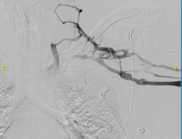
The patient did well, recovering much of his range of motion quickly with the help of physical therapy. He was taken to the angio suite and underwent venography shown below. There is an occlusion of the subclavian with outflow via collateral veins. Not seeing collaterals is as important as seeing a good angiographic result.
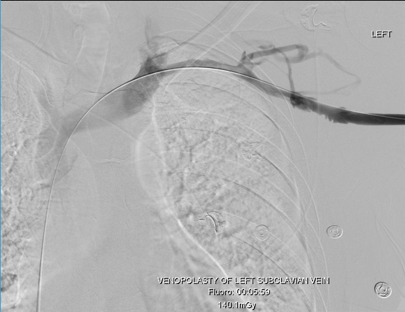
Venoplasty was done to 8mm -I try not be overly aggressive here, just to break the strictures that caused the balloon to have a waist in two sections. The final result is below, with the absence of the venous collaterals. If they were still present despite an angiographically satisfactory result, I would perform IVUS to see what the problem was. In no circumstances would I place a stent at this juncture -my plan is to keep the patient on anticoagulation for 3 more months.
First rib resection should be in the armamentarium of every vascular surgeon. I sense a rise in the diagnosis of this and this has been commented on the SVSConnect boards (link), possibly from a greater awareness of the signs and symptoms of the diseases associated with the thoracic outlet. Since coming to CCAD, there have been enough cases for it to become a routine which I did not expect.
The thoracic outlet, like the median arcuate ligament and the inguinal ligament, cause trouble for stents.
Looking at the thoracic outlet (above), the vein has a particularly narrow outlet under the hinge of clavicle and rib. I generally find it tragic to see a stent here because it limits the possibility of improvement with rib resection. Like the median arcuate ligament for the celiac axis and the inguinal ligament for the common femoral artery, stents get crushed in this position. If you think about it, it is probably our insistence on bipedalism that engendered these design flaws. Stents get compressed by the weight of the shoulder and arm (thoracic outlet), the mediastinum (median arcuate ligament), and the abdominal cavity organs (inguinal ligament) under these choke points, something the quadruped does not suffer.
Quadrupeds that come to mind, RIP GOT.
So is it barbaric, this open surgery? No. The barbarism is in offering patients an easy solution that will get that patient out the door happy, but not knowing (at best) or not caring (at worst) that the biomechanics do not favor any kind of durability.
Absolute Neutral Position is suprisingly universal
A body floating in space, a fetus in the womb, a dad lounging in his favorite chair, share the feature of weightlessness and represent the absolute neutral position (figure above) of the human which is the position of a relaxed supine quadruped -a dead mouse. Anything else is a stress position, including standing. Repeating motions outside of this relaxed pose or holding those positions away from this absolute neutral for long periods of time is a nidus for injury and pain. That is why most land animals sleep flat on the ground.
The Pain Operations
Operations to relieve pain are often the most gratifying to both patient and surgeon to perform successfully. This circumstance applies to the commonly performed procedures such as spine surgery, endometrial ablations, and varicose vein resections. When the pain is due to a rare set of circumstances, things are not so easy. Typically for rarer pain syndromes, two things need to coincide for the successful operation to happen. First is the patient must suffer while more common and potentially life threatening diseases are ruled out and even treated if these are found. This may take months or years. The second necessary condition is finding a physician who has seen the particular pain syndrome before and understand how to test for it and treat it. That meant the majority of people never get treated, or are shunted into the circle of shame as malingering, drug seeking, and mentally unstable. The opioid epidemic creates double jeopardy for these patients -they can become addicts as their pain is never successfully diagnosed and treated and they get labeled as drug seeking.
A random list of conditions that cause pain that should be on the mind after the usual things are ruled out. Also, vasculitis, autoimmune disorders, and foreign body reactions
All pain syndromes that can be successfully treated share common features that give you a degree of surety about the diagnosis, but at the end, there is a leap of faith on the part of both patient and practitioner because many of these operations have a failure rate ranging from 5-20 percent. First, the symptoms must be associated with sensory nerves, somatic or visceral. Second, there is a physical mechanism for that nerve to be inflamed from compression, swelling, or irritation that can be accounted for through history, physical examination, and imaging studies. Third, though not a constant, a major nerve trunk will be associated with a blood vessel, typically and artery, that is also affected by compression. Fourth, when swollen veins are the cause of pain, it has to be recognized that at an end stage the organ that the veins drain can also be affected.
The Pain Must Have a Testable Anatomic Basis
The somatic sensory nerves in the periphery are well mapped out and known since even classical times. The described pain should be consistent with a nerve. The best and easiest example is a neuroma that forms in an amputation stump. It triggers pain in its former distribution. It is palpable as a nodular mass. It is visible under ultrasound or cross sectional imaging. And it is easy to turn off temporarily with an injection of lidocaine, either under palpation or image guidance. If you can turn off the nerve and relieve the pain, it is likely that ablating or relieving the nerve of irritation will also relieve the pain. Such is the case in median arcuate ligament syndrome (figure below). The celiac plexus is caught under the median arcuate ligament and compressed. It causes a neuropathy that is felt in its visceral sensory distribution and the brain interprets these signals in the typical ways irritation of the stomach is interpreted -as pain, burning, nausea, sensations of bloating, and general malaise. These nerves can be turned off with a celiac plexus block and the effects tested by giving the patient a sandwich. When it works, the patient will say they will have had relief for the first time in years and operation to relieve the ligament compression and ablate the nerve can proceed. Same for many of the diseases listed.
Tight Spaces Impinging Nerves, Arteries, and Veins
Many of the tight spaces involving the nerves have accompanying arteries that are compressed. This results in injury to the artery in the form of intimal hyperplasia, post stenotic dilatation, aneurysm formation, and thromboembolism. Shared tight spaces that cause problems for nerves and arteries have the common features of fixed ligaments, adjacent bones and muscles, inflammation, and motion. These include the thoracic outlet, antecubital fossa, cubital canal, diaphragmatic hiatus at median arcuate ligament, inguinal ligament, popliteal fossa, carpal tunnel, obturator canal, mediastinum, retroperitoneum -basically anywhere nerve, compression, and motion occur. In some instances of median arcuate ligament syndrome, postures and breathing trigger the pain. Holding a child in an arm may trigger pain in neurogenic thoracic outlet. Or sitting while wearing tight jeans may trigger a burning pain in meralgia paresthetica. It is not uncommon to find damaged arteries in median arcuate ligament syndrome, thoracic outlet syndrome, and popliteal entrapment or thrombosed veins in nutcracker syndrome, May-Thurner Syndrome, and Paget-von Schroetter Syndrome. Because nerves are typically difficult to visualize, their compression may only be inferred by testing for compression in their adjacent arteries.
Dilated Veins and Swollen Organs and Visceral Pain
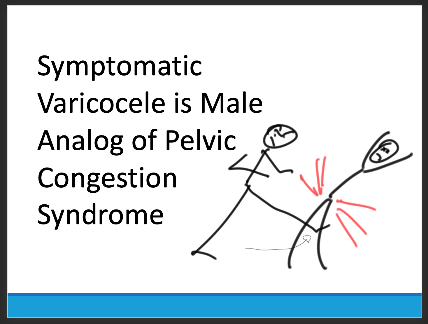
Venous hypertension is most commonly conceived of as varicose and spider veins of the legs and offer a model of pain when applied to other pain caused by venous dissension. The visceral sensory fibers veins and arteries trigger a very intense pain that localizes to the trigger. I have often witnessed this when I manipulate a blood vessel during local anesthesia cases. Visceral pain from swelling has a dull achiness that is localizable to my spider veins after a long day standing like a bruise (below). The swelling from varicoceles which I have also had feel nothing less than feeling the aftereffects of getting a kick in the balls -not the immediate sharp pain but imagine about 5 minutes after with the mild nausea, abdominal discomfort and desire not to move too much, and even a little flank pain. Imagine this occurring low in the pelvis with ovarian vein varices in pelvic congestion syndrome. This kind of swollen gonad pain afflicts many women whose pain is so frequently dismissed by male physicians because they have no context -well imagine getting kicked in the balls hard, wait about 5 minutes and that moment stretch it out to whenever you stand for a long period of time (below).
Actual Slide From Midwest Vascular Surgery Traveling Fellowship talk 2017, Chicago, IL, USA
When a limb is swollen from a thrombosis, the veins hurt and is similar to a bone pain from a fracture or a pulled muscle -that is how the brain processes the pain, but when the muscles and skin get tight from edema, the pain is sharp and dire. This is the same kind for pain from a distended left kidney from nutcracker syndrome or a spleen from a splenic vein thrombosis. These conditions can be modeled and predicted based on history and correct differential and confirmed with proper imaging -always.
Build a theory of the pain based on a testable proposition and set of nerves
That is the final message. These pain syndrome require some imagination and empathy to map and model. Predictive tests then can be performed on physical examination, functional testing, or imaging. Often, the adjacent artery is the only thing that can be reliably visualized and tested, knowing that it is the nerve that is compressed. Turning off the offending nerve with a block and relieving the pain is the most powerful argument for operating. It is building the argument for an operation that requires these objective data, but at the end, it does require some experience and faith. You have to believe in your patient and the science and when they coincide, you have to act.


 The celiac plexus is caught under the median arcuate ligament and compressed. It causes a neuropathy that is felt in its visceral sensory distribution and the brain interprets these signals in the typical ways irritation of the stomach is interpreted -as pain, burning, nausea, sensations of bloating, and general malaise. These nerves can be turned off with a celiac plexus block and the effects tested by giving the patient a sandwich. When it works, the patient will say they will have had relief for the first time in years and operation to relieve the ligament compression and ablate the nerve can proceed. Same for many of the diseases listed.
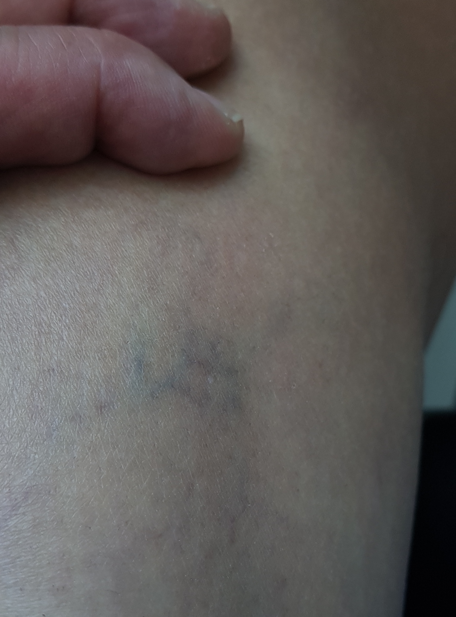 The swelling from varicoceles which I have also had feel nothing less than feeling the aftereffects of getting a kick in the balls -not the immediate sharp pain but imagine about 5 minutes after with the mild nausea, abdominal discomfort and desire not to move too much, and even a little flank pain. Imagine this occurring low in the pelvis with ovarian vein varices in pelvic congestion syndrome. This kind of swollen gonad pain afflicts many women whose pain is so frequently dismissed by male physicians because they have no context -well imagine getting kicked in the balls hard, wait about 5 minutes and that moment stretch it out to whenever you stand for a long period of time (below).