Generated from my prior posts on the biomechanical problems generated by a bipedal lifestyle, this podcast discusses those issues. This was discussed by Dr. Elen Feurriegel on her lecture series “Big Mysteries of Human Evolution” available on Audible where she describes the human spine as a stack of teacups and saucers supporting a bowling ball.
In George Orwell’s Animal Farm, under the dictatorship of the alpha pig, Napoleon, the pigs who represented the nomenklatura of the farm chant the slogan “four legs good, two legs better,” after initially declaring “four legs good, two legs bad” during their revolution. They clearly understood the luxury afforded by a bipedal lifestyle, because in rising on two legs, you get arms and hands which can do many things like caress a baby or wield a cudgel. What the pigs in the parable weren’t realizing were the consequences of a bipedal lifestyle.
When Lucy, the Australopithecine, bipedal hominid ran about on two legs, she did have the use of two arms and hands. Possibly an adaptation to living in tall grasslands with few trees, the ability to stand tall like meerkats, allowed the biped to see far into the horizon for big cats who probably loved the big brained hominid for the high calorie meal inside the hard skull -many fossils from this time show puncture marks from the incisors of medium to large cats.
The walking and running put heat stress on the brain, and the tool use which happened incredibly early and is observed in the chimpanzee, likely drove the selection for a larger brain (more neurons will allow for one to lose some neurons to heat stress but stay in the game), but it created likely the first problem for our ancestors -discharging a cantaloupe sized head through a pelvis that was small to begin with but now also reshaped for bipedalism. We still suffer from a childbirth process that no other mammal faces -birthing a less than fully cooked baby -a tradeoff for that giant head.
Standing also meant the load bearing was shifted 90 degrees with long term consequences. For our ancestors who only lived about 20-40 years if the chimps are correct, this wasn’t a big deal as arthritis and tendinitis didn’t preclude eating and breeding and didn’t affect them until they were old. But with modern sanitation and social structures, we are reaching 100 years and the majority of the problems of the integument -the bones and ligament, the low back pain, the sore knees, the ratchety hips, can all be explained by our bipedal lifestyle. Your arm is 30-50 pounds of meat and bone and supported only by muscles off your spine, and your blood vessels and nerves traverse a narrow passage through these muscles and your first rib. Your diaphragm with 5-10 pounds of heart, lungs, and blood sits on first branch artery off of your aorta. Your veins, designed to drain blood from your organs, have to do so with over a meter of static water pressure and your sump pumps only work when you are walking. Muscles and their tendons are stretched tight in the odd way that upright walking and running demands, compressing blood vessels and nerves. All of this weight is put on your feet which have to deal with up to a ton of pressure with running…
I’ve talked about this concept many times before but never had a chance to put it together like this talk. I may write an article. Looking back, I did this blog post (Link).
I am grateful to Ms. Mei Nortley and Mr. John Raphael for the invitation to give this talk.
When I was a young attending at the Allen Pavilion of Columbia Presbyterian Hospital, I was called into an operating room for a stat consult on a patient about to undergo a cholecystectomy. During the case, the IV had infiltrated and a bag of saline had filled the patient’s hand and forearm with saline, causing the hand to look like an inflated glove. The fingers were cool and white and the edema was firm but yielded to touch.
I elevated the hand and firmly squeezed the edema out of each digit, then gently massaged the edema from the hand onto the forearm. From there, I pushed the edema onto the arm. I then wrapped the hand up in an Ace wrap, and suspended it from an IV pole and returned to my case. Later, I returned and the hand was restored, warm, and perfused.
The lymphatics serve to move extracellular fluid (link). They can be overwhelmed much as drainage from a house can be overwhelmed resulting in puddles and ponds (link). This extracellular space has been “discovered” to be a new organ, but vascular surgeons have known about it for some time. Ultrastructurally, it is very close to a sea sponge with lattices of structural protein connecting cells to form tissues. And like a sea sponge, the salty water can be squeezed out or drained using gravity.
In olden times in central Europe, if you had chronic leg ulcers, you went to abbeys that specialized in their care. There, nuns would milk the edema out of your leg swollen typically from parasites and dress the leg and ulcer in linen cloth soaked in special oils. This is how Dr. Paul Gerson Unna came up with his eponymous Unna’s Boot, substituting Zinc Oxide paste which created a bacteriostatic environment.
Professor Paul Gerson Unna
Every year or so, I will be consulted for what I term a lymphatic emergency. A subset of this is phlegmasia. Whatever color you find -alba (white) or cerulea (blue) is really no matter -who really knows which comes first? It is an emergency in that the time clock for arterial ischemia -minutes to an hour for nerves, an hour to 6 for skeletal muscles, 6-12 for skin and bone, are all in play. The instinct is to go right to fasciotomy, but what you are usually doing is releasing the extracellular space, and the muscles are typically fine, even though their compartment pressures were very high.
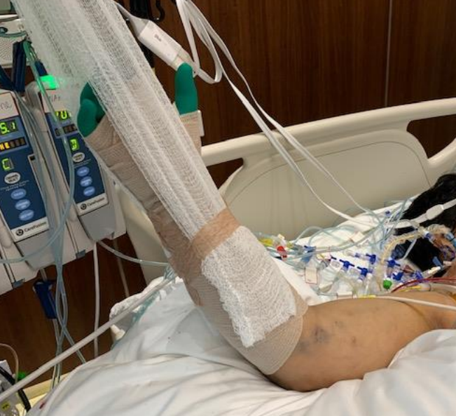
Take this patient who developed severe upper extremity edema in the recovery phase after a cardiac arrest.
The ICU staff noted the had discoloration about four hours after the arrest. There were no arterial pulses and the forearm and hand were rock hard, the finger tips ice cold. Compartment pressures measured using the arterial line and needle method didn’t drop after the initial flush of saline below 70mmHg. While I could have been justified in performing upper extremity fasciotomy and even trying thrombectomy in a critically ill, coagulopathic patient on multiple pressors, I could just as easily have been on solid ground for saying the life was more valuable than the dominant hand. Both would have been the wrong move.
I performed the nun’s milking maneuver mentioned at the beginning and lacking an Unna’s boot, I compressed and elevated the best I could with double gloving using a small sized glove and ACE wrap.
Notice the edema has segregated into the arm.
In the morning, taking down the dressing, and re-compressing, there was now a radial artery signal and the fingers were a much improved color. The pulse-oximeter waveform was near normal. As an aside -the pulse oximeter uses the same technology as the digital photoplethysmography for generating toe waveforms in the vascular lab -ie. a vascular lab at every bedside! We have collected and are analyzing the data on this for publication.
The pulse oximetry waveform is the same tech as digital photoplethysmography. Cotton cast padding (Webril) and Coban wrap is a good method of compression that avoids the problems with ACE wrapping.
It’s a hard thing to not run off to the operating room in most cases because that is how we are trained, but understanding how a patient got to that point is crucial in deciding if compression alone will work. If they call you from the ER about a patient with a swollen cold foot with diminished signals, you have to figure out the mechanism. Was it arterial occlusion, rest pain, and chronic dependency of the foot that resulted in this? Typically the swelling appears late. Was it heart failure and inability to walk, resulting in the patient sitting all day in a chair that is the cause? Was it pregnancy with a DVT? Was it the deadly sin of sloth? Only in arterial occlusion in a chronic presentation would compression be contraindicate. In this ICU case, the lack of arterial signal is secondary to the swelling, not the cause of it.
Elevation alone does not manage edema well. Only hanging upside down or being in water up to your neck…
Compression is a necessary component of treating lymphedema emergencies because elevation alone may be insufficient, particularly in the leg.
Wrapping a leg is a critically, undertaught skill. Also, never cover the knee cap.
Elastic compression is ubiquitously available as the ACE wrap, but they can shift and move and roll, causing zones of excess and not enough compression. TED hose and compression stockings are definitely helpful in long term management, but with legs, compression needs to go up to the knee joint, or up to the groin, never halfway or the edema will create a line of ischemia at the end of the stocking that blisters when the stocking is removed, and can progress to full thickness necrosis. Cotton cast padding and Coban, or an Unna’s Boot may be the safest in terms of avoiding skin injury.
ACE wrapping is never taught adequately, and for it to work well and avoid injury to the skin, the wrapping has to be reapplied several times a day. It should be a prerequisite for nursing and medical student certification, as edema is the most common vascular disease.
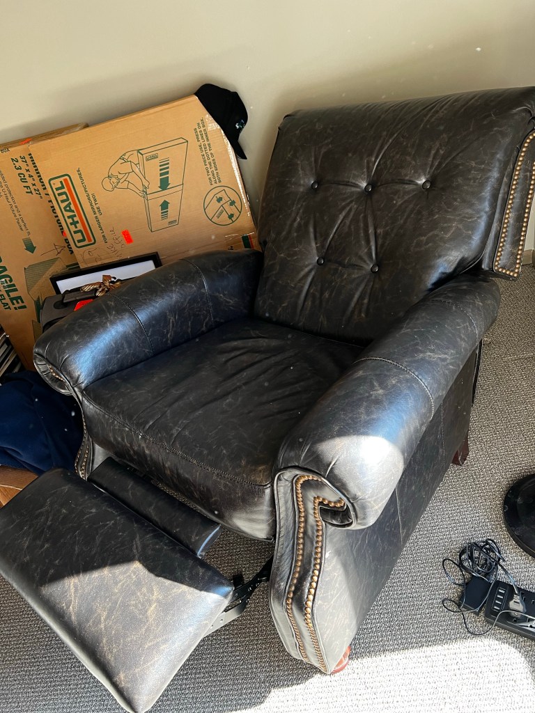
Moving into our new home after four years out of country, I welcome an old friend from storage, but also unfortunately a health hazard, only mitigated by being fully reclinable.
A recently published paper caused breathless worldwide headlines about a “new” human organ hiding in plain site — the interstitium. It had me smiling because vascular surgeons, the good ones, recognize it and have been managing it for a long time. The interstitium is described as the space outside the cells. The new interest in it is like people suddenly obsessing about the stuffing in sofas. It is the body’s contained negative space and it is the most important organ because it was the first. It has been there all the while.
The genome and its expression, the organism, carry the past like hoarders. Look at a skin cell, and you see a nucleus and a cell membrane, the hallmark of the eukaryote, and the mitochondria that it took captive in eons past when it was a sea bacteria that was eaten and refused to be digested. The next most important step in evolution was multicellularity and specialization of these cells. The earliest efforts started as clumps of cells, but clumps have a limit — every cell had to have exposure to the outside and eventually these became spheres with a hollow internal space. Here was the first interstitium — the first inside, the first not-outside.
To these first animals, segregating an internal space different from the outer sea had advantages. You can concentrate nutrients inside when the seas outside are plentiful and use these when they are not. Add some structure and you have an endoskeleton — we share this with sponges inside this interstitium. As the organism became larger, this sphere flattened and some became animals with one pore ingesting and ejecting and others with two holes. We fall into a lineage that found transiting food through a cylinder to be advantageous. The nutrients were digested and absorbed from the worm into this internal space. The interstitial waters needed to be mixed as food came not from the outside but from this internal protodigestive tract, to have currents and streams. This was done with the development of tubes lined with smooth muscles that beat, interspaced with one way tricuspid valves. This primitive circulatory system is seen in many of our spiny sea cousins like starfish and sea cucumber, and lives in us as the lymphatics.
The interstitium is the remnants of this primitive sea creature that we carry with us, carrying within this pouch of internal sea. The fluid that fills blisters is a kind of briny sea water. When you see an edematous patient, observe the level of this sea by seeing where the edema ends. See how easy it is to milk out this edema out of a hand or foot, just as it is to squeeze the water out of a sponge. Edema is so common that it is easy to forget that so many diseases cause failure of the lymphatics — the bilge pumps of the body, and that on this tide may come many other things that makes the problem worse. In other instances, it may be just high tide in Venice, right before all the sewage gets washed out into the Adriatic.
The interstitium, as much as it was the progenitor of the circulatory system, is likely the foundational element of the nervous system. The various ion pumps are highly conserved and are useful only when concentration gradients are stable. The bioluminescent jellyfish is testament to this. Without the interstitium, cross membrane voltage potentials cannot be maintained. It is the bioelectric spark that life motion. If a planaria, a flatworm, is to have a soul, it resides in the interstitium. It is the spiritual ether bottled inside us. The ghost in our machine swims our portable primordial sea.
These old parts and compartments are hiding in plain site. The lymphatics beat and spread some of the nutrients from the gut into the venous system in connections up at the base of the neck. Both have been superseded by the portal venous system and the circulatory system but the lymphatics persist because there was no reason to abandon it, but possibly it is critical to our existence. The interstitium must play a critical role in homeostasis in the same way that the older autonomic nervous system plays critical subliminal roles by being both a buffer and a store. Every cell in our body is in contact with this inner sea as much as the first cell was afloat in the primordial one.
The interstitium is the final contact point between each cell and the organism as a whole. Oxygen does not go from alveoli to the skin without transiting the interstitium. Just as we are only beginning to grasp the complexity of genetics and the heredity of epigenetics, we are just noticing the interstitium. Up to now, it is as if we have been studying the outlines and histories of Byzantium, Rome, and Carthage, in isolation without studying the depth, composition, and currents of the Mediterranean.