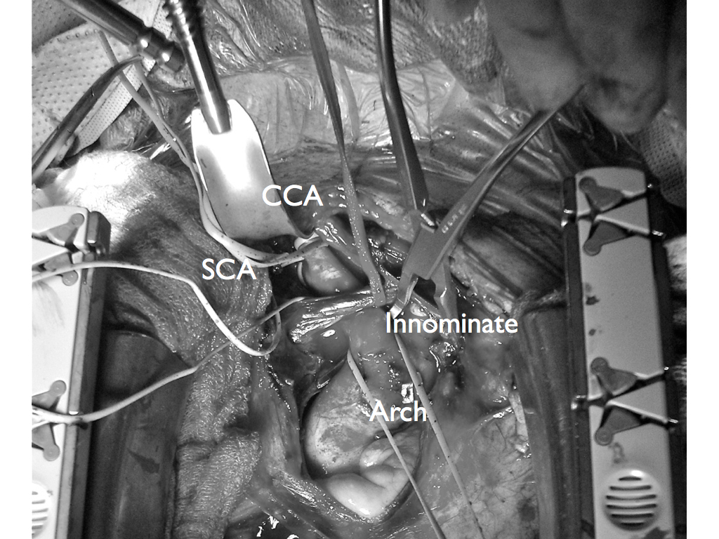
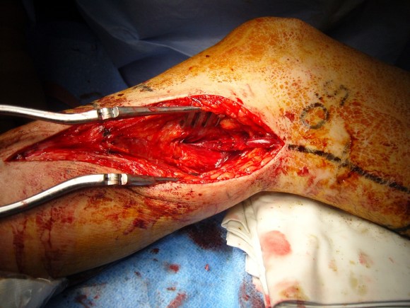
The patient, a 47 year old woman, was referred for syncope, but it was much worse than that. Excessive stress, standing, and thinking hard were described as causes of her syncope. Frequent headaches and lethargy lead to inability to keep a job. Several MVA’s resulted in revocation of her driver’s license. Added to that was a two to three pack a day cigarette habit. Her cardiac workup was negative, but it was noted that her carotid duplex was notable for a left ICA occlusion and left vertebral occlusion. Blood pressure in both arms was in the 70’s systolic while in her better thigh it was 90’s. She had an open right carotid system but the flows in the common carotid were attenuated. CTA of the arch revealed severe arch disease affecting the origins of her great vessels. The innominate artery was severely diseased to its bifurcation with a small <2mm channel of flow. She had a dominant right vertebral artery that was patent, and the right ICA had moderate disease at its origin. This was in 2009, and I entertained intervention, but wasn’t all that confident that kissing stents into the innominate was all that great of an option even though there are reports of innominate interventions in the literature. I had the opportunity to perform a handful of great vessel reconstruction with Ken Cherry during my fellowship and felt that this was an ideal case for an innominate endarterectomy.
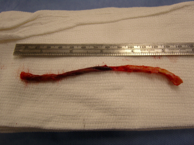
This is one of those rare and infrequent cases from vascular surgery history. The exposures is one of the grand vistas of vascular surgery. The arch, thankfully, was only calcified at the origins of the vessel and clamped well. The endarterectomy was not that much different from an aortoiliac endarterectomy with a fibrocalcific plaque and was extended onto the common carotid while the origin plaque of the subclavian was plucked cleanly. The phrenic and vagus nerves were protected. The patient was centrally hypertensive as found by a long femoral arterial line and was kept that way for the duration of the clamp. A bovine pericardial patch was applied and the sternum was closed over a mediastinal chest tube.
The recovery was impressive for the patient’s immediately improved state of consciousness, lack of lethargy, and improved cognition. She was herself impressed enough to quit smoking during that admission for good. Her right brachial cuff pressure now correlated well. She went home POD 5. When I last saw her 2 years later, she was employed and symptom free with continued patency of her repair, consistent with the earlier reports of this operation (Cherry et al. J Vasc Surg; 989;9:718-14).
From my archives, the CTA illustrates two points. First, tunneling can be done without taking down much of the retroperitoneum. This lesson came to me after taking a course in laparoscopic aortic surgery with Dr. Dion in Quebec City. The old BARD-IMPRA tunnelers with their bullet tips -the short gently curved one, is particularly well suited for tunneling from the groin to the aorta -if you have a hand on the retroperitoneal pelvis, it is very straightforward to guide the tunneler to the proper location. The other point is that the graft is applied proximally end to side with a leftward orientation. This combined with dissection of the retroperitoneum with a large Ligasure or harmonic scalpel lets you avoid the problem of having no tissues to close over the graft. You take down the retroperitoneum with a cuff of tissue of about 5cm from the duodeum. Normally, this can be bloody but with the energy devices, it is not. This provides excellent graft coverage. You just have to mind the IMV which may or may not have to be taken down. End to side is preferred because you preserve endovascular options, but in this case, the anastomosis was done end to end.
April 30, 2009 11:33 PM
Aortic Bypass for occlusive disease
The patient arrived with the history of severe claudication. He was a middle
age smoker whose job required walking several miles a day. This became
increasingly difficult until he was clearly limping at short distances. He was
also developing cramps in his legs at night, worse in his left leg.
On examining him, he had no pulses in his left leg from the groin down.
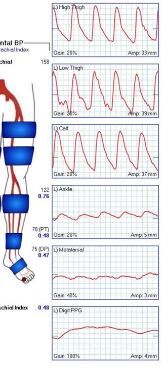
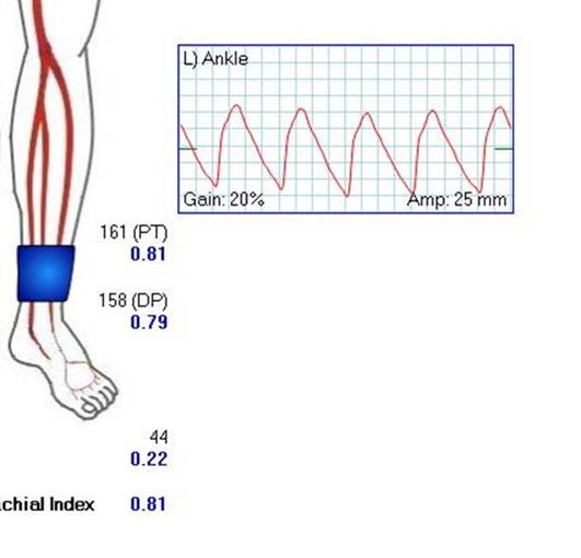
The pulse volume recordings (PVR’s, red lines) on the left clearly
demonstrate normal flows in his right leg with sharp upstrokes, dicrotic
notch, and shallow diastolic relaxation. The left leg had attenuated flows on
the pulse volume recordings with dampened, gradual series of mounds.
The flow was flat at the metatarsal level (foot). His ankle brachial index
( BI) on the right leg was 0.75 which was mildly depressed. The ABI on his
left leg was 0.43 which was severely depressed.
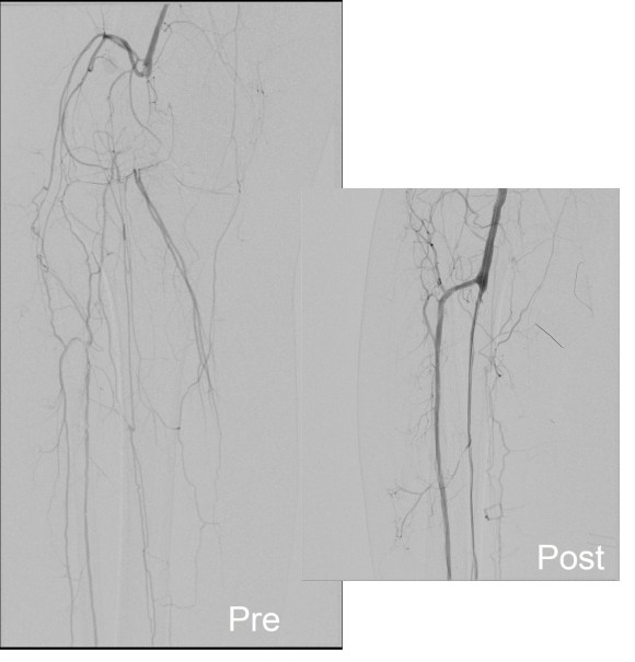
CT was performed (above left) showing that his left iliac system was
occluded. This is due to atherosclerosis which is a systemic disease. This
kind of blockage can occur in any organ, but it was most severe in this
patient’s leg. His right common iliac artery also had moderate plaque.
He underwent an aorto-right iliac and left femoral artery bypass with a
bifurcated graft (above right). This greatly improved flows in his left leg, with
his BI improving to 1.05 from 0.43. The PVR’s also reflect this improved
flow. The right leg, surprisingly, also had an improvement despite not
having a severe stenosis in his common iliac artery. The fact is, the
common iliac artery, but being heavily diseased over the length of the
artery, offered a hemodynamically significant stenosis despite being patent.
His BI on the right improved from 0.75 to 1.03.
The operation was done with minimal invasiveness in mind. The CT
allowed for planning of the abdominal incision directly over the part of the
aorta requiring operation. The groin incision on the left was created
obliquely as to avoid crossing the groin crease -which I believe increases
the chances for tension on the wound and subsequent infection. The graft was tunneled without mobilizing the sigmoid colon directly up to the bifurcation using an IMPRA tunneler -by placing the hand in the pelvis, the
tunneler can be felt and guided in the correct trajectory. The graft was a
Gelsoft Plus graft soaked in Rifampin. This antibiotic bonds to the gelatin in
the graft giving protection against indolent bacterial infections for about 3-6
months after the procedure -this is helpful especially with groin incisions. The operation took 2.5 hours and the patient went home within a few days.
The patient is now walking without pain and will be returning to work. He
has also successfully quit smoking which has a significant impact on his
risks of future heart attack, stroke, or peripheral vascular complication. His
relative youth (in his fifties) required that we give him a repair that would
give him the best chance at maintaining patency for many years. The aortic
bypass graft for occlusive disease has a proven track record with patency
measured in decades.
From my case files, this was a case which I performed in 2010 and published in a prior blog.
CCx: Patient is a 56 year old man with complaints of pain in right leg with walking short distances and discomfort in the foot at night.
HPI: The patient has had cramps in his right calf with walking about a block for over a year, but over the past three months, he has developed pain with walking less than half a block which is incapacitating. He has developed pain at night which wakes him and he has taken to sleeping with his right foot dangling off the edge of the bed. This has resulted in some swelling of that leg which makes it doubly uncomfortable to wear shoes. He works as a manager at a local big box store and walks constantly. He used to smoke but quit last week. He feels this has worsened the pain.
Past Medical History: Hypertension, dyslipidemia, acid reflux
Pulse volume recordings notable for moderately diminished signals right high thigh cuff.
CTA: Moderate atherosclerosis of infrarenal abdominal aorta and its bifurcation with severe plaque of the right common iliac artery and occlusion of the external iliac artery. There was reconstitution of the common femoral artery on the right via collaterals. The left common iliac artery was affected by a moderate (50-75%) stenosis due to low density plaque.
Impression: PVD with rest pain of right leg due to severe aortoiliac occlusive disease and occlusion of right external iliac artery.
Plan: After discussing treatment options, we decided to try a right external iliac artery remote endarterectomy with angioplasty and stenting of his common iliac disease. This was chosen over aorto-bifemoral bypass because he had limited time off from work and work did require that he lift more than 20 pounds.
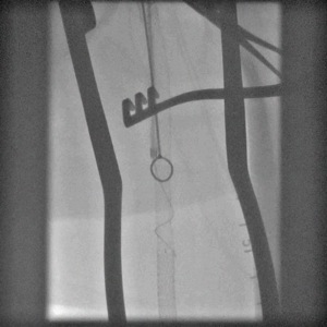
Up and Over Wire during remote endarterectomy ensures wire access if rupture occurs.
Operation:
Remote endarterectomy of right external iliac artery with aortography, bilateral common iliac artery angioplasty and stenting.
This operation was done via a single right groin exposure and percutaneous access of the left groin. The common femoral artery had severe posterior plaque which was the starting point of the endarterectomy. Up and over access of the right external iliac artery was achieved and a wire was passed across the occluded external iliac artery and into the right femoral system. With clamping of the common femoral artery, the wire was brought out and controlled with a Fogarty clamp -this allowed for excellent stabilization and control and possible emergent balloon occlusion in the case of a perforation.
A Vollmer ring dissector was sent over wire and plaque up the external iliac artery under fluoroscopy and dissection was stopped at the iliac bifurcation which was heavily plaqued. A Moll Ring cutting device (LeMaitre) was used to transect the plaque which was removed.
The right and left common iliac arteries were stented with self expanding nitinol covered stents and post-dilated. I chose this as I have had occlusions occur in the setting of diffuse TASC C disease with low density plaque -I suspect that thrombus propogates across open cells like weeds growing through chicken wire. The stents on the right were extended across the iliac bifurcation.
A completion angiogram is here to the right. The common femoral artery was repaired with a patch angioplasty (bovine pericardial patch, LeMaitre).
The groin was closed and the patient recovered and was discharged in a few days with excellent palpable pulses on the right and improved pulses on the left. He was without symptoms of claudication or rest pain in the right leg.
Discussion:
Remote endarterectomy allows for removal of plaque via a single groin incision, obviating the need for an abdominal exposure required in an aorto-bifemoral bypass. This minimally invasive technique is associated with a low complication rate and earlier return to full work status because the abdominal incision is avoided.
Smeets et al [reference] reviewed with 7 year experience with 48 patients and had a technical success rate of 88%. One patient died due to a myocardial infarction within 30 days of the operation. The complication rate was low. 6 patients required coversion (retroperitoneal flank exposure) for additional arteriotomy (3 patients) and bypass (3 patients). The primary and assisted patencies shown to the right were acceptable with a secondary patency of 94% at 3 years.
These cases require more surveillance than an aortobifemoral bypass. Intimal hyperplasia does occur in random loci in the SFA remote endarterectomy and this should apply to the external iliac artery. I chose the title because the external iliac artery biologically behaves like the superficial femoral artery in relation to endovascular patencies and not like the common iliac artery or aorta -probably because it shares a common embryology with the SFA, not the CIA. It is a troublesome artery that is often overlooked by vascular surgeons when femorofemoral bypass is performed for occlusive disease -the supplying external iliac artery though patent is usually diseased and has a small lumen. With a fem-fem bypass, both legs are supplied often through an artery with the caliber of a child’s drink straw. I have seen the donor leg become symptomatic through what is termed steal, but in fact reflects the hemodynamic inadequacies of a diseased external iliac artery.
I feel that 5mm is the minimal lumen caliber for an external iliac artery, and a 4mm lumen in an adult will clearly show a hemodynamic effect particularly after exercise or application of vasodilators in the endo suite. Stenting an occluded external iliac artery though technically feasible even in this case is not a durable solution in my experience. This operation allowed the patient to return to work without an extended convalescence.
I think removing the plaque offers advantages over stenting to the inguinal ligament. The common iliac stents have superior potency to external iliac artery stents and moving the stent point to the CIA and not stenting the EIA in my experience has better long term potency.
The patient is a baby who had undergone cardiac catheterization prior to repair of tetralogy of Fallot. Postoperatively he developed acute limb ischemia and was found to have an occlusion of his left common femoral artery. This was unusual, as typically, these babies tolerate catheterization and a high rate of CFA occlusion without ill effect. Usually treatment with heparin for a period of time is sufficient. In this child, there was clearly severe ischemia. What was different about this child was that his mother and her two siblings suffered from severe Raynaud’s disease, suggesting in this baby, vasospasm played a role. The foot was cold and not moving, and there were no signals from the groin down. The baby was taken to the OR for a CFA exploration and thrombectomy, as I felt the likelihood of infarction in this baby was high. The common femoral artery was exposed via an oblique incision and it was about 2mm in diameter, translucent, and very elastic. There was thrombus and I performed a longitudinal arteriotomy. There was minimal thrombus and a gossamer dissection flap. Inflow was easily established. There was no backbleeding.
Repairing the artery was initially not straightforward. The available nearby vein was even smaller for lack of blood flow and in spasm, and primarily repairing it was not a good option. There is literature describing patch angioplasty, but I felt there had to be a faster, better solution that did not require 8x magnification. Then it struck me that the artery was so pliable that it was every easy to mobilize a length of it from under the ligament.
My inspiration was the Heineken-Mikulicz pyloroplasty.
Once the laterally oriented stay sutures were in, interrupted (always) 8-0 sutures created a nice repair. The blood flow was high resistance, but the outcome was immediate. The baby had had over two days of ischemia, and I did go ahead and perform fasciotomies of the leg. There was dead muscle in all the compartments, but with VAC therapy, the baby healed within a few weeks, and a year later came in walking without a limp!
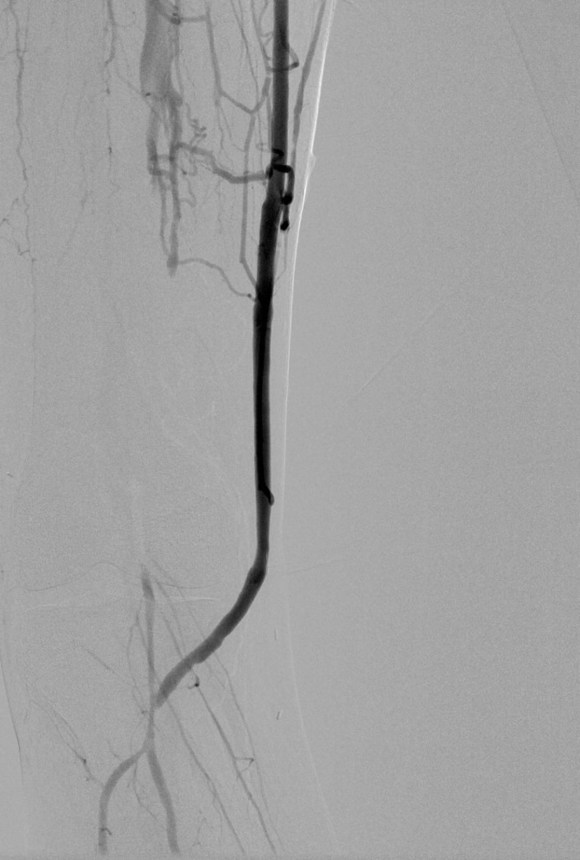
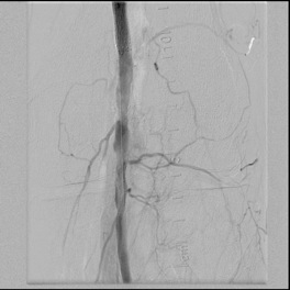
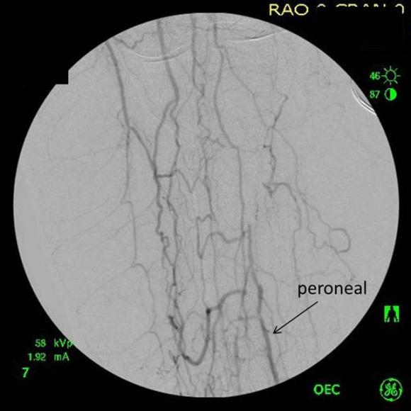
The patient is an elderly woman who had severe rest pain due to popliteal artery and tibial occlusion. She had no leg veins and sparse arm veins which would have to be spliced to achieve a femoro-peroneal bypass. Her preop CTA showed a patent SFA and proximal popliteal artery occluding above the joint and reconstituting only the peroneal artery. I planned for a retrograde popliteal remote endarterectomy tibioperoneal trunk endarterectomy via a below knee exposure with patch angioplasty of the arteriotomy, the bailout would be a short arm vein bypass from the above knee popliteal artery to peroneal artery.
Arteriography was performed via a left common femoral access and is shown below.
The popliteal artery was occluded and there was a very small peroneal artery that continued down the leg with seeming occlusion of the anterior and posterior tibial arteries. The popliteal and anterior tibial artery were exposed through a below knee incision taking care to avoid venous injury which can be troublesome source of bleeding. The tibioperoneal trunk down to the bifurcation and origin of the peroneal artery was exposed through the same incision. And anterior tibial artery origin was controlled with a vessel loop largely out of habit although it seemed clear it was occluded, as were the proximal popliteal and tibioperoneal trunk branches. The arteriotomy was created from the tibioperoneal trunk to the popliteal artery and endarterectomy was performed from distal to proximal to create a starting point for the ring dissectors used in remote endarterectomy. The anterior tibial plaque branched off much like an external carotid artery plaque and I decided to see what would happen if I did an eversion endarterectomy. I was able to mobilize a short length of the artery and was able to pull as I endarterectomized around the plaque and it thinned very nicely and came out with a gossamer end point. More gratifyingly, the backbleeding was excellent –this was controlled with the vessel loop very nicely. The retrograde popliteal endarterectomy was performed as described in another post in another case –link. The artery was then patched and completion arteriography was performed.
What was fascinating was it seemed I had reopened not just the pop-peroneal axis but the anterior tibial artery was also open, very dramatically so. The patient also had a bounding dorsalis pedis artery pulse. She recovered and went home two days later and in three years of followup while I was still in Iowa, she remained widely patent, maintained on Coumadin anticoagulation.
Popliteal endarterectomy for localized popliteal artery disease has been described (reference) with decent short term patency and successful limb salvage. Nasr et al. performed their endarterectomy via a posterior exposure. I think I recall coming across this in a book chapter from the old Wylie textbook which is long out of print. I think that the anterior tibial artery never lit up well because it was part of a highly developed collateral network, but it was patent all along. Duplex which was not done, would have given a better indication of its patency. I think that the patency of the popliteal endarterectomy is related to its relative shortness and in this case, the added outflow cannot hurt.
Reference
Nasr H et al. Popliteal endarterectomy for localized popliteal artery disease. Ann Vasc Surg 2014 (in press).
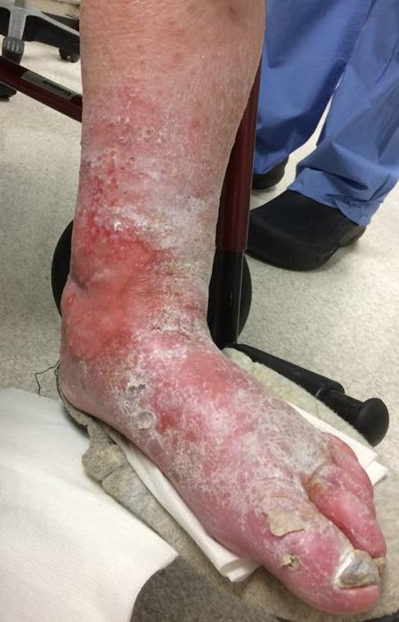
The patient is a very pleasant elderly lady who had a prior EVAR complicated by graft limb thrombosis treated with thrombectomy. She recovered from that but subsequently developed ulceration of her left ankle. She had been sleeping in a chair because it hurt her to sleep flat –her leg and foot would burn with pain. A wound care center had tried an Unna’s boot, but it caused her worse pain, and the ulcer increased in size. At admission, she had an exquisitely tender, edematous leg and ankle with a large ulcer weeping edema fluid. There were no palpable pedal level pulses.
I admitted her for workup and treatment of a mixed etiology arterial and venous ulcer.
These are patients for whom rest pain is relieved by avoiding recumbency, but with prolonged sitting, as in this lady, edema accumulates and starts to leak, creating an ulcer of the venous type, in the medial ankle (gaiter) region. These don’t resolve without addressing the underlying cause which is the arterial insufficiency. Fixing the arterial insufficiency then allows for leg elevation and compression. For the trainees, venous ulcers almost uniformly heal with Unna’s boot therapy. Elevation should relieve discomfort in venous ulcers. Neither of these occurred and raises the suspicion of arterial insufficiency.
At admission, her PVR’s showed severe popliteal/tibial level occlusive disease. CTA was performed and it showed the common femoral and superficial femoral arteries to be patent but plaque occluded the popliteal artery and origins of the tibial vessels.
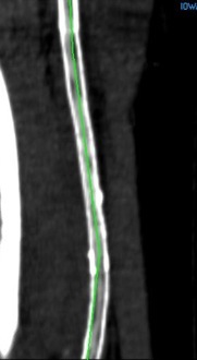
The only patent runoff was via her peroneal artery. Centerline evaluation of the CT scan was performed, with manual centerline created through the occluded segment of popliteal artery. I find this useful for planning endarterectomy and bypasses, and with attention to detail, images that are the equivalent to tibial angiograms come to life. This is a centerline through the femoropopliteal to peroneal system.
Vein mapping revealed a paucity of good vein –only a short segment in the proximal thigh on the left and for a short segment on the right. Stress testing revealed that she was good to moderate risk. Isolated popliteal occlusive disease with poor tibial runoff, while feasible for intervention, is not likely to be durable. Multisegment vein bypass on the other hand, using at least three segments, meant a long operation for this frail old lady and a prolonged recovery. I felt that popliteal endarterectomy and distal SFA remote endarterectomy offered a good option for revascularization, with either a patch repair or a short bypass to the peroneal artery. The backup plan was composite vein, but it was unlikely to be needed because the plaque was not the calcium pipe type plaque that does not endarterectomize well.
The patient was positioned on the table supine. The short segment of proximal greater saphenous vein was harvested –it was of suitable caliber, but below its first major tributary point, the veins was thick walled and small. The total length was about 10 cm. The below knee popliteal space was opened and the popliteal through tibioperoneal trunk bifurcation was exposed. Antegrade puncture of the common femoral artery allowed for arteriography and it showed the occlusion at the knee with reconstitution of the peroneal artery.
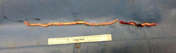
The popliteal artery was opened and endarterectomy of the occlusive plaque was performed. Retrograde remote endarterectomy (EndoRE) with Vollmer rings was performed to the mid superficial femoral artery where on the CTA the calcified plaque ended. The technical point about retrograde EndoRE is that the ring catches as the plaque gets larger more proximally, and has to be swapped out for a larger ring. Ultimately a 7mm Moll Ring Cutter was used to cut the plaque (picture below, arrow to more proximal SFA plaque).
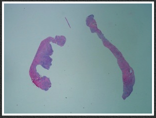
The plaque, because it is larger the more proximal you go, came out with some difficulty via the below knee popliteal artery. This is not a great concern if it won’t come out –you merely have to cut down on the SFA in the thigh to fish out the plaque. In this case, it was not necessary, and it came out in several pieces, facilitated by the cutter which was used to graft the plaque in segments to retrieve it. Unfortunately, I don’t have a picture from this case of the plaque, but I have inserted a popliteal endarterectomy plaque image below from an prior case of popliteal endarterectomy.
This restored pulsatile flow to the below knee popliteal artery. Opening the artery down to the tibioperoneal artery revealed the artery to occluded and I took the endarterectomy to the peroneal artery origin and everted a short segment of posterior tibial plaque. The peroneal artery was large and would accept flow readily, so I chose to bypass to it using the short segment of saphenous vein that I had harvested for a possible patch or short bypass. The vein was reversed and anastomosed in the usual manner. Arteriograms are below.
The flows were multiphasic. I attempted to cross the posterior tibial occlusion but ended up with contrast extravasation, therefore stopped with this repair. The patient’s wounds were closed and ulcer cleansed and compressed. In the week postop, she healed her ulcer and her two short incisions, and felt good enough to go home with homecare. Her noninvasive studies and duplex confirmed the patency of her revascularization, and there was a multiphasic signal in her posterior tibial artery as well as peroneal.
In the handful of patients I have managed this way, either with popliteal endarterectomy and patch or short (micro) bypass, they have stayed patent past a year, but do require surveillance. Because of her frailty and unsteadiness of gait, I chose not to anticoagulate with Coumadin which is my usual practice, but have her on Plavix and aspirin.
The patient was originally bypassed for ischemic rest pain due to femoropoliteal occlusive disease. He had an occlusion of his popliteal artery with reconstitution of his below knee popliteal artery. I performed a femoro-posterior tibial artery bypass with reversed saphenous vein.
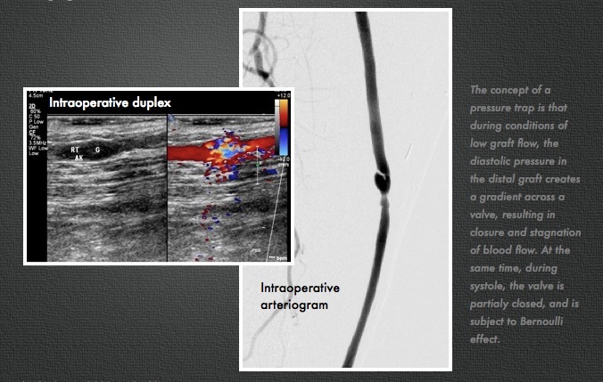
In followup, he developed at worsening stenosis in the distal graft at around 10 months post-op. Arteriography showed a moderate stenosis due to a valve that appeared to close. It was reported retained valve cusps which was strange because the vein was reversed. The flow was noted to be sluggish in the distal runoff, without a distal anastomotic stenosis. He was taken to the operating room, and this valve station was exposed and it had a severe stenosis due to valvular hypertrophy. This was repaired with patch angioplasty. At that time, I thought that this had developed due to a dynamic stenosis. No other stenoses were seen at that time.
In followup, after an initial period of two months without symptoms, the patient developed claudication that worsened. Graft duplex showed a severe stenosis in the mid graft. This was a valve station that was proximal to the previously treated valve station.
I took him to the operative endovascular suite, and arteriography showed a severe stenosis (image above, right) due to a hypertrophied valve. .
On review of the literature, I find that Tullis, Strandness et al. found 11 of 66 (17%) reversed saphenous vein bypass grafts had functional valves, with 50% of these developing >50% stenosis, with a mean time to recognition being 10 months [ref 1]. In a followup study, Strandness’s group found that valve related revisions composed 16.7% of all graft revisions in reversed vein bypass grafts, while none of the revisions for in-situ grafts were valve related. No specific valve features, they concluded, could be identified as a high risk [ref 2].
Robiscek et al [ref 3] performed intraoperative flow studies and found that under conditions of low graft flow, pressure traps occurred in reversed vein grafts. When diastolic pressure in the vein segment distal to the valve is greater than the systemic diastolic pressure, the valve closes.
This patient does have sluggish outflow due to tibial arteries that are severely disease. I believe this causes a pressure trap, but I don’t believe it is a sufficient condition for development of valvular hypertropy resulting in a stenosis.
I think that a second factor is fluttering of the valves due to turbulence. The valve cusps are in a widened segment of vein, and there is naturally turbulence that occurs -this may cause the valve leaflet to flutter like a flag in a stiff wind and cause intimal hyperplasia.
Outflow disease causes valve closure in reversed vein bypass grafts. These valves are also susceptible to turbulent flow.
The concept of a pressure trap is that during conditions of low graft flow, the diastolic pressure in the distal graft creates a gradient across a valve, resulting in closure and stagnation of blood flow. At the same time, at the onset of systole, the valve is partialy closed, and is subject to Bernoulli effect.
This is supported by the pathology which shows hypertrophy of the resected valve cusps.
This model would predict another stenosis at a more proximal point at some later date if the patient has another valve station.
While I still reverse the vein when I bypass, I am cognizant of vessel sizes and won’t reverse if there is too great of a taper in the vein diameter. I also think that not reversing to diseased or single vessel tibial runoff may be a way of avoiding this problem.
Completion angiography fits into the range of things that many of us were taught to do because it might help avoid the problem of early graft failure. I remember a time in the nineties when vascular surgery was synonymous with terrifyingly long bypass operations that sometimes worked. Back in that preinternet era, all day bypass operations were capped at the end with a flat plate arteriogram. As with all things archaic and historic, I firmly believe that our trainees should feel comfortable with this type of on-table arteriography because not every place will have a corridor of rooms with robotic c-arms. I feel that each trainee should feel comfortable wheeling in a portable c-arm, assembling it, turning it on, put in patient information, and perform a study. But I digress. The completion arteriogram clearly has a role in bypass surgery, but I question its usage as a “I do it all the time” routine. When anything is written in stone, it immediately takes on a hallowed, sanctified aura, usually taken on during M&M’s when the person at the podium intones beatifically looking skyward, “the completion arteriogram showed no abnormalities.” Science is about questioning the status quo and backing up practice with evidence.
The purpose of the arteriogram is to evaluate the anatomy for treatable lesions. Screening for these lesions can be just as easily performed with handheld pulse Doppler and if needed, duplex ultrasound. In my experience, the triad of pink toes, palpable pedal pulses, and multiphasic signals in the distal anastomosis is more than enough evidence to start drying up and closing. In this particular case shown in the picture above, the anastomosis looked pristine, but the signals were weak and monophasic in the distal anastomosis despite palpable pulses. Arteriography reveals the reason below, but frankly, the arteriogram was dispensible even in this case (trainees –reason why?). In fact, arteriography takes care of the surgeon more than it does the patient. Tan et al [J Vasc Surg 2014;60:678-85] for the Vascular Study Group of New England, including my friend Dr. Alik Farber, reviewed the VSGNE database and found that a strategy of compulsive completion studies which included angiography or duplex ultrasonography, did not improve short term or 1 year graft patency.
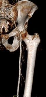
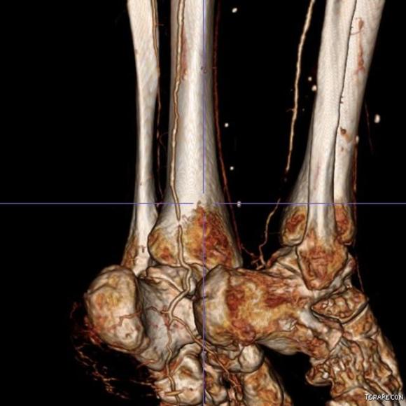
I have used many different flavors of image post processing software including Osiris, Vitrea, and now Aquarius, aka TeraRecon. But I notice that outside of endovascular planning, people rarely use the virtual 3D reconstructed images (the pretty pictures) for anything other than posting images for publication in JVS, and even there I think we have reached saturation.
I have found 3D reconstruction to be especially useful for open surgical planning, and that is by doing two things. First, on viewing the 3DVR data, I reorient and center on the surgeon’s perspective, using left button to rotate the picture around the zero at the center of the screen, and the right mouse button to grab the whole image and recenter as necessary.
Surgeon’s eye OR view
I then window-level in tissue density -this is done by pressing both the right and left mouse buttons, but you can choose this off the menu.
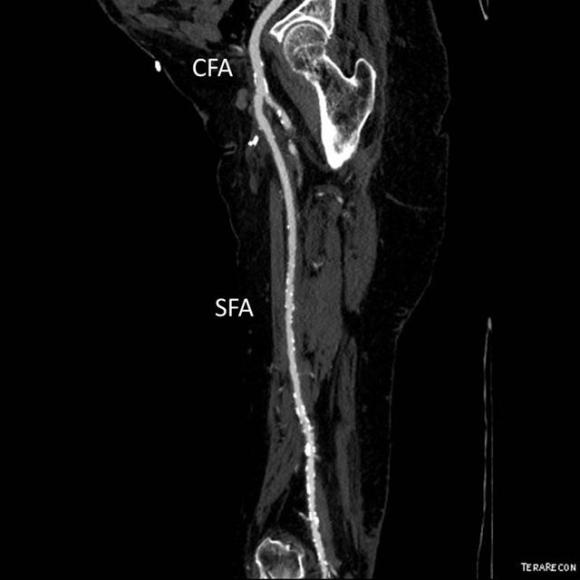
I can plan the incisions and exposures from any angle -in this case, I can see the saphenous vein and its relative proximity to the CFA to perform an in site bypass to the AK POP. And I see the loci of the tributaries that I may need to ligate.
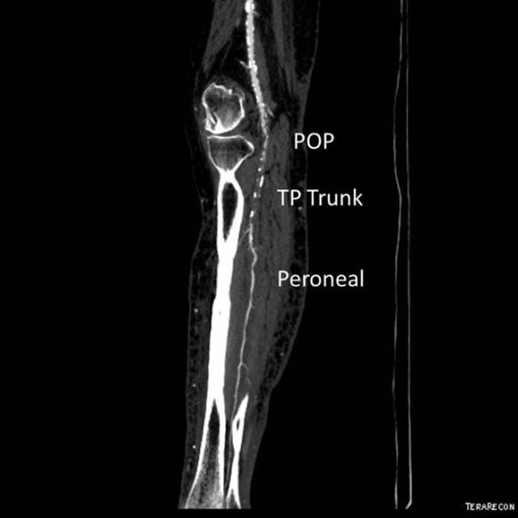
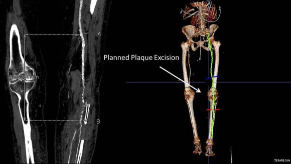
When conduit is limited, or PTFE or cadaver vein is being considered, in the setting of occluded SFA stents, I have found that it is possible and perhaps preferable to attempt removal of these stents using remote endarterectomy.
The CTA, particularly with 3D reconstruction, is helpful in planning these cases.
The additional material needed is fluoroscopy and endovascular skills. Directing a subintimal Glidewire helps free the stent and aid passage of the Moll ring dissector.
Adding cold saline seems to help shrink the stents. A plaque free distal end point allow the stents to be removed with a gentle tug.
Completion distal end pointStent removalSpecimen -4 occluded nitinol stents placed into TASC D lesion
I put these patients on coumadin anticoagulation. Surveillance is needed for recurrent stenoses -typically these occur randomly as focal TASC A stenoses, and likely represent remnant medial tissues that have caused intimal hyperplasia. This may be particularly amenable to treatment with drug eluting balloons. Failure as thrombosis typically is limited to the treated vessel without the embolism seen when PTFE grafts fail. Failure tends to occur in smokers. Inability to pass the dissector is usually seen in patients with heavy calcification -diabetics, renal failure, and I would avoid attempting remote endarterectomy in these patients. When the dissectors fail to pass, cutting down and directly endarterectomizing the vessel and resuming remote endarterectomy is feasible. The common femoral artery is repaired with a patch. I try to avoid having to place a distal stent and when a tapered end point, as in a successful carotid endarterectomy, is achieved, usually unnecessary.
Parts of this was presented at Midwest Vascular in 2008.


 .
.


 Completion angiography fits into the range of things that many of us were taught to do because it might help avoid the problem of early graft failure. I remember a time in the nineties when vascular surgery was synonymous with terrifyingly long bypass operations that sometimes worked. Back in that preinternet era, all day bypass operations were capped at the end with a flat plate arteriogram. As with all things archaic and historic, I firmly believe that our trainees should feel comfortable with this type of on-table arteriography because not every place will have a corridor of rooms with robotic c-arms. I feel that each trainee should feel comfortable wheeling in a portable c-arm, assembling it, turning it on, put in patient information, and perform a study. But I digress. The completion arteriogram clearly has a role in bypass surgery, but I question its usage as a “I do it all the time” routine. When anything is written in stone, it immediately takes on a hallowed, sanctified aura, usually taken on during M&M’s when the person at the podium intones beatifically looking skyward, “the completion arteriogram showed no abnormalities.” Science is about questioning the status quo and backing up practice with evidence.