 Completion angiography fits into the range of things that many of us were taught to do because it might help avoid the problem of early graft failure. I remember a time in the nineties when vascular surgery was synonymous with terrifyingly long bypass operations that sometimes worked. Back in that preinternet era, all day bypass operations were capped at the end with a flat plate arteriogram. As with all things archaic and historic, I firmly believe that our trainees should feel comfortable with this type of on-table arteriography because not every place will have a corridor of rooms with robotic c-arms. I feel that each trainee should feel comfortable wheeling in a portable c-arm, assembling it, turning it on, put in patient information, and perform a study. But I digress. The completion arteriogram clearly has a role in bypass surgery, but I question its usage as a “I do it all the time” routine. When anything is written in stone, it immediately takes on a hallowed, sanctified aura, usually taken on during M&M’s when the person at the podium intones beatifically looking skyward, “the completion arteriogram showed no abnormalities.” Science is about questioning the status quo and backing up practice with evidence.
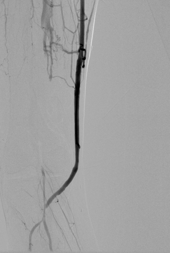
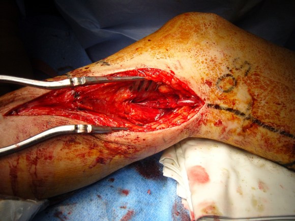
The purpose of the arteriogram is to evaluate the anatomy for treatable lesions. Screening for these lesions can be just as easily performed with handheld pulse Doppler and if needed, duplex ultrasound. In my experience, the triad of pink toes, palpable pedal pulses, and multiphasic signals in the distal anastomosis is more than enough evidence to start drying up and closing. In this particular case shown in the picture above, the anastomosis looked pristine, but the signals were weak and monophasic in the distal anastomosis despite palpable pulses. Arteriography reveals the reason below, but frankly, the arteriogram was dispensible even in this case (trainees –reason why?). In fact, arteriography takes care of the surgeon more than it does the patient. Tan et al [J Vasc Surg 2014;60:678-85] for the Vascular Study Group of New England, including my friend Dr. Alik Farber, reviewed the VSGNE database and found that a strategy of compulsive completion studies which included angiography or duplex ultrasonography, did not improve short term or 1 year graft patency.