Last December, I received an email from a LOCUMS company begging for Christmas coverage at a nearby hospital. It offered an eye watering $330/hr for coverage during Christmas week and weekend. A friend posted about this on X, and it was edifying because no vascular surgeon gets this as regular pay, but it reveals the current market price of an on-call vascular surgeon, at least as much as travel nursing, and the willingness of hospitals to pay for it, reveals the true value of an experienced nurse. If you wonder why it takes so long to see a specialist in 2026, the answer is in the fact that very few people were biting for this offer to be cast so far and wide.
The fact is that no hospital can do things with sharp objects without running into the need for a vascular surgeon.
References
Ojeda LM, Arcila SM, Nunes VA, Duarte CM, Papi M, Jacobs DL, Malgor EA, Malgor RD. The value of vascular surgeons in modern health care systems: A systematic review and meta-analysis. J Vasc Surg. 2025 Oct 28:S0741-5214(25)01865-8. doi: 10.1016/j.jvs.2025.07.062. Epub ahead of print. PMID: 41167378.
Kim Y, Weissler EH, Williams ZF, Mohan S, Coleman DM. Defining the Value of Vascular Surgery Service at a Tertiary Academic Medical Center. Ann Vasc Surg. 2024 Dec;109:198-205. doi: 10.1016/j.avsg.2024.06.040. Epub 2024 Jul 24. PMID: 39059626.
Powell R, Brown K, Davies M, Hart J, Hsu J, Johnson B, Makaroun M, Schanzer A, Shutze W, Weaver F, White J; SVS Valuation Work Group. The value of the modern vascular surgeon to the health care system: A report from the Society for Vascular Surgery Valuation Work Group. J Vasc Surg. 2021 Feb;73(2):359-371.e3. doi: 10.1016/j.jvs.2020.05.056. Epub 2020 Jun 23. PMID: 32585182.
Johnson CE, Manzur MF, Wilson TA, Brown Wadé N, Weaver FA. The financial value of vascular surgeons as operative consultants to other surgical specialties. J Vasc Surg. 2019 Apr;69(4):1314-1321. doi: 10.1016/j.jvs.2018.07.035. Epub 2018 Oct 24. PMID: 30528406; PMCID: PMC8386947.
Today, I got up at 630, made some coffee, and Zoomed in on our morning report at our main campus hospital. I have a patient there who I will be operating on tomorrow and wanted to know the current status of the patient. Once the report was over, I brushed my teeth and drove into my hospital which is a regional community hospital. I had an angiogram for a patient who was having a problem with blood flow to the leg. The cath lab was ready to go at 0800, and I was done by 0900, where I quickly ran over to to my office for clinic. My fellow who was doing her community rotation helped me with the angiogram, and then came over to clinic where I saw 27 patients from 0900 to 1600hrs, two of them virtually. At 1600hrs, I had a hospital committee meeting where I am the chief of surgery for my community hospital, and at 1700, was done. I ate a snack as I finished up some paperwork, and got on another Zoom meeting of my institute with over a hundred people to have an update meeting. I then drive to my golf club, and got on the range and hit golf balls for 30 minutes, then got on the putting green and practiced for another 30 minutes. Then I drove home and had dinner with my family. I watched highlights of a football (American) game I recorded over the weekend, while reading email, then sat down to write this before I showered and went to bed.
This week I have 9 cases scheduled -several angiograms and interventions, a leg bypass, a few fistula creations, and a laparoscopic procedure (I’m one of the few vascular surgeons who do laparoscopic surgery). As I sit in bed, I listen to a journal article read to me by the voice of Gwyneth Paltrow (it’s AI) -I find it easier than actually reading the thing, and then I watch a few TikToks, read Reddit, and then go to sleep around 2300h. Cycle starts again in the morning, but will wake at 0530 to get to the main campus hospital to perform an operation. Arriving at main campus on a Wednesday, we have a combined grand rounds with the whole Surgery Department prior to operating.
Lifestyle On weekends, when I am not on call, I still catch up on my patients from a report from my trainees or my nurse practitioner who makes rounds. I even do this sometimes when I’m out of town. Usually, I play competitive golf with members at my club -the more pressure the better. I find competition to be relaxing. Afterwords, I come home and write, read a little, and watch sports depending on the season or golf. My writing is sometimes work-related, sometimes in my journal. I kept a personal blog for over ten years on golfism.org. I am working on a novel -have been for a decade but not making much progress. I read mostly nonfiction but will listen to audiobooks of science fiction -currently marching through all the Dune prequels written by Brian Herbert, the son of Frank Herbert, the author of Dune and its original sequels. I am working on a grand unifying theory of circulation.
Procedures As a vascular surgeon, I perform operations in the traditional open fashion, and endovascular procedures which are a done with imaging from x-ray. Occasionally, I do laparoscopic surgery. The open surgical procedures include operations on the aorta and its branches, and on arteries in the legs, arms, and neck. I also work on veins throughout the body. The patient arrives with a set of conditions, a prior history, and an examination, and given a problem, you evaluate it with various tests which can be blood tests, vascular tests, imaging studies like X-ray, Ultrasound, Vascular Lab Studies, CT scans and MRI’s. This is called the workup -getting data to plan a procedure. Knowledge of anatomy and physiology and biomechanics of flow are crucial to put together a plan that will be successful in treating the disease with low complication rate and good durability. The procedures require a great deal of planning and often I include my colleagues within my department and those in other specialties to get their insights for making a plan that accounts for the reason for operation, plan for operation, contingency plans, and recovery in the hospital, and healing outside the hospital. You can see some of these cases on my blog, vascsurg.me.
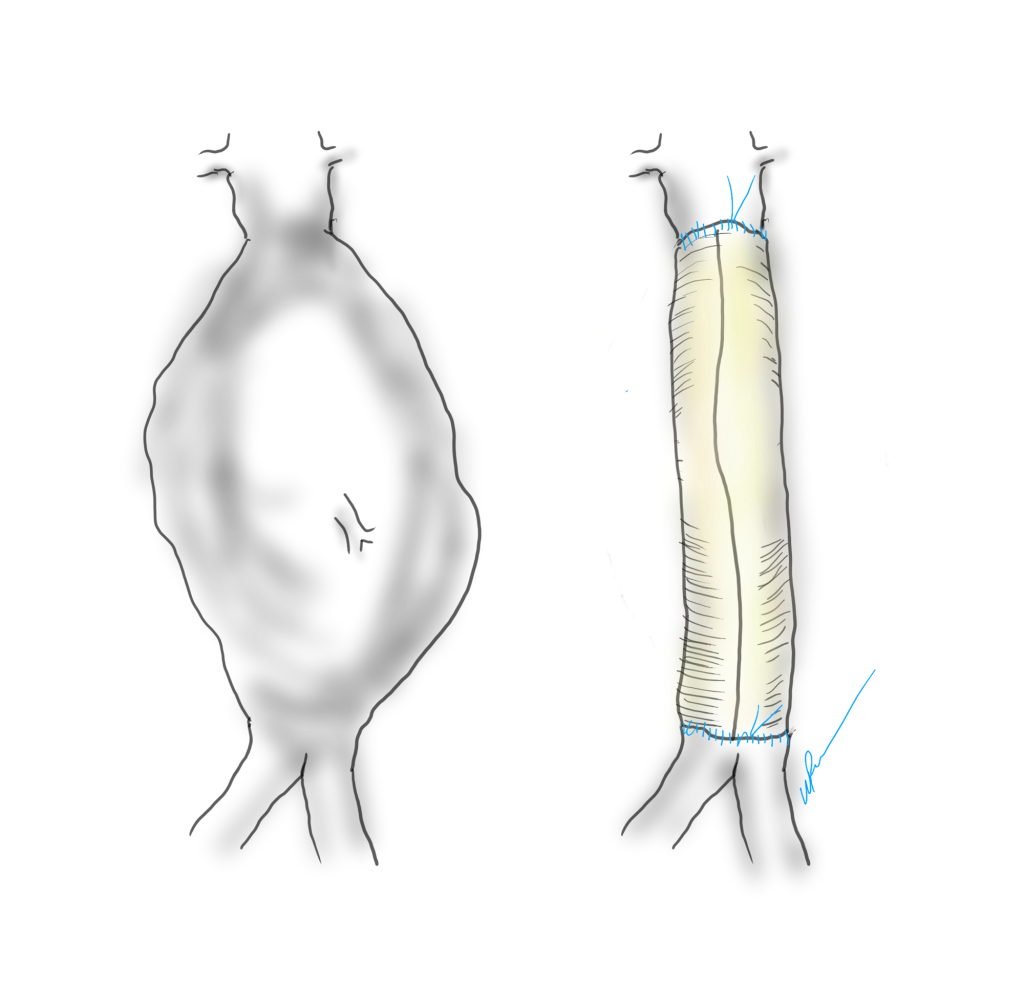
The image above shows a common femoral artery aneurysm presenting as a pulsatile mass in the right groin. The first image on left is an arteriogram (a sketch of one) that I would get prior to surgery. The patient is also suffering from pain in the right leg due to a lack of blood flow because his superficial femoral artery (SFA) is occluded and his profunda femoral artery (PFA) is open but has a blockage at its origin where the aneurysm ends. I plan the surgery and execute it. During surgery, things may pop up -good things like finding an otherwise pristine SFA filled with plaque. Removing the plaque, it becomes a great conduit for replacing the aneurysm and avoids using an expensive graft which can become infected -your own tissues fight off infection better than graft.
In the F1 Movie, Brad Pitt’s character describes a sense of pure driving, being in the flow, being completely at peace on the road. The best moments in surgery, I reach a flow state where actions follow one after the other. It’s a form of spiritual ecstasy, to be completely focused and present. Even better is having the patient do well -to be able to walk without pain and the fear of possibly losing a leg or dying.
Who should not do vascular surgery. By definition, anyone not trained in vascular surgery. Successful vascular surgeons come in all shapes and sizes, but they share common traits -grit, focus, some intelligence, and hand-eye coordination. That would mean those who give up easily, have trouble with focus, are unintelligent, and have poor dexterity should not go into vascular surgery. The saddest cases are when the desire to be something does not match up with the reality. It is possible for non-vascular surgeonsto make a living doing a focused practice around varicose veins for example, but a good vascular surgeon is hard to create. Also, you should not do this for money or prestige, there are easier ways to get money or prestige.
Who should go into vascular surgery. Anyone who thinks they might like it should certainly look into it. The best way is to directly observe a vascular surgeon at work. That is the whole purpose of the rotations in medical school. Sadly, many medical schools do not offer much time in a surgery rotation and vascular surgery exposure is inconsistent. Our society has been working hard for over a decade to improve this and we are seeing it in the excellent applicants to our training programs. The best candidates are driven people with a track record of academic excellence, but the qualities that make a good surgeon are harder to define. Desire alone is insufficient and sadly academic excellence, while it will get you into the door, doesn’t predict who will be a great surgeon. There has to be grit -an ability to persist despite hardship. There has to be a nimble mind that can solve problems quickly. And there has to be the physical hand skills that define surgery but somehow have been dropped from the initial evaluation of candidates for surgery.
Who should not go into surgery. Based on my answers above, those quick to give up, are unintelligent, and poorly coordinated should not go into surgery. I would add to this lazy, dishonest, and sociopathic. No criminals please.
There is no perfect answer to this. I knew a fellow who did not score well on tests and was rejected from medical school five years in a row, but eventually got in and completed a residency in a surgical subspecialty and has a very successful practice. While he was being rejected from medical school, he spent five years in the lab, and he could do open heart surgery on dogs very well, was coauthor on numerous papers, and his surgical skill was excellent -like if you were stuck taking tennis lessons from a professional for five years but never playing an actual game. There are also many examples of people who were told too late that they were no good for surgery.
What you should not do is listen to just a single person who has a poor opinion of you. You should examine the situation and decide if there is some truth to the issue, but you need at least three opinions. For example, I would like to be a professional golfer. I can get at least three people to tell me honestly that this is a bad idea. I would like to be a writer. I can get at least three people to tell me honestly this is a good idea. You get the picture. In medical school you will rotate and work with many people and you will have grades and feedback. You need to get honest opinions as you move forward. You need to study hard and get great grades because no matter what you do, your patients will be depending on you.
Recently, an AI was fed the world literature on AAA repair and asked about guidelines and superiority of open versus endo repair. It concluded that the past twenty year, endografting has only benefitted the physicians and the device companies (this was present at VEITH). I recommend open to patients likely to benefit from it. I recommend EVAR same way. They are not equivalent especially when patients end up getting insurance denials. I hope it isn’t too late to turn this boat around and train surgeons on open techniques that seem to have been abandoned in many parts of the world.
There was an OpMed article on Doximity (https://www.doximity.com/newsfeed/1946e8dd-eddc-4eb4-aad6-46fe59c86da5/public) which reports that 69% of 58,000 physicians surveyed said they would provide emergency care. That number is depressingly low at first view but can be answered by asking how many of us are ATLS, ACLS, or BLS certified? A quick search fails to give a result, although various pro CPR groups have on their websites that all caregivers should be trained in BLS. The darker question is how often do fully trained and certified physicians choose to withhold care and hide their identities?
I can give you a quick answer. Most doctors will sit on their hands when the PA announces “is there a doctor on the plane?” hoping that someone else will raise their hand. Back when I was a second year surgical resident, I took a vacation with my wife to London and Paris. On the flight, over the Atlantic, the cabin crew asked for any medical assistance. Before I had a chance to contemplate the question my wife jumped up and pointed at me and shouted “He’s a Doctor!”
I was in shorts and hoodie, with a baseball cap. Back then in my late twenties, I looked about 15 years old. The British Airways stewardess looked at me dubiously, then looked around behind me to see if any other hands were raised. <sound of crickets>
She escorted me up the stairs to first class and in one of the giant chair-and-a-half recliners was a pale fellow in a nice suit, diaphoretic, dyspneic, and maybe a little drunk. He couldn’t speak well but was awake and maintaining his airway. His radial pulse was thready and weak. I pressed the button that fully reclined him into a bed, not a little jealous.
“Are you having chest pain?” <head shake>
“Do you have pain anywhere?” <head shake>
“Are you diabetic?” <¯\_(ツ)_/¯>
Cold, clammy, dehydrated, drunk -hypoglycemia was my diagnosis. I asked the stewardess if they had any tubing, a funnel, and orange juice -because that is how you deliver sugar to someone who can’t protect their airway. I was an enthusiastic PGY2 and the orange juice enema was one I was eager to roll out. She looked at me funny and handed me a large black leather suitcase -the kind you see sniper rifles disassembled and packed. In it was a pretty thorough crash cart with defibrillator, airways, Mac blades and handles, bag mask, IV’s, bags of saline, and boxed syringes of code meds including D50. Oxygen was available. It was British Airways first class after all.
I looked around and saw no great place to hang an IV, so I grabbed the D50, horse needle and all, and found his cephalic vein and injected the whole vial. The change was instantaneous -the eyes which were spinning beachballs, stopped wobbling and focused. All that was missing was that Apple Macintosh “bongggg” sound. I gave the fellow a gauze and instructed the stewardess to give him orange juice spiked with sugar.
“Shall we land?” asked the stewardess. The neighboring passengers, all dressed as if for a fancy cocktail party, looked at me with eyes that said, “We really need to get to London.”
“Where are we?” I asked.
“Over Reykjavik. The captain needs to know now.”
I look at my patient, and he had unreclined himself in his fancy leather loveseat, and shook his head. “Thanks. I’ve got to get to London for a meeting.” He was going to be fine. I recommended he see someone for his diabetes (which he confessed to neglecting), and I walked downstairs and back to my seat in steerage.
A older couple (I’m sure they were middle-aged like I am now) was next to us and the lady smiled, and the man leaned over and asked, “how did it go?”
“Hypoglycemia. Are you a doctor?” I says.
“Why yes, a cardiologist. We’re going to London for a conference!” he chirped. I think he caught that I was giving him an accusing look, and added, “you’re wife volunteered you so well, so enthusiastically, I figured you had it well in hand. Good job.”
I sat down and my wife immediately asked, “did you ask them to upgrade us?”
“No.” That is the advantage of a wife when she isn’t volunteering you for missions, she’s looking out for your interests. I was going to make some grand statement of my purpose in life, but was interrupted. The stewardess came up with a brown bag full of tiny bottles of liquors, spirits, and whiskeys, which made me very happy, but my wife just rolled her eyes.
“You would have been happy with a cookie,” she hissed. “Why didn’t you ask for an upgrade? What’s wrong with these people?” And I thought the same, for a different reason. Seated all around me were likely cardiologists headed to London for that conference. Just counting bald heads, there were at least twenty.
Now, nearly thirty years on, I don’t blame those fine folk for not being quick on the draw. I am sure one of them would have stood up eventually, but the last thing you want to do on vacation is work, and what I did upstairs in first class was not much different from the work I was doing every other night on call (this was 1995).
Now, in the middle of my career, there isn’t much that gets my blood running, so I empathize with the festive, sanguine attitudes of the many physicians probably on the plane with me, headed for a nice holiday and conference in London. Some happy fellow jumps up and takes care of the problem, so no need.
Also, I’m not shy in crowds or stressful situations. Everyone in first class was watching me get venipuncture with the D50 syringe and the horse needle which was easy because the fellow was so thin. At that point in my life, screaming HIV positive crack addicts fighting you while getting central lines and spinal taps were the norm. I suppose I couldn’t fault someone more bookish and scholarly for not standing up right away. I assume 69% would have.
I’ve been called about a half dozen other times on planes. It used to be my wife volunteering me, but over the years, even she has taken on a bit of a glazed attitude. The last one a few years ago was a poor fellow whose wedding ring was causing an ischemic finger, made worse by traumatic attempts at removing the ring. Soap and rubber bands fixed him. It barely elicited an eye roll from the spouse who did not volunteer me that time. It was one of those cheap airlines in the American Southwest and I got nary a thanks.
I have never contemplated the medical malpractice ramifications of rescuing someone, saving a life. I assume something like sea-law prevails up in the air, where the captain can marry folks and push them off gang planks, where decency, need, and common sense prevails over tort law. Unfortunately, I have never seen another black suitcase since that first time on British Airways, and the pre-9/11 days of carrying a pocket knife are long gone, making emergency surgeries and fashioning of MacGyvered medical devices impossible. The idea of embarking on supporting the life of someone when the last time you ran a code was in medical school may be too much to ask someone, and doing the wrong thing may be worse than doing the right thing badly.
Did you know you can fix a tension pneumothorax with a pen, a rubber band, and a condom, with the appropriate knife and fortitude, and maybe a tiny bottle of vodka.
But isn’t that why we went into medicine? To save a life is to save the world, the Talmud tells us, and we can be heroes, if just for one day.
The patient is a woman in her forties who works hard and smokes cigarettes to find stress relief. The year prior to presentation, she began to get cramps in her calves while she walked the halls of the building she cleaned, and this became unbearable. A consultation at our hospital revealed moderate to severe diffuse atherosclerosis without a dominant lesion but notable small distal aorta and iliac arteries with a 50% stenosis of the left iliac origin. Recommendations were to quit smoking and exercise. She found this difficult to achieve and went to another hospital nearby.
There, 6 months prior to presentation, she began complaining of painful cyanosis of her toes which was described as blue toe syndrome. These outside studies were not available. She was taken to the OR and her common iliac arteries were stented. This gave her relief, but the soon pain returned three months later -her stents had occluded. This was treated with more stents, extending them proximally into the aorta and distally in the case of the right across the iliac bifurcation. This afforded her relief for three more months until one weekend she found herself unable to walk again for more than minimal distances, and she took herself to my hospital, University Hospital, Cleveland Medical Center.
On examination, she was a large woman with no femoral pulses, but signals could be obtained in her popliteal and tibial arteries. Her PVR’s showed inflow disease and poor flow at the feet.
Her baseline CTA in workup of her claudication the year prior to getting stented shows the aorta and iliacs, while open, are small, with aortic lumen diameter reaching 10mm and common iliac lumen diameter at 6mm with diffuse atherosclerosis (below).
Aorta scanned winter prior to index intervention showing small aorta diameter of 10mm and diffuse narrowing of common iliac artery, 50% stenosis left CIA orgiin. Intervention was not scheduled. Patient went elsewhere and underwent intervention .
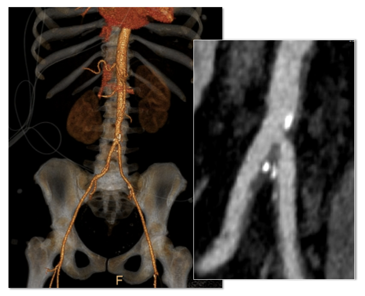
The CTA on presentation shows bilateral stent occlusion. A closer look shows the second set of stents extending the original stents both proximally into the aorta (raising the bifurcation) and distally into the external iliac and across the internal iliac origins (white arrows). The internal iliac arteries, despite the stents and on the right thrombus in the stent, supply flow to the external iliac arteries which have not thrombosed.
The treatment options were
Exercise and risk factor modification
Reintervention
Axillo-bifemoral bypass
Aortobifemoral bypass
Aortoiliac endarterectomy and patch angioplasty, stent removal
Although exercise and risk factor modification should be part of the treatment regimen, the best time to institute this was before her first intervention. With the long segment occlusion of her stents, coverage of the right internal iliac artery and occlusion, and acuity of her symptoms, this is no longer feasible.
It reveals a certain kind of bias when we prescribe walking exercise to those who can’t afford gyms or equipment, and whose neighborhoods are unwalkable.
Reintervention, having failed once, will not be durable. Even with anticoagulation, any recanalization -thrombolysis, thrombectomy, balloon angioplasty, atherectomy, lasering, and restenting, would not be durable.
It is likely the patient is frequently vasoconstricted and this is exacerbated by smoking. While never diagnosed with Raynaud’s, she did give a history of easily have numb, cold fingers and toes in the chilly winters in Cleveland. Even normal spectral Doppler signals will show pauses in flow in the peripheral arteries. Combined with any hypercoagulability and injured lumenal surfaces from interventions, and stents will go down.
An Aside on Small Aorta Syndrome in Women
One of the advantages of being a village elder is you remember forgotten concepts that guided treatment “back in the day.” The small aorta syndrome defined as having an aorta smaller than 12mm in diameter is one of those. Best described as not having enough pipe -imagine a small caliber fuel line throttling an engine. For all the muscles involved in standing and walking, there is a minimal diameter necessary for function.
Small aorta syndrome stands up to objective testing. A patient with a small aorta but otherwise patent lower extremity arteries, can present with claudication and demonstrate drops in ABI with exercise. These are typically female smokers with elevated BMI. Along with their small aortas, their external iliac arteries will be small, and I used to wonder if some critical period of inactivity in their early years failed to grow these arteries, or if this process of normal growth and remodeling is retarded by smoking.
Small aorta was a common indication for aortobifemoral bypass (ref). Unlike some abandoned indications for operation like “4.5cm AAA” and “asymptomatic 60% internal carotid artery stenois,” it had a testable finding of drop in ABI with exercise, but its acceptance has waned in the advent of the endovascular era. In a purely open era, I think there was greater emphasis and awareness on engineering the hemodynamics. While endovascular interventions simplify treatment, just stenting a small arteries usually doesn’t fix the problem as illustrated in this case. That is because there is a maximum size that the arteries receiving the stents will allow.
The iliac artery and aortic bifurcation will only tolerate so much upsizing with stents before rupturing. The interventions are constrained by the size of the adventitia. What is also ignored is the concept of elasticity -the 7mm lumen through a reopened and restented artery provides more resistance to flow than a 7mm artery restored via endarterectomy. All stents decrease elasticity of the circuit and decreases flow in a pulsatile circuit because of the increased impedance. Bovine pericardial patches on the other hand add elasticity. Endarterectomy restores elasticity. .
Enough Pipe
In the early 2000’s, I used to live in an pre-war apartment in Riverdale down the hill from Drs. Takao Ohki and Frank Veith. The apartments above and below me all shared this same feature -poor water pressure, because during a restoration twenty years before, the owner used the wrong, smaller size of pipe for this line of apartments. The taps would run, but if more than one apartment ran the shower or dishwasher, the taps would drip. The apartments would claudicate. The pipes were all patent, but inadequate. Not enough pipe. This patient endowed with small vessels, grew a large body, and smoked, and her muscles needed more pipe to support the added load. Not enough pipe.
So is the solution an aortobifemoral bypass? It is the board answer and a durable one, but it shares with axillobifemoral bypasses the risk of groin infections, particularly with a large body habits (below). The outflow arteries, all patent, are small and likely subject to vasoconstriction. My choice of ABF graft in this patient is a 14x7mm bifurcate which is on the small side, but I would be afraid that a 16x8mm graft would be too large on both the aortic and iliac side, resulting in mural thrombus formation.
A vertical groin incision will create a 3 inch deep canyon in the fat to get to the CFA
Axillo femoral bypasses, aside from the groin issues, suffer from poor long term durability and is not a great choice for a 40 year old. Her axillary artery was 6mm and sourcing flow to the lower torso from that is never great. Also, supplying a long 8 or 10mm graft would recapitulate the original problem of a small aorta. Not enough pipe.
For me, the best option would be to remove the stents and restore the distal aorta and iliac arteries to their original elasticity and slightly larger than original diameter. I would then be able to reopen flow to the occluded right internal iliac artery. Not just enough lumen, but enough and correct pipe.
Technique: Exposure
Exposure is predicated on the planned extent of the endarterectomy, place for clamping, and plans for aortobifemoral bypass if the endarterectomy results in poor adventitia. In a woman, the iliac bifurcations are easier to reach. A midline laparotomy is the incision of choice here. Let me digress here about the laparotomy. Over the three decades since the launch of laparoscopic surgery and subsequently endovascular surgery, the midline laparotomy has gotten an undeserved bad rap. Laparotomies are well tolerated and should not be viewed as a rare bailout or outright failure of laparoscopic therapy. Rather, it is still the gold standard exposure.
The infrarenal aorta to the right external iliac artery is exposed as well as the common iliac. In this patient the sigmoid mesentery was fatty and did not readily expose the iliac bifurcation so a separate exposure of the distal left common iliac artery was performed by mobilizing the left colon.
The aorta above the bifurcation was prepared for clamping. This involves circumferential exposure as I prefer a transverse aortic cross clamp. The lumbar arteries are clamped with bulldogs or aneurysm clips. The right external iliac well beyond the stent is controlled and the internal iliac is exposed and controlled. On the left the internal and external iliac arteries are expose and controlled.
The patient is heparinized clamps applied, and I make the arteriotomy with a 15 blade cutting down to the stent. The aorta is cut to a point about a centimeter below the clamp. The external iliac is cut to where there is patency of the artery and the plaque is mild. The endarterectomy is performed in the same way one does a carotid or femoral, with care to find the correct endarterectomy plane outside the plaque and good end points where the plaque adheres well. The internal iliac plaque on the right was chronically occluded but was successfully removed via eversion resulting in back bleeding. I sound the artery with a dilator to make sure a dissected plaque isn’t occluding it then reclamp.
The left common iliac artery is opened via a separate arteriotomy as I find tailoring a Y-shaped patch laborious. The arteriotomy is extended under the sigmoid mesentery and then moving the left colon medially the arteriotomy is finished slightly beyond the external iliac origin. The endarterectomy is finished short of the iliac bifurcation and any narrowing at the bifurcation is treated with the patch.
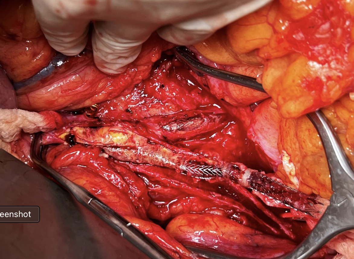
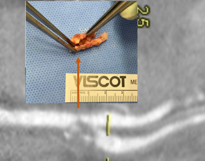
The specimen shows that the adventitia remains separated from the stents by the plaque. I rarely use tacking sutures as I feel a properly performed endarterectomy results in no plaque or well adherent mild plaque.
The patch needs to be thoughtfully applied. An overly large one will billow, and at worse case, create an artificial aneurysm. For example, for a 7mm iliac artery, the circumference is 22mm. Adding an 8mm patch with 1mm suture bites results in a 26mm circumference and an 8.3mm end diameter. Narrower is okay, but much larger will result in size mismatch that the body compensates for by laying mural thrombus. A long 8mm wide patch can be cut from a large swatch of bovine pericardium, remembering to add a slight angulation onto the iliac artery.
This operation avoids the groins with exposure or access into a small artery with a large sheath. A 4mm artery with 12.6mm circumference receiving an 8F sheath receives a 2.6mm hole, a 40% defect across the anterior hemicircumference of 6.3mm. This is not trivial, particularly because the arteries are often atretic after prolonged occlusion, may tear with a closure device, most of which are off IFU for such a small vessel. Avoiding the groins altogether is a great benefit to this type of procedure.
A postoperative CTA showed wide patency of the restored aorta and iliac arteries.
At followup several months after the procedure, the patient was walking well without claudicating and was ready to return to work. PVRs showed excellent flows down to the toes.
Hemodynamic Engineering
A surgical trainee has to develop a sense for flow. Looking at a circuit, she has to ask “how does blood get from point A to point B?” Merely providing a pipe does not mean a cure. For example, replacing a blocked artery with a steel pipe would provide flow but it has a hemodynamic impact that is different from the native vessel. Flow stoppages during the cardiac cycle that is modulated in a normal artery by the elasticity of its wall. While we don’t deliver steel pipes, we do something similar in ballooning heavily calcified arteries, or stenting them. ABF with prosthetic bypass offers a safe, broadly available method of treating this, but is fraught with problems for patient who develop groin infections or occlude a bypass for the reasons previously mentioned. Endarterectomy and patching with bovine pericardium allows for more precise restoration which would be a laudable goal in a young patient.
And my final point is this. This patient can yet undergo aortobifemoral bypass. Ironically, even larger stents may be safely placed than was previously possible. One of the principles laid out by Dr. Jack Wylie in his peerless surgical atlases was of leaving a patient in a condition to allow for future necessary operations. For a forty year old patient with many decades left, this is a critical concept.
This case represents the second aortoiliac endarterectomy I performed to remove failed stents. The third just happened today with resection of failed CERAB stents which I did with my chair and fellow Mayo Alum, Dr. Jae Cho. I think that there is a room for this operation which should not remain in the history books.
At the VEITH Symposium, which I attended briefly last week, while I foraged for lunch and sought out friends, I wandered into a crab trap (diagram above). Or more specifically, the WL Gore exhibit hall (below).
The coffee and bevarages featured all day, and the steak buffet at lunch, draws people in, like the smell of chicken to a crab, and once you have a plate of food, you then are kind of committed to moving forward into the conference room where they have a video feed of aortic symposium and tables to gobble your lunch.
Like a hand reaching into a crab trap to retrieve the catch, the reps wander in and chat you up, but thankfully only if they know you, which is fine because any hard sales tactic would trigger a fight or flight reflex that would ruin the generally chill atmosphere. There are exits to the left because, you know, fire codes, but they are small, and going out the way you came in risked bumping people juggling plates of their lunch, cups of their coffee. So you go in, sit down, and nibble, watch someone you vaguely know up on the big screen who just decided to go full head shave bald (why is that a thing?), check your phone and find out your friends are in another trap on the other side of the center. And their doors are closed, invitation only. Silly crabs.
Median arcuate ligament syndrom (MALS), also known as celiac axis compression syndrome (CACS) and its eponym Dunbar Syndrome, is manifest as epigastric abdominal pain and a compendium of symptoms, arising from chronic compression and inflammation resulting from compression of the celiac plexus between the median arcuate ligament and the celiac axis.
Graphic showing the pathoanatomy of neurogenic MALS (from ref 1). The repeated trauma to the celiac plexus results in inflammation and nerve injury with transmission of pain and neuropathic sensations.
The diaphragm muscle descends from the neck during development (the phrenic nerve originates from C3-C5 nerve roots), and in perhaps up to 25 percent of individuals, drapes across the origin of the celiac axis, and sometimes anchors further down impinging on the SMA or renal artery origins.
While a significant number of patients have this coverage of the celiac axis origin, not everyone has pain. Some whose celiac axis is compressed develop post-stenotic dilatation. For some of these, there is damage to the celiac axis resulting in intimal injury, dissections, thromboses, webs. Turbulent flow causing post-stenotic dilatation in the celiac axis can proceed to aneurysm formation. Downstream in the splenic and hepatic artery and its branches, turbulent flow can engender tortuosity (lengthening) and aneurysms (widening). This disease subset of celiac axis compression should be termed aMALS (arterial median arcuate ligament syndrome).
A question was asked at this year’s VEITH Symposium as to whether post-stenotic dilatation due to median arcuate ligament compression could be considered an aneurysm. The answer given was no, but I think it would be yes in the above example.
Both arterial and neurogenic manifestations of celiac axis compression are under the same ICD code of I77.4, referring to both celiac axis compression syndrome and median arcuate ligament syndrome. While I would never suggest more ICD codes, there should be a differentiation similar to the other compression syndrome, thoracic outlet syndrome (TOS). The pain-based syndrome, which is more common, should be termed neurogenic MALS, or nMALS, and the arterial disease secondary to celiac axis compression should be termed arterial MALS or aMALS. The treatment of nMALS is surgical ablation of the celiac plexus along with median arcuate ligament release, done via open, laparoscopic, and robotic techniques. The treatment of aMALS is the treatment of the arterial complications of celiac axis compression and should involve median arcuate release and treatment of the arterial pathology with either open or endovascular techniques.
Case Presentation
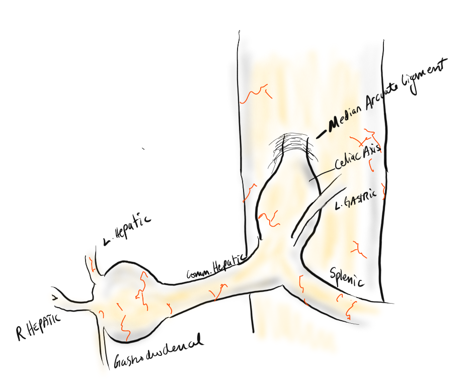
The patient is a middle-aged man with several months of right sided abdominal pain, mostly in the right midaxillary line at the costal margin, right upper quadrant abdominal pain, and right sub-scapular pain. He did not have gallstones, and had no gastrointestinal complaints. He is hypertensive and was on a single agent which he took in the mornings. His pain began during the day and crescendoed in the evening. His prior visits to the emergency room had revealed a hepatic artery aneurysm and celiac axis aneurysm. In the ED, his examination was significant for pain and mild tenderness in the right upper quadrant of his abdomen. He underwent a CT scan.
Common Hepatic Artery Aneurysm, 2.4cm with celiac axis ectasia to 14mm, median arcuate ligament compression of celiac axis
The CTA showed compression of the first centimeter of the celiac axis by the median arcuate ligament of the diaphragm and mild post-stenotic dilatation to 14mm. At the terminus of the common hepatic artery, where the hepatic bifurcated was a 2.4cm aneurysm with mural thrombus. With blood pressure control, his pain remitted.
The trainees and I had a lively discussion as to indications for repair and whether this constituted a symptomatic aneurysm. As I have stated in past posts, all pain has a nerve and a mechanism for pain. Abdominal pain and its points of referral are well known going back to the 19th century and encapsulated in Cope’s Early Diagnosis of the Acute Abdomen, whose most recent steward, Dr. William Silen just passed this September. Processes involving the gallbladder and nearby hepatic artery refer to the right upper quadrant abdomen, right chest, right shoulder and scapula which was where the patient’s pain was. And it improved with controlling his hypertension. There was no question to me the aneurysm was symptomatic, likely from strain on the aneurysm.
The question then devolves to whether this is to be done endovascularly or open. While it seems straightforward for me, I have realized at large meetings there will always be some endovascularist proposing something. For me, to exclude pressure from the aneurysm and avoid rupture, the aneurysm had to be isolated from the blood flow and pressure. Ideally, this would be done with tiny covered stents. There are no 7mm x 4mm stents bifurcation stents.
Hypothetical bifurcated small stent system -does not exist, would not work.
Embolization of the hepatic aneurysm, which is done for the splenic, offers hazard of hepatic ischemia. Despite what is written in the textbooks about the portal venous system providing most of the perfusion of the liver, you have to remember there is only portal flow when there is food. Acutely losing one of the hepatics, even clamping it for a time, reverberates as a spike in the LFTs, along with attendant systemic inflammatory response. While the liver, like spleen, can recover and regrow, you mess with it at your great peril. Based on the CTA, closing the hepatic artery with coils and plugs will likely be tolerated as hepatic flow would continue via the gastroduodenal artery which is not small, but there is no guarantee that the aneurysm wouldn’t be pressurized yet by the prominent GDA (if you disagree please feel free to comment).
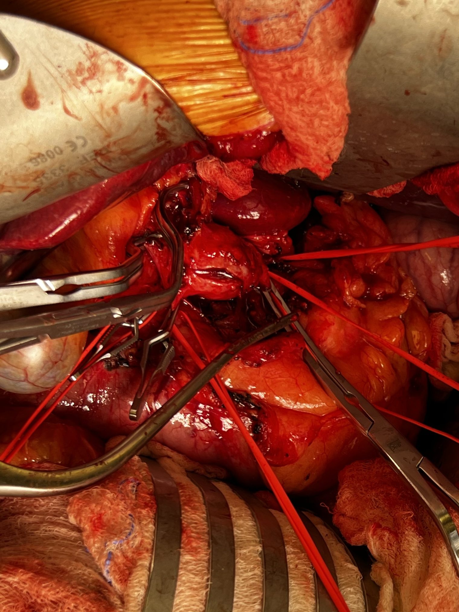
He was prepared for surgery with echocardiography (normal) and lab testing (normal LFT’s, CBC, BMP, INR), and taken to the OR. A chevron incision was made to broadly expose the area. The median arcuate ligament was exposed and released -there was dense tissues proximal to the dilated celiac axis. The aneurysm was dissected out and the small branches were carefully dissected out and controlled. It is easy to injure the branch hepatic arteries which can constrict on dissection.
A suitable length of saphenous vein was harvested and prepared. The three vessels diagrammed above did not present themselves suitable for a single Carrel patch so I sewed end to end to a patch incorporating the right hepatic and gastroduodenal arteries, and performed a sequential side to end anastomosis to the left gastric artery.
The patient recovered well and was discharged home on POD#5, and in followup had no further symptoms.
Discussion:
The differentiation of arterial and neurogenic manifestations of MALS is an important refinement of our understanding of this disease, which I believe to be a byproduct of our bipedal lifestyle. The lordotic curvature of the spine, necessary to balance our upper torso on a vertical spine, pushes the spine forward and applies tension to the median arcuate ligament, along with other structures such as the duodenum and left renal vein in superior mesenteric artery syndrome and nutcracker syndrome, and the left iliac vein in May-Thurner Syndrome.
This compression is not only enough to narrow the celiac, but injure the artery by crushing. Stenting here does not do well because of the external compression and even after release, the artery may be damaged and require repair.
The chevron exposure heals well and is well tolerated and offers perfect exposure. While I was doing it, it occurred to me that a laparoscopic bypass is technically possible, and may be preferred to the long incision. Recent multi-institution study of MALS treatment would suggest laparoscopic approach offers a lower complication rate compared to open surgery (ref 2.)
The critical thing is having more surgeons recognize the compression that occurs in the abdomen and manifests in disparate and unconventional ways. The key is tying pain to a lesion, a mechanism, a nerve, just the way Cope’s does.
References
Weber JM, Boules M, Fong K, Abraham B, Bena J, El-Hayek K, Kroh M, Park WM. Median Arcuate Ligament Syndrome Is Not a Vascular Disease. Ann Vasc Surg. 2016 Jan;30:22-7. doi: 10.1016/j.avsg.2015.07.013. Epub 2015 Sep 10. PMID: 26365109.
DeCarlo C, Woo K, van Petersen AS, Geelkerken R, Chen AJ, Yeh SL, Kim GY, Henke PK, Tracci MC, Schneck MB, Grotemeyer D, Meyer B, DeMartino RR, Wilkins PB, Iranmanesh S, Rastogi V, Aulivola B, Korepta LM, Shutze WP, Jett KG, Sorber R, Abularrage CJ, Long GW, Bove PG, Davies MG, Miserlis D, Shih M, Yi J, Gupta R, Loa J, Robinson DA, Gombert A, Doukas P, de Caridi G, Benedetto F, Wittgen CM, Smeds MR, Sumpio BE, Harris S, Szeberin Z, Pomozi E, Stilo F, Montelione N, Mouawad NJ, Lawrence P, Dua A. Factors Associated With Successful Median Arcuate Ligament Release in an International, Multi-Institutional Cohort. J Vasc Surg. 2022 Oct 25:S0741-5214(22)02443-0. doi: 10.1016/j.jvs.2022.10.022. Epub ahead of print. PMID: 36306935.
In no particular order, I list these problematic situations that are outsized in their ability to take a case sideways.
Ischemia syndromes in the unconscious. The unconscious tell you nothing about their pain and follow no commands. Therefore, vigilance and a low threshold for operating are what will save the patient if they are salvageable. Objective evidence of flow -examination, handheld pulse Doppler, duplex ultrasound, CT angiogram, exploration and visual inspection, must be obtained. The typical scenarios are dissections of the ascending thoracic aorta, polytrauma patients, and patients on ECMO. By the time the dissection is repaired and the patient is off pump, they may be long past the 6 hour threshold for irreversible ischemia for gut or muscles. The patient involved in a rollover MVA who had their femur fracture reduced after ten hours waiting on the add on schedule should have their compartments assessed visually through fasciotomies. Patients on ECMO via femoral access must by practice have distal perfusion cannulation. Assessment for ischemia need to start at admission for the unconscious patient with assessments of flow and function. Waiting until markers of cell death are apparent on blood tests is not the right approach unless the patient is DNR.
Operations in redo or irradiated fields. Preparation and coordination is key. Most vascular surgeons have a plan for controlling arteries and veins in these settings, but a common scenario is in trauma or oncologic surgery. I don’t know if anyone has done this, but the idea comes to me that if there is concern for oncologic invasion of a major artery -an aorta or iliac, it would be reasonable to place a wire, balloon, or stent graft across that area with solid seal zones to allow for free dissection and resection of any involved artery.
Central venous rupture during venoplasty for hemodialysis access with an open fistula. Instead of venous pressures, with a fistula attached, arterial pressure is driving the leak. A leak of an SVC can lead to a fatal cardiac tamponade. Because the heart fails to fill, CPR is futile. The only thing can be done once this has occurred is to be prepared to a. ligate the fistula, b. Drain the pericardium either with needle pericardiocentesis or left anterior thoracotomy. Better still is preparing for SVC venoplasty by balloon occluding the fistula prior to inflating the balloon in the SVC.
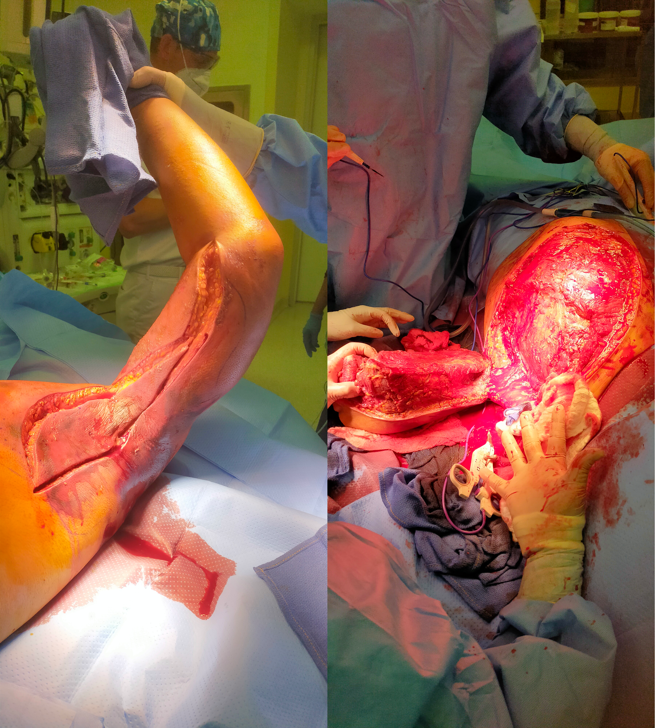
Rapidly progressing skin infections. It is amazing how fast necrotizing infections can progress. I’ve seen simple infections of a finger spread to the whole arm over the course of an hour or two in the waiting room of an emergency room. There are forgotten anecdotes of medical students dying after nicking their hands in gross anatomy. I saw a concert pianist lose her arm after getting a thorn from her rose garden. The image below is of a forequarter amputation I had to perform on a young man with a fulminant infection of the muscles of the left upper extremity undergoing a forequarter amputation after an overnight of misdiagnosis as a cellulitis at an outside facility. It grew among other things Candida auris, a terribly frightening organism and spread to his chest wall and ribs, resulting in death.
5. Iliocaval venous injury, particularly small tributaries going under aorta or around its branches. While not pressurized, they have tremendous flow like a hole in a plastic bag holding a goldfish, and without precise control, you are as likely to widen the hole or make more holes as you try to suture the holes. I’ve had some success using the Park clamp (link). You can’t buy one but you are free to have one made by your local smith. Otherwise, you need to keep your finger on the hole while you call in help, usually in the form of more vascular surgeons to get exposure and the vein properly clamped for repair.
I recently had lunch with Dr. PJ O’Hara, emeritus professor, and former partner of mine from the Cleveland Clinic. We hadn’t met since 2018 at the VAM in Boston, while I was still in Abu Dhabi. It was a recent case I did that caused me to reach out. I won’t be posting that recent case in detail today -it was a patient who had had multiple aortoiliac interventions for aortic bifurcation disease, but who closed up their stents within a few months of intervention. Rather than subject that patient to another round of interventions, I chose aortoiliac endarterectomy because the prior interventions failed to address the basic problem of the undersized aorta and iliac arteries.
The last case that Dr. O’Hara did before retiring was an aortoiliac endarterectomy which I assisted with, nearly a decade ago. During that case, Dr. O’Hara mentioned a video he had put together for an SVS meeting. He was kind enough to give me a copy share.
Aortoiliac endarterectomy -forget thee not!
The modern application of this technique is in the removal of occluded aortoiliac stents. The aorta and iliac arteries are restored, and yes, stents can go back in if needed.
A quick survey of some of my contacts at major centers reveals that this technique is rapidly becoming forgotten as its practitioner retire or revert to teaching the technically easier aortobifemoral bypass (ABF) graft. I hope to revive this because I know there are many patients who have challenging anatomy for ABF but potentially could undergo plaque and stent removal and restoration of their aorta and iliac arteries.
CTA tends to overread stenoses which was in the 60-79% range on duplex
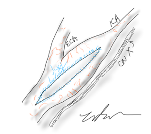
The patient is a middle aged executive who complains of bouts of aphasia triggered by intense conversations and business meetings. It first occurred while driving to Dubai on a conference call. Since then, they occurred several times a week, typically triggered during meetings where he needs to think and speak. Casual conversation and cognition does not seem to trigger this. Workup revealed a heterogeneous plaque affecting the left ICA with velocities in the 60-79% range. CTA confirmed this plaque. MRI failed to show any stroke or other lesions. Neurology evaluation showed normal exam. The patient underwent endarterectomy, and had a normal recovery. In followup, he denied any further episodes of aphasia.
Standard endarterectomy with patch
Aphasia, the loss of function in the language centers, typically of the left brain, although in a minority, it may live in the right hemisphere, is terrifying manifestation of stroke. This case, if examined superficially, is nothing special in that TIA’s associated with a reasonable culprit lesion went away after elimination of that culprit lesion. To me, it was fascinating because it represents a possible case of brain claudication.
The human brain is believed to have evolved to its large size in conjunction with bipedalism, social hunting and gathering, and climate change in the Great Rift Valley favoring a savannah over forests, that created heat stresses on the brain, favoring the development of sweating and redundancies in brain tissue. The advent of fire and cooking enhanced available calories to feed this enlarged brain’s metabolic needs. When the metabolism isn’t supported through adequate blood supply, the brain tissue dies. Rarely, it blinkers on and off, and even more rarely, this occurs in the motor strip triggering today a neurologic evaluation including a carotid duplex that brings these patients to our attention. The fascinating question for me is, does increased metabolic demand in the form of complex thinking result in a supply-demand mismatch much as seen in exercise induced angina or claudication? If it can, can we test for it?
The tests we have available are hemodynamically based. At its simplest, after carotid angiography, an occluding balloon can be inflated to test for symptoms. This is an archaic test and I do not do it. There are nuclear medicine, PET CT, and MRI tests that use pharmacologic agents to induce hypotension, but again, for this patient, it wouldn’t apply. This patient needed the equivalent of a treadmill in the MRI machine. Maybe having him read a dry, technical treatise on neurobiology taped to the MRI tube?
I went to the OR with the indication of TIAs associated with a >50% lesion, but I did tell the patient that it was possible his thinking-induced aphasia would not remit. Thankfully it did.