Categories
amputation aortoiliac occlusive disease (AIOD) BKA graft infection limb salvage PAD tibial revascularization

Never say never

fem-at-bypass.jpgIf you work long enough, you will not only see everything, but you may end up doing something that you say you would never do. You will be confronted with a scenario that would test not just your skills but also your boundaries. The adage, never say never is a warning that all of us may face a choice -to remain rigidly consistent with some earlier proclamation or to excuse a little hypocrisy for the sake of the patient.

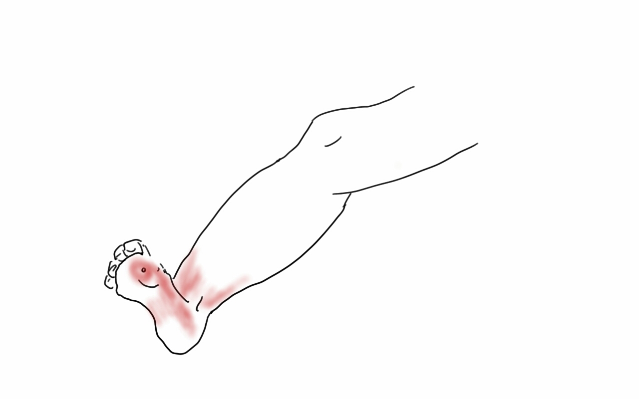
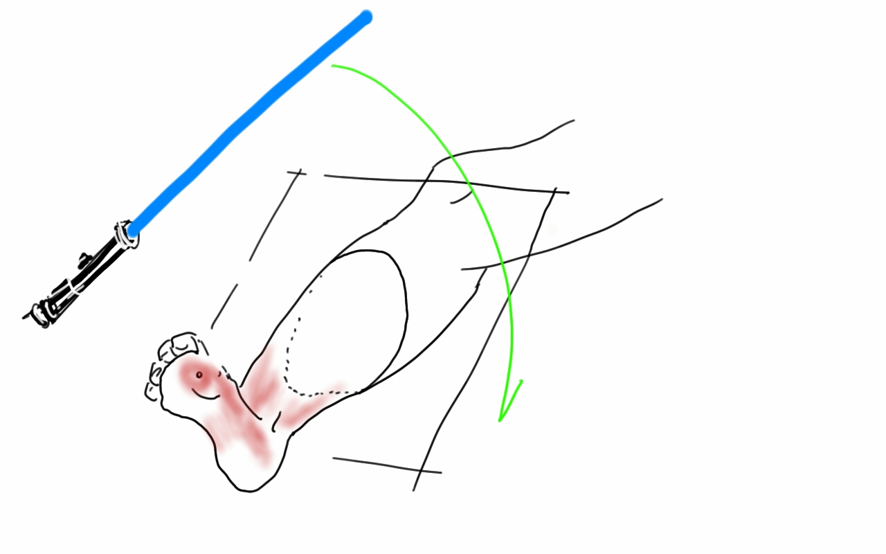
At one point in recent years, I saw a patient who had an axillary artery to anterior tibial artery bypass with PTFE (figure above). That was a kind of marvel to me, but my initial response was a bit of a sneer.

“Who does this?” I thought.

Giving it some thought, the rationale could have been to spare the patient from a hip disarticulation as the patient had had failed revascularizations and was occluded from the infrarenal aorta to the anterior tibial artery on that side. If you see such a thing, it sparks wonder as it feels both wrong and splendid at the same time because some surgeon had the audacity to pull it off. By the time I saw it, the patient had avulsed the proximal anastomosis, infarcted their leg to their thigh, and was headed for a hip disarticulation, four years after the creation of the bypass. Four years of patency!

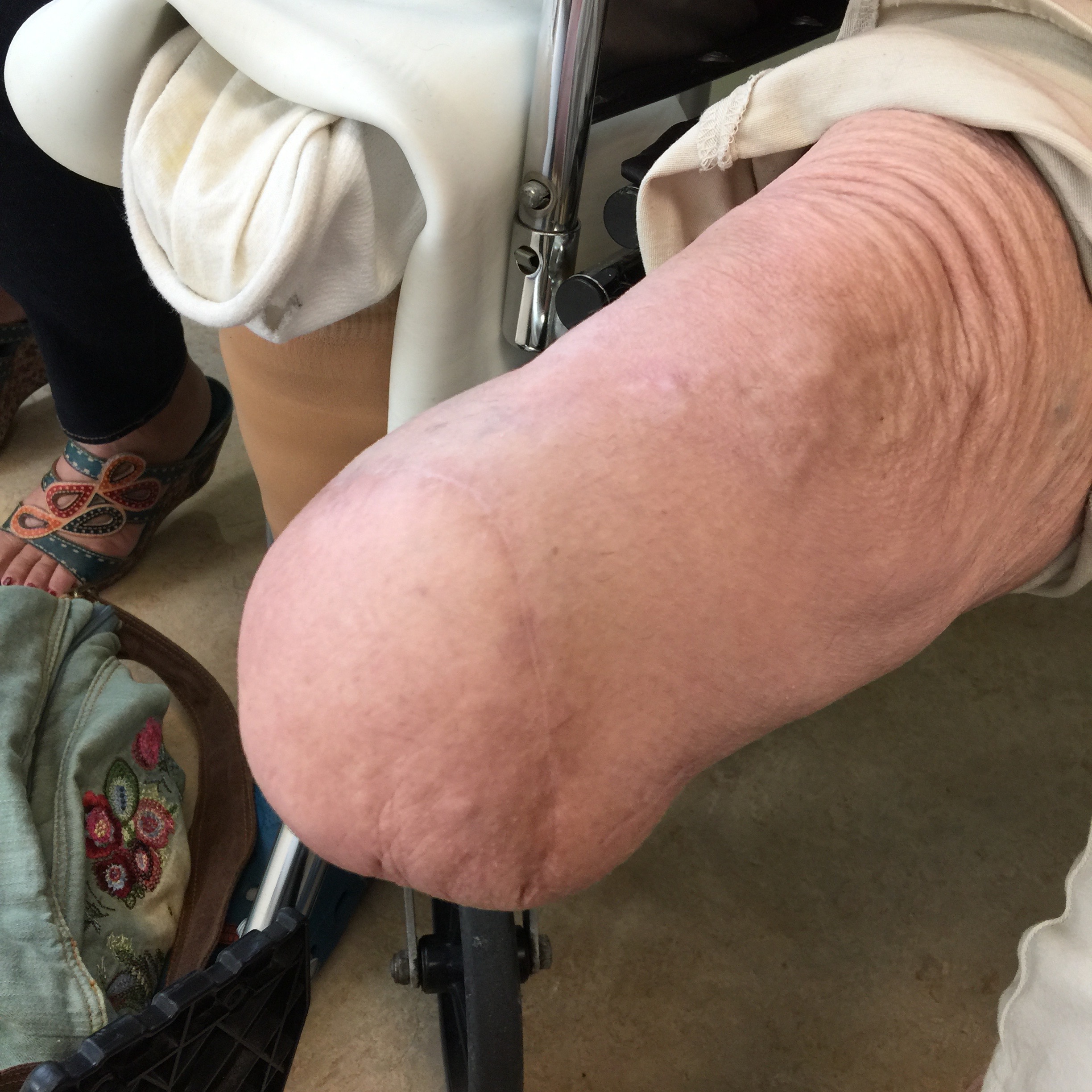
It was no wonder I remembered this case when this middle aged man presented to our clinic with a gangrenous right third toe. He had diabetes, hypertension, CAD with prior PTCA, prior acute mesenteric ischemia with bowel resection with an SMA stent, CHF with moderately reduced EF, CKD, and aortoiliac occlusive disease treated in past with aorto-bi-iliac bypass, left to right fem-fem bypass complicated by graft infection requiring resection of the fem-fem bypass, with subsequent development of rest pain on left leg and gangrene on right leg. He had been told at his home institution that he required eventual bilateral hip disarticulations. At the time of consultation, he was minimally ambulatory, limited by severe pain. He had been this way for over a year.

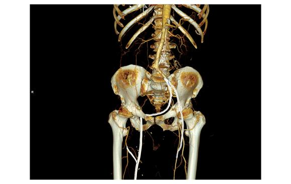
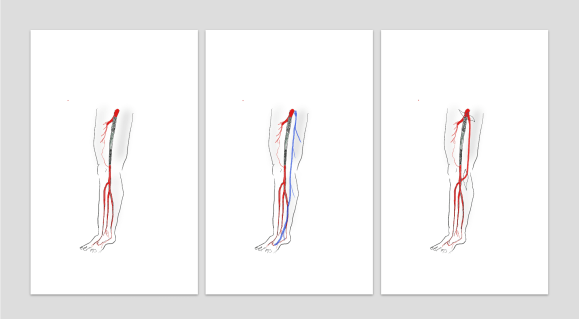
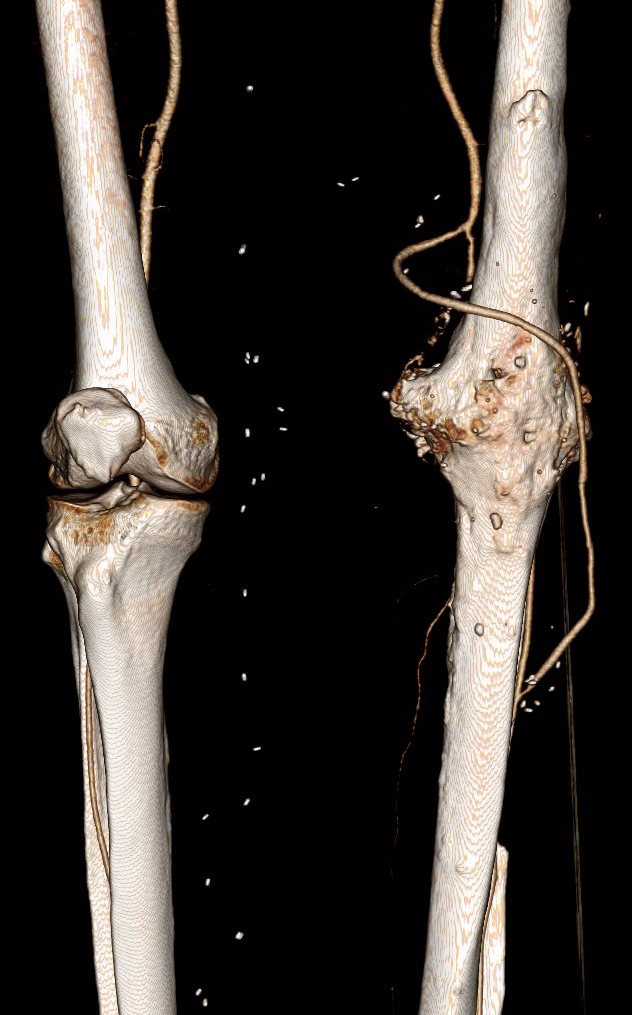
On examination, he had heavy scarring in both groins from prior open incisions healed by secondary intention, a midline laparotomy incision. He had weak bilateral axillary and brachial artery pulses. He had no pulses in either leg. The right foot had gangrene of the distal phalanx of the third toe. The both feet were anemic and painful -the left foot had more dependent rubor. Pulse volume recordings were flat in both legs. TCPO2 was in the 20-40mmHg range at the thighs bilaterally suggesting reduced potential for healing an above knee amputation. Vein mapping showed no suitable saphenous vein in either leg. CTA (figure below) showed both external iliac arteries to be occluded or absent and the common femoral arteries to be occluded or missing bilaterally.

AngioRunOff 1.0 B20f

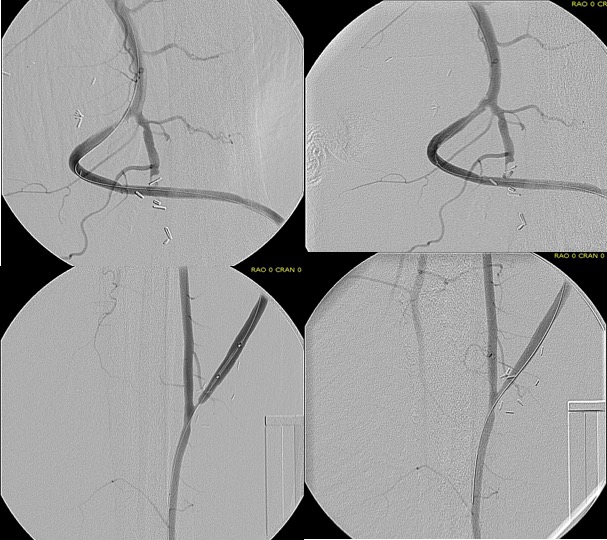
The left femoral bifurcation was preserved and the left SFA was patent into small underfilled tibial vessels. On the right, there was an isolated segment of profunda femoral artery that reconstituted from pelvic collaterals. The right below knee popliteal artery reconstituted and had underfilled but patent three vessel runoff (figure below).

AngioRunOff 1.0 B20f (4)
posterior view of right popliteal reconstitution

The patient was admitted for workup and treatment. Heparin drip was started. In the setting of rest pain, I find that heparin drip improves circulation and symptoms even though it shouldn’t. I don’t have a great explanation for this, but it does, and I would welcome comments. To better assess how much operation the patient could tolerate, a cardiac risk assessment was performed. He was deemed a moderate to severely elevated risk due to his EF of 35% but had a normal nuclear stress test.

The options I presented to the patient were
1. Hip disarticulations
2. Bilateral above knee amputations with a wait and see approach to hip disrticulation
3. Sympathectomy
4. Axillo-profunda or popliteal bypass on right and Axillo femoral bypass on left
5. Ilio-right popliteal and left femoral bypass.
6. Thoracic or supraceliac aorta to right profunda and left femoral bypass

Hip disarticulation is the bogeyman of leg amputations done for peripheral vascular disease. When done for trauma or cancer in young people, the ability to rehab and walk again is excellent. When done for tissue loss in elderly, non-ambulatory patients, the reported mortality of the operation rises to above 50%. It is usually posed as a lead in to comfort measures. The above knee amputations were not likely to heal despite the neither here nor there findings of the TCPO2 which is only good when the results or normal or dismally low. Sympathectomy is an option for those without options, but this patient still had options, I felt.

Any revascularization relies on the choice of inflow, outlow, and conduit. In endovascular revascularizations, the conduit is the previously occluded vessels, but in this instance, because of the infected grafts, there was neither continuity, nor a good option even if there was as the common femoral artery is a terrible recipient of endovascular therapy. The options then devolve to choosing an inflow. The axillary arteries are technically easiest to access and manage and form the basis of treatment of high risk patients requiring limb salvage who have no endovascular options. The axillofemoral bypass is given a bad reputation of having a poor patency, but the key is the quality of the vessels and the number of potential tension, compression, and kink points. I think the reason why the axillary to anterior tibial bypass lasted for four years in the first patient had to do with his immobility, and the pristine nature of the anterior tibial artery -the only patent vessel below his umbilicus. Here to, the inflow disease appears to have spared his right popliteal artery and his left superficial femoral artery.

The only compromise with an axillary artery inflow is the amount of potential flow. In a patient with a 6-7mm axillary artery, the amount of flow going to both an arm and a leg, and a lower torso, would greatly exceed the flow capacity of that vessel. The infrarenal aorta on this man is graft and is relatively inaccessible due to the prior laparotomy for acute mesenteric ischemia, signalling the high likelihood of adhesions. The supraceliac aorta is an excellent inflow source and I have had good results dissecting it out laparoscopically as it is often deep and narrow an exposure to try to dissect open -While the retroperitoneal tunneling can be tricky, it is not insurmountable and good bypasses can result (link).

IMG_3242

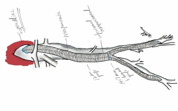
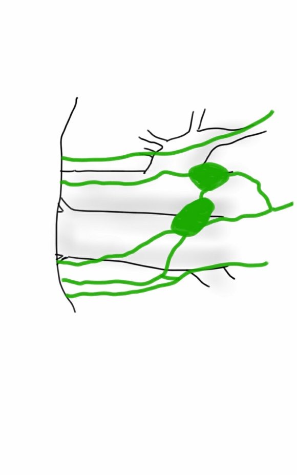
My eye focused on the left iliac graft which perfused the internal iliac artery on that side. The graft was generous, and likely a dilated 8mm graft, and could be exposed via a left lower quadrant retroperitoneal exposure (the transplant exposure). This would allow me to to then tunnel to avoid the terribly scarred groins. On the right side, the obdurator canal could be traversed into the postioer compartment of the thigh -a graft could be sent to the below knee popliteal artery with a side graft to the tiny profunda femoral artery. On the left, the graft could be tunneled laterally near the insertion of the sartorius muscle and onto the superficial femoral artery. All of the incisions would be made in virgin skin, the only redo dissection being digging out the left iliac graft while avoiding injury to the ureter.

So I proposed a ilio-popliteal bypass. Not quite an axillo-tibial bypass, but almost there. There was some karmic balance being restored by my taking decision. It would be with PTFE all around. I quoted a 5-15% risk of major morbidity and mortality, lifelong anticoagulation, and right third toe amputation. The patient agreed.

Sketch001
sketch of iliopopliteal and iliofemoral bypassrs

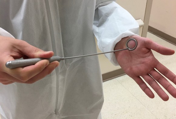
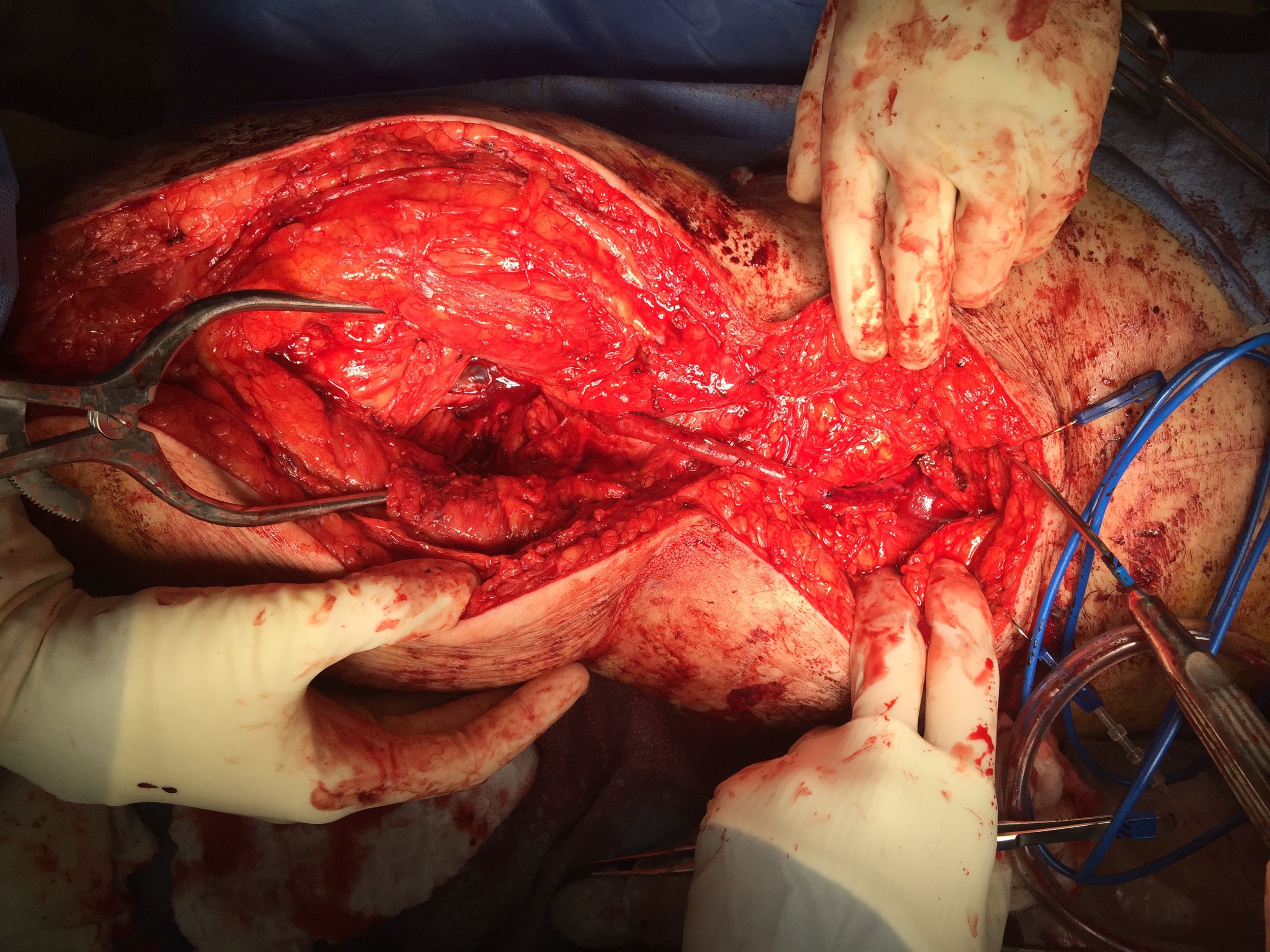
The operation was done in a hybrid suite, as should all limb salvage cases. The retroperitoneal dissection was challenging because of the heavy scar tissue around the well incorporated iliac bypass, but with patience, a clampable 3cm segment was achieved. I am a big fan of Wylie hypogastric clamps because they stay out of the way when placed in a tight narrow spot, and for that same reason, I prefer the Cherry supraceliac aortic clamp. They were designed by my mentor, Ken Cherry, and his mentor, Jack Wylie for this kind of operation. A few venous bleeders were easily handled with my ring compressors (below), and I hope to continue this chain of innovation, but I digress.

park clamp.jpg
a Park clamp

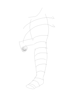
The bladder was dissected off the pubis to allow the graft to be tunneled to the right pelvis. A counter incision in the right lower quadrant abdomen and a mid thigh incision mobilizins the anterior compartment muscles to the posterior compartment allowed me to tunnel across the obturator foramen. The obdurator vessels need to be avoided or there will be bleeding. this mid thigh incision allowed exposure of the profunda femoral artery more proximally and allowe the graft to be tunneled anatomically to the below knee popliteal artery for anastomosis. A jump graft was taken off this graft on the thigh to the profunda femoral artery which was small and diseased -no more than 2mm in size. An axillary bypass to this profunda would be doomed to the compromised patency rate published for ax-fem bypasses giving them a bad name. The left superficial femoral artery was exposed and provided outflow to the left iliofemoral bypass which was tunneled far laterally under the inguinal ligament to avoid the scar tissue where the common femoral artery was.

There was immediately multiphasic signal in both feet on release of clamps. After closure of all the incisions and dressings, the right middle toe was amputated.

The patient recently came back for an 8 month followup. His grafts remained patent and he was walking without limitations. Given the high quality of the inflow (large iliac graft), and the amount of decent outflow -the right popliteal and profunda, the left superficial femoral and retrograde to the profunda, gives the patients some surety of longevity for his grafts. These grafts will need lifelong surveillance.

I have since opened my mind a bit about that axillary artery to anterior tibial artery bypass. When given the choice between comfort measures for an otherwise nonambulatory but alert patient and hip disarticulation with its attendant high risk, a bypass from a large axillary artery to a large, relatively disease free anterior tibial artery is not the worst thing that could happen. For that patient, it gave them 4 more years with their family, which in any measure, is priceless. To that patient’s family, that surgeon was a savior.

Categories
amputation aortoiliac occlusive disease (AIOD) BKA bypass Commentary complications limb salvage Lymphatic techniques tibial revascularization training Uncategorized Wounds

Exovascularist’s Dilemma: Where Is Our LIMA to LAD

During our daily morning huddles, peopled by cardiologists and cardiac surgeons, one thing impresses me more than anything else. The assembled interventional cardiologists, world class and renown, they who can place a stent in any part of the body, will defer to the unassailable superiority of the LIMA to LAD bypass over any existing intervention. I am always a little sad that the analog for this, the vein bypass in the leg does not get the same love. The open surgical bypass of the leg is the great straw man at international symposia. It is fast becoming a diminishing and curious habit of a fading generation.

The acknowledged superior hemodynamics and patency of the bypass is diminished in the literature by pooling patency loss with other factors such as amputation, heart attacks, and death. Some vascular surgeons dogmatically cling to habits learned in training that favor complications, making themselves their own worst enemies both in the literature and in the marketplace. These bad habits involve long incision length, closure techniques that do not anticipate edema, and wound orientation that makes failure more likely.

Operations require far more support and resources to succeed than do interventions that soon go home. Brilliant operations alone will not heal the patient. It is pathways and postoperative care infrastructure and staff that prevent these secondary complications -the very complications that keep the leg vein bypass from being as respected, if not loved, as the LIMA to LAD.

The postoperative care of these patients devolves to management of leg edema. No medical or nursing school adequately teaches the basic science nor management of edema, which is the most common vascular condition

The incisions are too long in the classic vein bypass. When you create and then close an incision, the inflammation drives the accumulation of fluid in the extracellular space – creating edema. This postoperative edema, poorly managed, results in complications that leave the patient hobbled with time lost to healing wound complications, pain, and excess limb weight. Additionally, vein bypasses usually involve groin exposure and the delicate lymphatics that coalesce there are perturbed or destroyed during exposure. Postop, this damage and the inflammation rapidly overcomes the capacity of a lymphatic system. The traditional vein harvest also involves cutting through deep layers of fat. The fat is typically closed by broad sutures that create areas of fat necrosis -potential fodder for bacteria. The best ways the complications of long and deep incisions is to avoid them altogether. The calculus of the operative moment – “I must see the vein,” must include the vision of a patient losing months to wound therapies to heal a gaping, necrotic, infected wound. I recommend skip incisions or adopting in-situ bypass technique with endovascular management of fistulae. Or corral your cardiac PA to harvest the vein segment in the thigh after mobilizing the vein in the leg with the endoscope.

The incisions are often closed with Nylon sutures and skin clips which can become potential foci of infection. With edema, they create zones of ischemia around them, killing skin and creating entry points for skin flora as the skin expands under an unyielding clip or suture. Placed under a pannus, these sutures or clips fester in an anaerobic environment. Closure should adhere to anatomy. The body relies on connective tissue planes to keep itself together. In the groin, these are Scarpa’s layer and the dermis. They should be closed with absorbable monofilament in a buried interrupted fashion at the dermis with a final running subcuticular layer of 4-0 absorbable monofilament. Steristrips or glue at the skin finishes the job. If you use sutures, particularly at the distal anastomotic site, take care to realize that you have about 12 hours before the skin dies in the best of circumstances, and less with microangiopathy of diabetes and ESRD. Squeezing out the edema before closure with a sterile Esmarch or short counterincisions or even a large one to allow for tension free closure over an anastomosis will prevent wound complications over your graft.

The classic longitudinal groin incisions that cut across the inguinal crease divides a tension point -that crease is like a cord that supports the pannus that is slung over it and when divided and then closed with a stitch, that stitch then bears the weight of that pannus every time the patient sits up or stands. If you are observant, wound necrosis typically starts at the groin crease under a surgical clip or suture. Incisions in the groin should be obqlique and parallel to this crease, or if you can, even inside this crease. When these wounds are closed, the natural lines of tension are in line with the incision rather than orthogonal to it. The natural forces keep the wound shut.

This is only the first step. The next is keeping the wound clean and dry for at least 5 days. At the Mayo Clinic, where I trained, the nurses up on 5 Mary Brigh were trained to blow dry the groin wounds every few hours on cool setting and redressing the wound with dry gauze. You can get something close to it by ensuring the wounds painted with betadine, allowed to air dry, and dressed with dry gauze. If there is a constant leak of fluid, you have a serious problem as there is too much edema in the leg, or the wound isn’t closed, or there is a lymph leak. It needs to be actively dried out or you get a wet, macerated, infected wound like a grenade went off in the groin.

They don’t teach compression wrap techniques in medical or nursing school

The simplest thing to avoid lymph leaks is to not make them. Cutting near lymph nodes is hazardous, and once below Scarpa’s you have to orient your dissection directly over the femoral artery. Stray horizontally and you will undoubtedly cut one of the 4 to 10 invisible lymph channels.

They are invisible but detectable -after you break them, you will see a constant wetness in the wound. Think about injecting a cc or two of Lymphazurin (Isosulfan Blue, for those not allergic to Sulfa) into the intertriginous space on the same foot and you will see the lymph channel in bright blue, or stare carefully at the likely spots for a lymph leak and clip it, burn it, Ligasure or Harmonic scalpel it.

Lymph leak identified from saphenectomy incision (for CABG)

So how did we get to a rather dry discussion about edema? Wound complications are tremendously debilitating and offset any benefit from vein bypass operations. These long incisions become terrible big wounds if not prevented. It takes the concerted effort of a team and particularly nursing in actively managing edema. And at the end, the patient too must be included in this discussion. For the vein bypass of the leg to get the same respect and love as the LIMA to LAD bypass, surgical wound complications must become never events.

Water goes downhill
Categories
AIOD amputation aortoiliac occlusive disease (AIOD) BKA bypass complications EndoRE innovation PAD remote endarterectromy techniques Wounds

Arterial Restoration -Something New, Something Old

Something that I recently promised Dr. James S.T. Yao, I will be working to publish on my stent removal and extended remote endarterectomy cases and techniques. Meanwhile, here is a talk.

Categories
amputation bypass Commentary PAD trauma

When Better is Better Than Good

original bypass

The dictum that better is the enemy of good is one of the old chestnuts carried around surgery training forever. It is an admonition against an unhealthy perfectionism that arises from either vanity or self doubt, and in the worse cases, both. The typical scenario is a surgeon trying to make a textbook picture perfect result and finding the patient’s tissues lacking, will take down their work to make it better, and repeat this process while the patient and everyone else in the room lingers.

Trying to avoid this, many surgeons will try to avoid any difficulties -the bad patch of scar tissue, irradiated body parts, areas of prior infection. But the mental contortions involved in avoiding “perfect” can result in actual physical contortions that in the end don’t pay off in good enough. I have not been immune to this, and I don’t think any physician or surgeon can honestly say they haven’t experienced some variation on this.

This patient is a younger middle aged man who in his youth experienced a posterior dislocation of his left knee, resulting in an arterial transection. This was repaired with an in-situ graft. Subsequently, he had complications of osteomyelitis and had his knee fused after resection of his joint. He did well with this bypass for several decades, but it finally failed several years ago, and a new one was created (image above).

Rather than directing the graft in line as in the previous one, this was was taken from a medial exposure of the femoral artery and tunneled superficially around the fused knee to coil lateral, ending in the anterior tibial artery.

This graft in turn thrombosed and was lysed by the outside surgeons and underwent serial interventions of proximal and distal stenoses at the anastomoses. The patient, when I met him, was contemplating an above knee amputation as a path to returning to work as a nurse in a rural hospital.

While there should be no reason long bypasses should do any less better than short bypasses, I do have to say these things about this patient’s bypass:

  1. No vein is perfect and the longer your bypass, the more chances you will have that a segment of bad vein will end up in your bypass
  2. Turning flow sharply can cause harsh turbulence. Turbulence can cause transition of potential energy into kinetic energy which acts to damage intimal, resulting in intimal hyperplasia.
  3. Thrombosis is a sure sign that your graft is disadvantaged, and the longer the period of thrombosis, the longer the intima “cooks” in the inflammatory response that accompanies thrombosis, making the vein graft even more vulnerable to subsequent intimal hyperplasia, thrombosis, or stricture.
  4. A high flow, small diameter vein graft entering a larger, disease free bed results in more turbulence but also Bernoulli effects that cause the graft to close intermittently, vibrating like one of those party favors that make a Bronx Cheer (a Heimlich valve). This is the cause I think of the distal long segment narrowing on this graft.

This patient was decided on amputation when our service was consulted, and after reviewing his CTA, I offered balloon angioplasty as his symptoms were primarily of paresthesia and neuropathic pain. I used cutting balloons and got angiographically satisfactory results.

intervention

The patient, although he admitted to feeling much better, was sad. He relayed that he had felt this way several times before, only to have his life interrupted by pain and weakness signaling a restenosis.


A direct graft would require about 10 centimeters of vein
 
It was only a month later when I heard the patient had returned with the same symptoms. He wasn’t angry nor full of any “I told you so” that frankly I was muttering to myself. Reviewing his CTA, he had restenosed to a pinhole. The vein, to use a scientific term, was “no good.”

The other interesting finding was that he had an abundance of very good vein. Following surgical dictum, his original and subsequent surgeons had used his vein from his contralateral saphenous vein. His right leg, fused at the knee, lacked a good calf muscle pump action. While there were no varicose veins, the greater and lesser saphenous veins were large and generous conduits, at least by 3DVR imagery, confirmed on duplex (image below, white arrows).

veins
3DVR showing presence of potential conduit

The extant arteries were smooth and plaque-free. I decided to harvest his lesser saphenous vein and through the same incision expose his distal superficial femoral artery and tibioperoneal trunk. While I anticipated some scarring, I was confident that the sections of artery I wanted to expose were easy to access because of some distance from the fused knee.

IMG_6478
On left short saphenous vein was harvested then same exposure used to expose TP Trunk

The picture shows the exposure and reversed vein graft in-situ, using the segment of lesser saphenous vein. As in prior experience in redo surgery, you can never know if a dissection will be easy or hard simply based on fear or concern for breaking something. It’s not until you start bushwacking –carving through scar and dealing with extraneous bleeding will you learn whether it was easy or hard. You can only be certain it was necessary. The only hitch was the femoral artery while well exposed, was buried in scar, and I chose not to get circumferential control as I was fairly deep, and had avid backbleeding from a posteriorly oriented collateral that required a mass clamp of the deep tissues.

Will this work better? Don’t know but it has a good chance, and I think a better chance. It is a large vein oriented in a straight path over a short distance going from good artery to good artery. This is better theoretically than a long meandering bypass with smaller vein. 

Categories
amputation BKA techniques

An Easy Way to Perform BKA -Treat it Like Plastic Surgeon

IMG_6438

At one time, I was performing below knee amputations in the same way that most textbooks prescribe. This patient presented with severe foot and leg infection with exposed tendons and cellulitis secondary to advanced diabetic neuropathy and arteriopathy. Sketch12683544

My sketch above showed the situation which we treated with two stage guillotine and final BKA. At the final stage, standard technique involves creating the classic flap shown below.

Sketch12683710

This is a tricky flap to make well because there are many variables -the amount of muscle left, the thickness of the subcutaneous fat, the vascular supply, etc. The biggest criticism I have of this flap is that it is prone to edema particularly in the meaty posterior flap, putting great stress on the skin sutures or staples. This then creates fat or skin necrosis in the posterior flap, or muscle necrosis resulting in wound infections and hospital readmissions, which are particularly at risk for need for further leg amputation in these patients with diabetes. I have to confess, while I could figure out how to cut the flap in the end, it was really hard to teach, and if it is hard to teach, you have to figure out a better way.

Here is the better way. The idea goes to the concept that the weight is borne in a distributive ring around the stump. The muscle on the tibia does not create a heel to bear weight on and so serves only the purpose of delivering blood to the overlying skin. So you only need the gastrocnemii and Achilles tendon for fascial closure, and maybe a little soleus. The elimination of posterior compartment muscle bulk greatly reduces the tension on the fascia and skin.

The incision can be simplified by cutting the flap along an oblique 2D plane -a light saber cut!

Sketch12684316

The posterior flap is reduced to just Achilles and a layer of soleus muscle, but otherwise, the internal bone and muscle organization is cut as below:

Sketch12684559

The skin at this point is never touched with a surgical instrument, only the subcutaneous later or fascia. Using interrupted absorbable heavy gauge monofilament (single strand of the double looped PDS used for abdominal closure is plenty of suture), interrupted buried sutures are placed in the fascia from middle outwards.

Sketch12684859This closes the fascia and allows the skin to be closed without tension using only occasional interrupted dermal sutures and a running 3-0 or 4-0 absorbable monofilament.

Sketch12685023

The skin is dress with a single layer of Xeroform and fluff gauze and gently a compressive dressing is applied from stump to above the knee, while keeping the knee cap exposed -the knee cap is prone to skin necrosis with ACE wraps that are left unattended.

The wound’s ability to heal is now largely a function of keeping the suture line tension and edema free. Edema is the great killer of these flaps and the problem with sutures or staples here is that edema creates zones of skin necrosis under these staples that then results in a postop wound breakdown and infection. With these simple steps largely copied from our plastic surgical confreres, I have been able to create very functional stumps with low morbidity.

IMG_6438

This patient is 3 months from her 2 stage amputation and is walking on her prosthetic leg and is very happy. Notice, the dog ears have contracted!