Categories
Commentary

VESS Meeting -Vail, CO 2015

IMG_3767

VESS is a special medical society whose mission is to foster academic activity and collegiality among young vascular surgeons. Dr. Vikram Kashyap, the outgoing president, gave an engaging talk about changing with the times, referencing three powerhouse rock groups -Rush, The Police, and the Red Hot Chili Peppers, three of my favorite groups.

Dr. Max Wohlauer presented our case report on “Osteosarcoma masquerading as an axillary artery pseudoanuerysm.”

IMG_3804

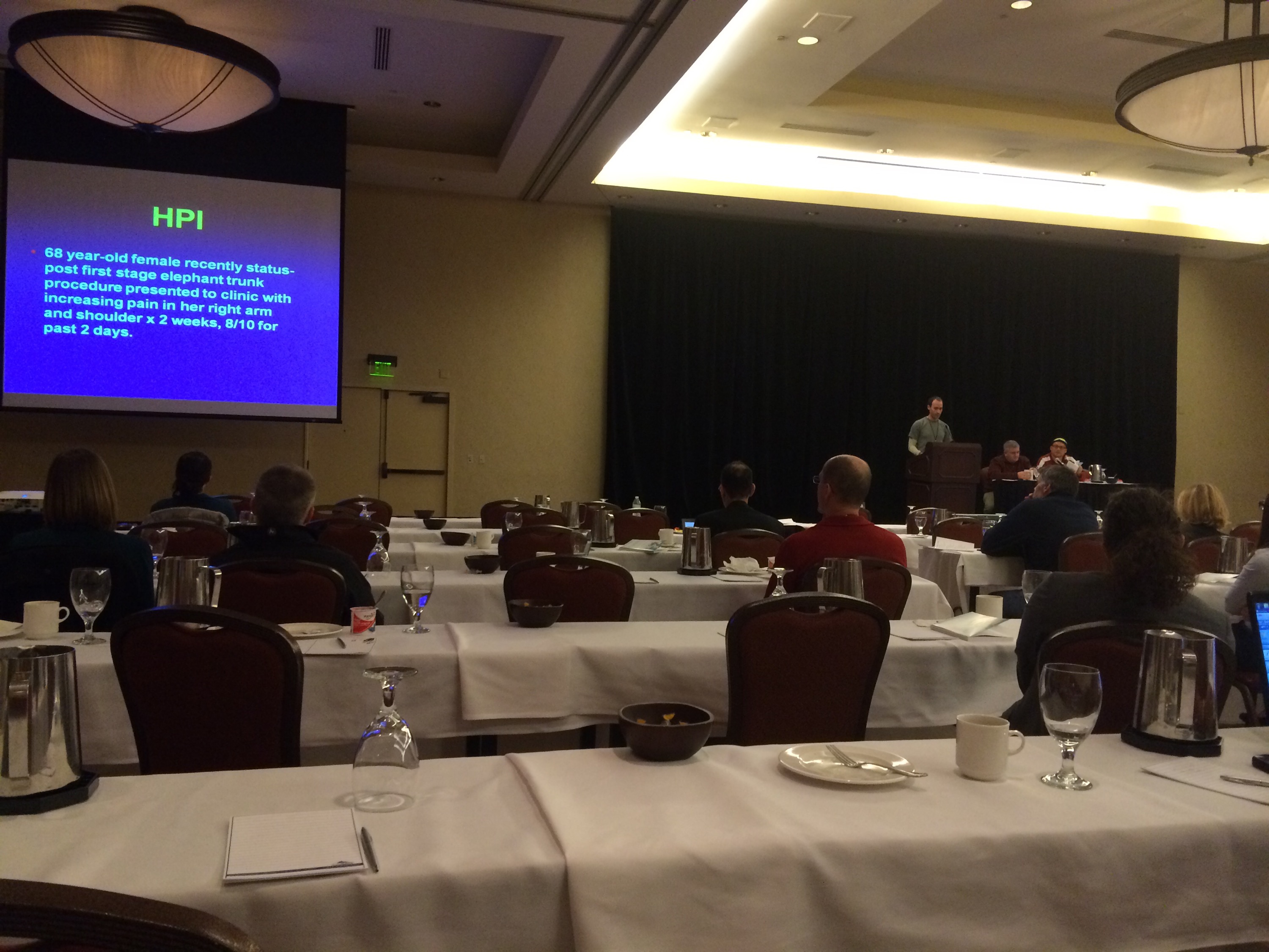
The case report ended the Friday morning session and was well received.

IMG_3805

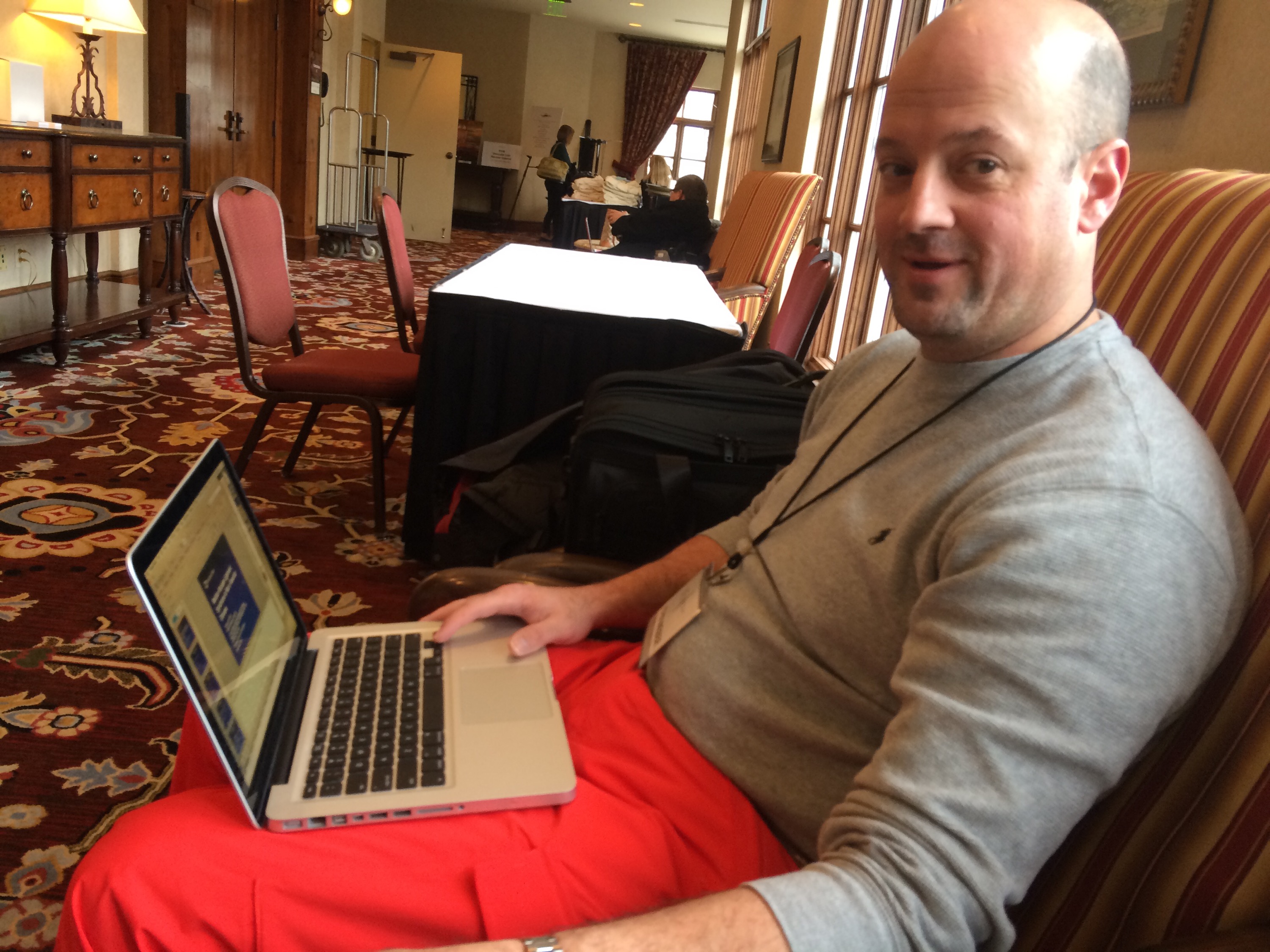
Dr. John Weber opened the Friday afternoon session with “Median arcuate ligament syndrome is not a vascular disease.” Shown above prepping for the talk, he is also in the conference dress code of being ready for the slopes.

The talk was also well received and not surprising to most who agreed. In a conversation I had with Dr. Kenneth Cherry, he mentioned that he and Dr. Jeb Hallett had concluded as much decades ago.

The scuttlebutt in the conference was about the NY Times article about the scandalous overuse of femoral stents by some physicians. It is a matter of course that the first line of therapy for mild to moderate claudication is a combination of risk factor modification and exercise therapy -something not mentioned by the writers as something emphasized by Vascular Surgeons who as a rule are against plying stents on asymptomatic or mildly symptomatic patients.

The other thing was the tremendous demand for vascular surgeons, and the success of the 0-5 training programs which represent the future of vascular surgery. Dr. Karl Illig professed as much in the Q&A of a report from Wake Forest. Surprisingly the majority (100%) of 0-5 graduates in 2013 went into academic practice.

Categories
TEVAR

Aortic Zones

TEVAR zones

Categories
techniques TEVAR training

Kitchen-top Thoracic Stent Graft

IMG_1237

Before manufactured thoracic stent grafts were approved for use, you had to make your own. I think that even in theory you should have this in your mental locker, because it is pretty straight forward to accomplish. The patient was a homeless man who got struck by an SUV while crossing Broadway merely blocks from Columbia Presbyterian. The specifics are lost to time, but he was found to have among his multiple injuries a tear in his thoracic aorta at the ligamentum arteriosum. Cardiothoracic surgery felt that he was far too high a risk to undergo open repair. I was on call, and when I looked at this patient’s scans, I realized that he might survive with a stent graft across the tear, but the only suitable grafts were short aortic cuffs intended for infrarenal repair with short delivery systems. Being young staff, I called our site chief at that time, a grizzled veteran, for some advice about making stent grafts.

IMG_1232IMG_1234
The process is simple enough, and discovering it is like finding out that a seemingly complicated dish has an exceedingly simple recipe. The process starts with an iron and an ironing board, with which you press flat a Cooley graft of 32 or larger diameter. The Cooley graft is a fine weave graft that has pressed cylindrical folds that allow you to collapse it like a Slinky toy. Ironing between two sheets of paper allows you to avoid overheating the fabric.

IMG_1233
Once flattened and stretched, it is now ready for placement of stents. The stents shown here are Gianturco stents which typically are constrained with a monofilament and has barbs. The barbs are removed with needle nose pliers. 5-0 monofilament suture is used to secure the stents in the graft. More spacing allows for the graft to accommodate tortuosity, but the graft may bunch up in the sheath. The top and bottom stents should be within 5mm of the graft edge –this way you will remember that at deployment.

IMG_1235
For this case which required only one stent, three were made and they were autoclaved. Loading into a large sheath of 24F is done over a catheter to preserve a wire channel. The graft is pushed in using the umbilical tape or silk suture technique referenced in Oderich’s paper about reloading modified stent grafts.
Because of the large deliver system, a conduit was required and sutured end to end into the common iliac artery –I no longer do this unless there is a problem with severe plaque requiring endarterectomy. The graft was deployed by push-pull technique with the heart rate slowed pharmacologically. The patient stabilized from this, took several months to recover from his other injuries but was discharged and lost to followup.

IMG_1236IMG_1237
Is this knowledge helpful? In 2015, debatable, but in 2003, it saved a life.

Categories
Lymphatic techniques Wounds

Extremity lymphatic leaks -a rare complication

2015/01/img_1225.jpg

This patient had had an endoscopic saphenectomy for coronary bypass at another institution and had developed a wound infection that eventually healed, but developed a pore at the scope site that continued to drain clear fluid soaking an ABD pad hourly. This had been cauterized and sutured without improvement.

2015/01/img_1227.jpg

The patient was taken to the operating room for dye lymphangiography and resection of the leak. The dye used was isosulfan blue which is lipophilic and is taken up by the lymphatics from the extra cellular space and pumped centrally within minutes of injection. The lymphatics are easily visible and the leak can be resected and the source vessels can be oversewn.

2015/01/img_1228.jpg

The suction cannula is on top of a lymphatic that was feeding the pore and was oversewn. I have had this occur in the forearm as well after a braciocephalic fistula creation.

2015/01/img_1229.jpg

The would was closed primarily. The lymphatics are evolutionarily the most primitive of our vasculature. They are gossamer threads often of single cell layer that have openings into the extracellular space and draw in fluid past bicuspid valves that bracket smooth muscle cells that periodically contract -a very primitive heart. The heart evolved from this mechanism.

It’s believed that multicellular organisms gained an advantage by maintaining an inner sea that was more organized and concentrated with nutrients than the surrounding sea, and to benefit all of the cells, it was necessary to circulate this inner ocean. The extracellular space is as porous as a sponge because that is what the ECM evolved from. Holding a wet sponge up in the air and watching the water go down with gravity is what happens when you stand up -edema. It is also why you can patiently and gently massage the extravasated saline from a displaced IV out of a hand and forearm. When the lymphatics stop pumping, the protein in the fluid eventually comes out of solution forming an insoluble solid -same thing that happens in cheese making when you add acid to milk -the hardened tissues of chronic lymphedema.

The pumping action is so efficient that injecting an amp of isosulfan blue into the web space between the toes subcutaneously will travel up the leg in about 5-10 minutes. There is a list of contraindications and precautions for its use, and hypersensitivity and anaphylaxis is reported to occur in up to 2 percent of patients.

This patient had primary closure and stopped leaking, after suffering from nearly a year of leakage.

Categories
AAA EVAR techniques training

Homemade laparoscopic suturing station -video

Tinkering in the basement, because bringing a laparoscopic tower and simulation setup into my house would be difficult.

Categories
Carotid PAD techniques

Innominate Endarterectomy -what the Satinsky clamp is for

Innominate.001

The patient, a 47 year old woman, was referred for syncope, but it was much worse than that. Excessive stress, standing, and thinking hard were described as causes of her syncope. Frequent headaches and lethargy lead to inability to keep a job. Several MVA’s resulted in revocation of her driver’s license. Added to that was a two to three pack a day cigarette habit. Her cardiac workup was negative, but it was noted that her carotid duplex was notable for a left ICA occlusion and left vertebral occlusion. Blood pressure in both arms was in the 70’s systolic while in her better thigh it was 90’s. She had an open right carotid system but the flows in the common carotid were attenuated. CTA of the arch revealed severe arch disease affecting the origins of her great vessels. The innominate artery was severely diseased to its bifurcation with a small <2mm channel of flow. She had a dominant right vertebral artery that was patent, and the right ICA had moderate disease at its origin. This was in 2009, and I entertained intervention, but wasn’t all that confident that kissing stents into the innominate was all that great of an option even though there are reports of innominate interventions in the literature. I had the opportunity to perform a handful of great vessel reconstruction with Ken Cherry during my fellowship and felt that this was an ideal case for an innominate endarterectomy.

This is one of those rare and infrequent cases from vascular surgery history. The exposures is one of the grand vistas of vascular surgery. The arch, thankfully, was only calcified at the origins of the vessel and clamped well. The endarterectomy was not that much different from an aortoiliac endarterectomy with a fibrocalcific plaque and was extended onto the common carotid while the origin plaque of the subclavian was plucked cleanly. The phrenic and vagus nerves were protected. The patient was centrally hypertensive as found by a long femoral arterial line and was kept that way for the duration of the clamp. A bovine pericardial patch was applied and the sternum was closed over a mediastinal chest tube.

The recovery was impressive for the patient’s immediately improved state of consciousness, lack of lethargy, and improved cognition. She was herself impressed enough to quit smoking during that admission for good. Her right brachial cuff pressure now correlated well. She went home POD 5. When I last saw her 2 years later, she was employed and symptom free with continued patency of her repair, consistent with the earlier reports of this operation (Cherry et al. J Vasc Surg; 989;9:718-14).

Categories
Journal Club

January 2015 Journal Club

Congratulations to Dr. Francisco Vargas for his December Journal Club presentation. January’s club meeting will be on the 20th.

Dr. Moqueet Qureshi: Open vs. Endo Popliteal Artery Aneurysm Repair

Dr. Deanna Nelson: VIASTAR Trial

Dr. Hazem El-Arousy: Cryografts in Infrainguinal Bypass

Categories
Journal Club

Journal Club Reformat

Screenshot - 12222014 - 07:54:40 AM

The December Journal Club was held last week with excellent attendance. The winner of the best presentation prize was Dr. Francisco Vargas who critiqued the CaVent trial. Many thanks to Leo Godlewski of Hanger Orthotics for sponsoring the club’s evening.

The score sheets had a misprint in the scaling factor. The scaling factor is highest for the substance of the critique (x3) and the next highest is the presentation substance (x2). Scoring was corrected for this.

To emphasize the importance of an in-depth understanding of the paper being presented and to keep the journal articles topical, the rules are being modified. First, the selected journal has to come from the past two years. The second modification is that every presentation has to end with a table of related or similar papers and their conclusions or results. These should be a listing of landmark papers or first of its kind papers, and the discussant should be prepared to comment on them. If there are too many papers, the discussant should be prepared to discuss a few landmark papers on the topic or discuss a meta-analysis or Cochrane review article on the topic.

The presented paper should be graded on the following scale:

  • Level 1 – High Quality – Randomized controlled trials with high power. Meta-analyses of multiple RCT’s.
  • Level 2 – Moderate Quality – Evidence from one well designed experiment. RCT’s with low power. Prospective cohort study.
  • Level 3 – Low Quality – Evidence obtained from well-designed, quasi-experimental studies such as non-randomized, controlled, single-group, pre-post, cohort, time, or matched case-control series.
  • Level 4 – Very Low Quality – Evidence from well-designed, non-experimental studies such as comparative and correlational descriptive and case studies. Evidence from case reports and clinical examples. Expert opinion.

Also, in the critique, a recommendation should be made and this recommendation also graded with the following scale:

A The USPSTF recommends the service. There is high certainty that the net benefit is substantial. Offer or provide this service.
B The USPSTF recommends the service. There is high certainty that the net benefit is moderate or there is moderate certainty that the net benefit is moderate to substantial. Offer or provide this service.
C The USPSTF recommends selectively offering or providing this service to individual patients based on professional judgment and patient preferences. There is at least moderate certainty that the net benefit is small. Offer or provide this service for selected patients depending on individual circumstances.
D The USPSTF recommends against the service. There is moderate or high certainty that the service has no net benefit or that the harms outweigh the benefits. Discourage the use of this service.
I The USPSTF concludes that the current evidence is insufficient to assess the balance of benefits and harms of the service. Evidence is lacking, of poor quality, or conflicting, and the balance of benefits and harms cannot be determined. Read the clinical considerations section of USPSTF Recommendation Statement. If the service is offered, patients should understand the uncertainty about the balance of benefits and harms.
Categories
MALS

Tissue from a MAL release (median arcuate ligament) is nerve

This was removed during a laparoscopic median arcuate ligament release. It was a hard white band compressed under the ligament and itself compressing the celiac axis. During the release, I grabbed a piece of these fibers and sent it for pathology under the preliminary diagnosis of celiac plexus, and it was. Described as “typical for a peripheral nerve…mostly Schwann cell nuclei in between nerve fiber.” Other micrographs in the specimen had ganglionic fibers but our pathologist wasn’t able to locate it. This is an important piece of the pathoanatomy because I believe that this is the nidus of the pain associated with median arcuate ligament syndrome, not a regional ischemia that can only occur if the celiac axis is an end artery, which can really only happen after a major exenteration like a Whipple procedure.

Categories
TEVAR

A Fossil of the Kennedy Administration

From my archives, January 23, 2010,


A Fossil of the Kennedy Administration


The patient was sent to my clinic with the diagnosis of a saccular thoracic aortic aneurysm of 5x10mm. He is a 78 year old man who was working at restoring a vintage John Deere tractor when he developed chest pain. He underwent a cardiopulmonary workup which included a CT which showed a 5x10mm saccular aneurysm of the inner curve of his aortic arch opposite the origin of the left subclavian artery. This same CT showed no pulmonary embolism. His past medical history is significant for hypertension and he has never had surgery. His only hospitalization was in 1962 when he had a head-on collision which caused rib and collar bone fractures -he was the driver and he bent the steering column. 


His examination was normal. He had had a complete cardiac workup including a stress test which was also normal. He persisted on having this intermittent midsternal chest pain.


CTA images: above and below. The CT shows a small area of contrast fill consistent with a focal 5x10mm saccular aneurysm of the inner curve of the aortic arch. 


My impression is this is a residual saccular aneurysm of the thoracic aorta in the ligamentum arteriosum, related to the head on collision from time of the Kennedy administration. The injuries and the steering column damage suggest significant transfer of energy. It may have been the cause of his pain. We discussed surveillance with medical therapy (to include anti-inflammatories for any tractor lifting injuries) versus TEVAR. Ultimately we chose TEVAR.


This entry was posted on my old Medscape Blog, The Pipes Are Calling, with great comments and a poll. The poll results show a majority favoring medical therapy and watching. The only problem was that this patient was still having chest pain without a good explanation -pulmonary embolism and coronary artery disease were ruled out. The patient also lived two hours away from a hospital and would be lifting old tractors and their parts because that is what old farmers in Iowa do for fun –they also never truly retire. I think it also shows a general skepticism of surgical solutions –this feeling is based on historical bias with open surgery rather than contemporary results using percutaneous techniques.


The saccular aneurysm is an inherently unstable one as the angles on the surface of the aorta caused by a sac create areas of focused high wall stress. Rupture is usually fatal and the mortality rates for emergency surgery exceed 50%.


The other consideration was the relative risks of intervention have decreased with endovascular technique. Where mortality and morbidity of open repair on the proximal thoracic aorta ranged from 5 to 10%, with endovascular techniques, these fall to under 2-5%. I have been placing these thoracic endografts percutaneously and have had good results with low complication rates.


I performed percutaneous TEVAR of this saccular aortic aneurysm. The patient recovered well and went home after two days. Two years later, his CTA (below) shows complete resolution of the saccular aortic aneurysm. His chest pain never recurred.