
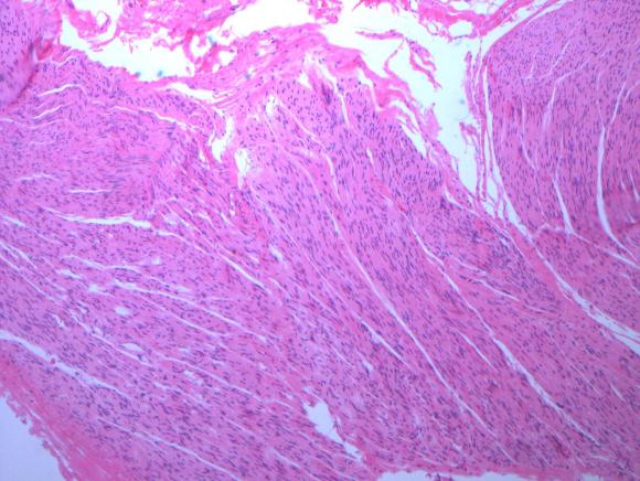
This was removed during a laparoscopic median arcuate ligament release. It was a hard white band compressed under the ligament and itself compressing the celiac axis. During the release, I grabbed a piece of these fibers and sent it for pathology under the preliminary diagnosis of celiac plexus, and it was. Described as “typical for a peripheral nerve…mostly Schwann cell nuclei in between nerve fiber.” Other micrographs in the specimen had ganglionic fibers but our pathologist wasn’t able to locate it. This is an important piece of the pathoanatomy because I believe that this is the nidus of the pain associated with median arcuate ligament syndrome, not a regional ischemia that can only occur if the celiac axis is an end artery, which can really only happen after a major exenteration like a Whipple procedure.
