Categories
PAD techniques TEVAR

External iliac remote endarterectomy in lieu of a conduit for TEVAR

IMG_1281

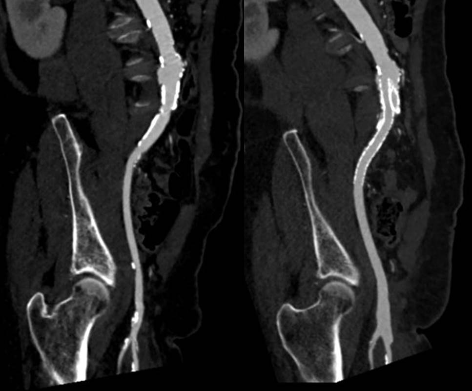
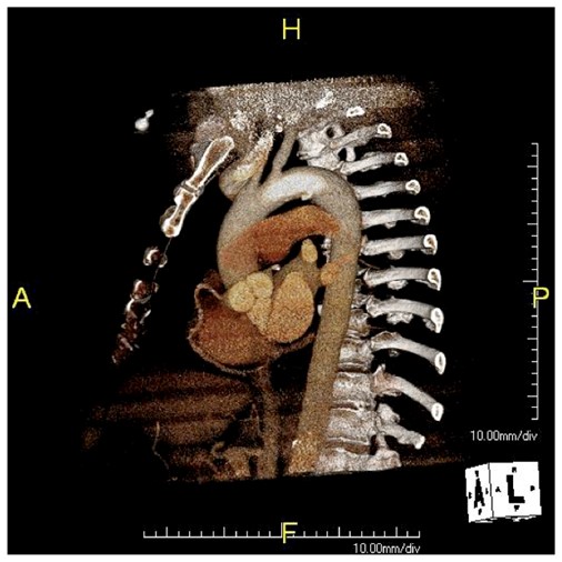
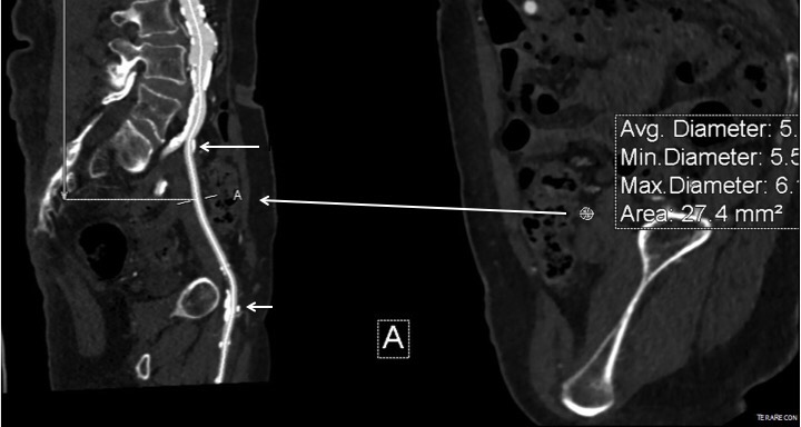
The patient had diffuse atherosclerosis with small luminal area even in areas without calcified plaque. It predicted inaccessibility for the 22 French sheath required to deliver the 32mm C-TAG device to be placed for a symptomatic type B thoracic aortic dissection associated with a small but expanding proximal aneurysm.

IMG_1277

My options included direct aortic puncture, an aortofemoral conduit, or an endoconduit. The aorta was heavily calcified and the bifurcation was narrowed by circumferential plaque down to 6-7mm at its narrowest and the left iliac had a severe narrowing due to this plaque. The common femoral artery was severely diseased with a lumen diameter of 4mm due to heavily calcified plaque.

I have come to favor direct aortic puncture over conduits, but the heavily calcified aorta and the absence of safe areas to clamp made me think about other options. My experience with endoconduits has been limited to revising problems of endoconduits from elsewhere, but others report it as a feasible option.

The problem with a long artery narrowed with irregular plaque and even intimal thickening is that it will readily expand to accommodate a large sheath but removing it involves the frictional resistance of the whole artery and typically the “iliac on a stick” avulsion involves the whole length of external iliac artery, likely because the common iliac is anchored by the aortoiliac plaque, the smaller diameter of the EIA, and the longer more tortuous path offering greater resistance in the EIA compared to the aorto-common iliac segment.

IMG_1272

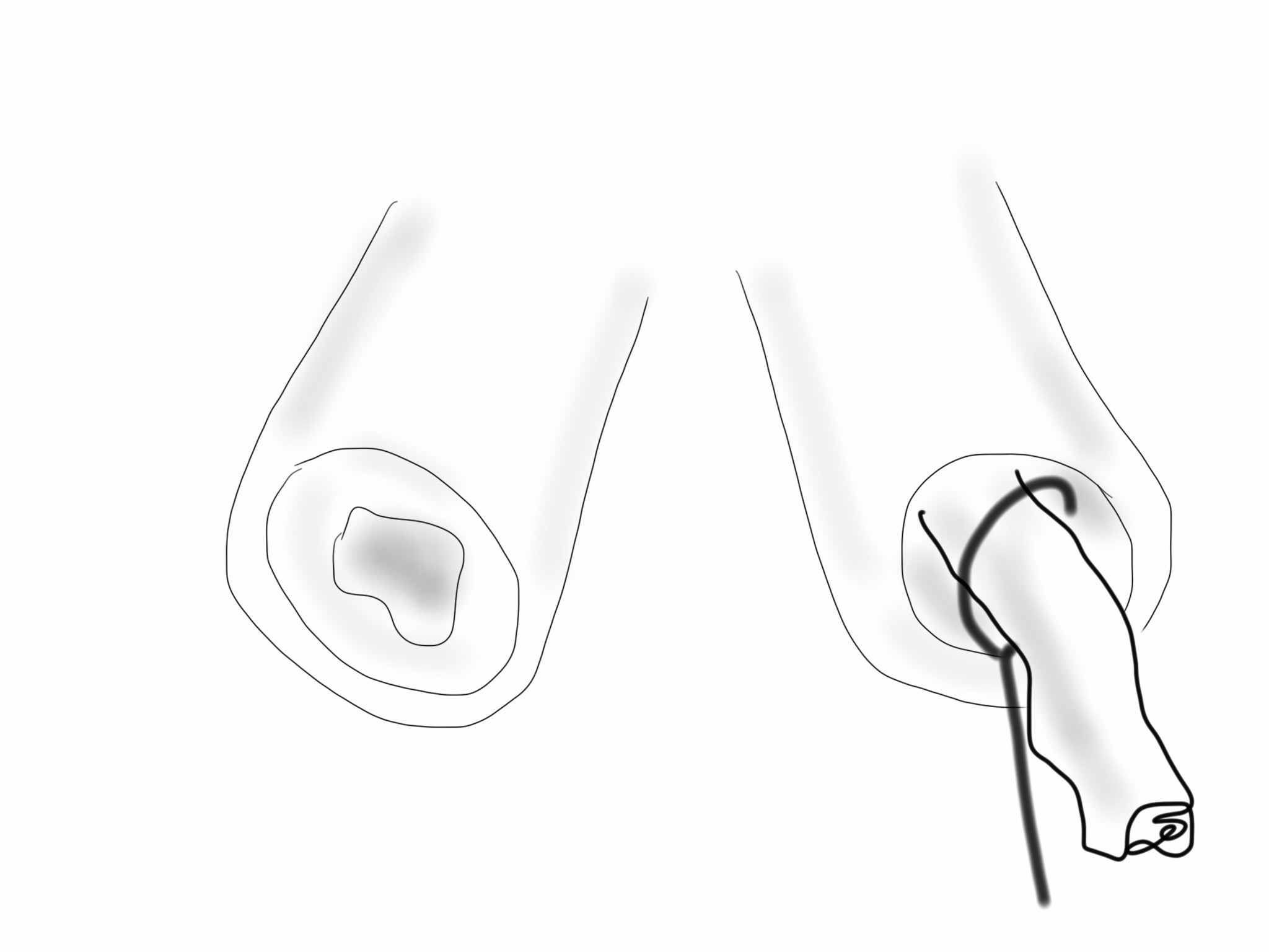
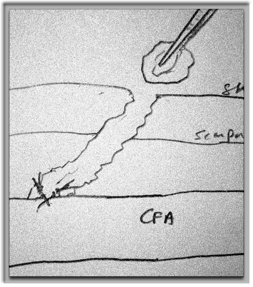
Remote endarterectomy, a technique involving endarterectomizing an artery through a single arteriotomy, offers the possibility of increasing the lumen of even a mildly diseased artery and reducing the frictional coefficient, assuming the remnant smooth adventitia is less resistant than rough irregular intimal plaque.

IMG_1275

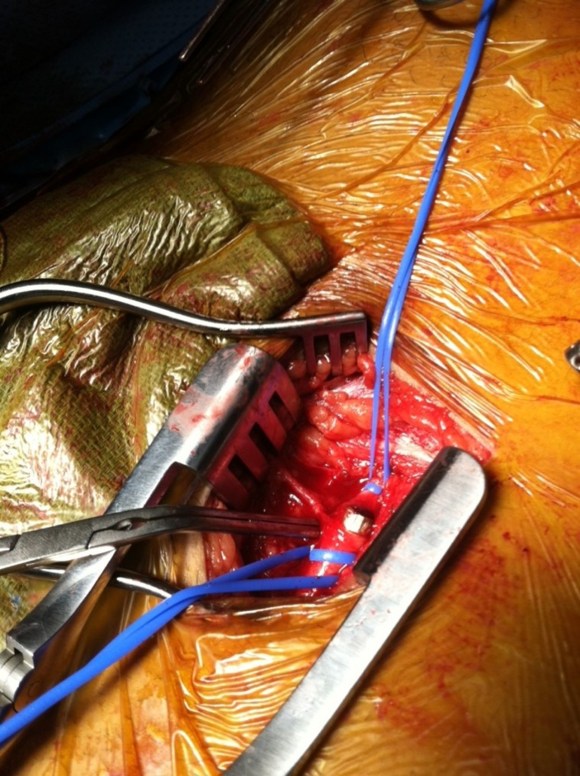
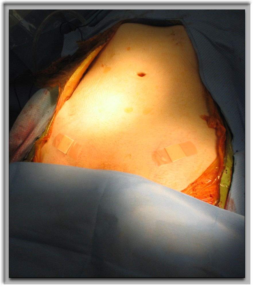
The plan was to expose the right common femoral artery and endarterectomize it and gain wire access from the R. CFA. A wire would be placed on the left iliofemoral system to protect it for later kissing iliac stents. A right EIA remote endarterectomy would be performed, and then the right aorto-common iliac segment would be balloon dilated to 8mm.

IMG_1278

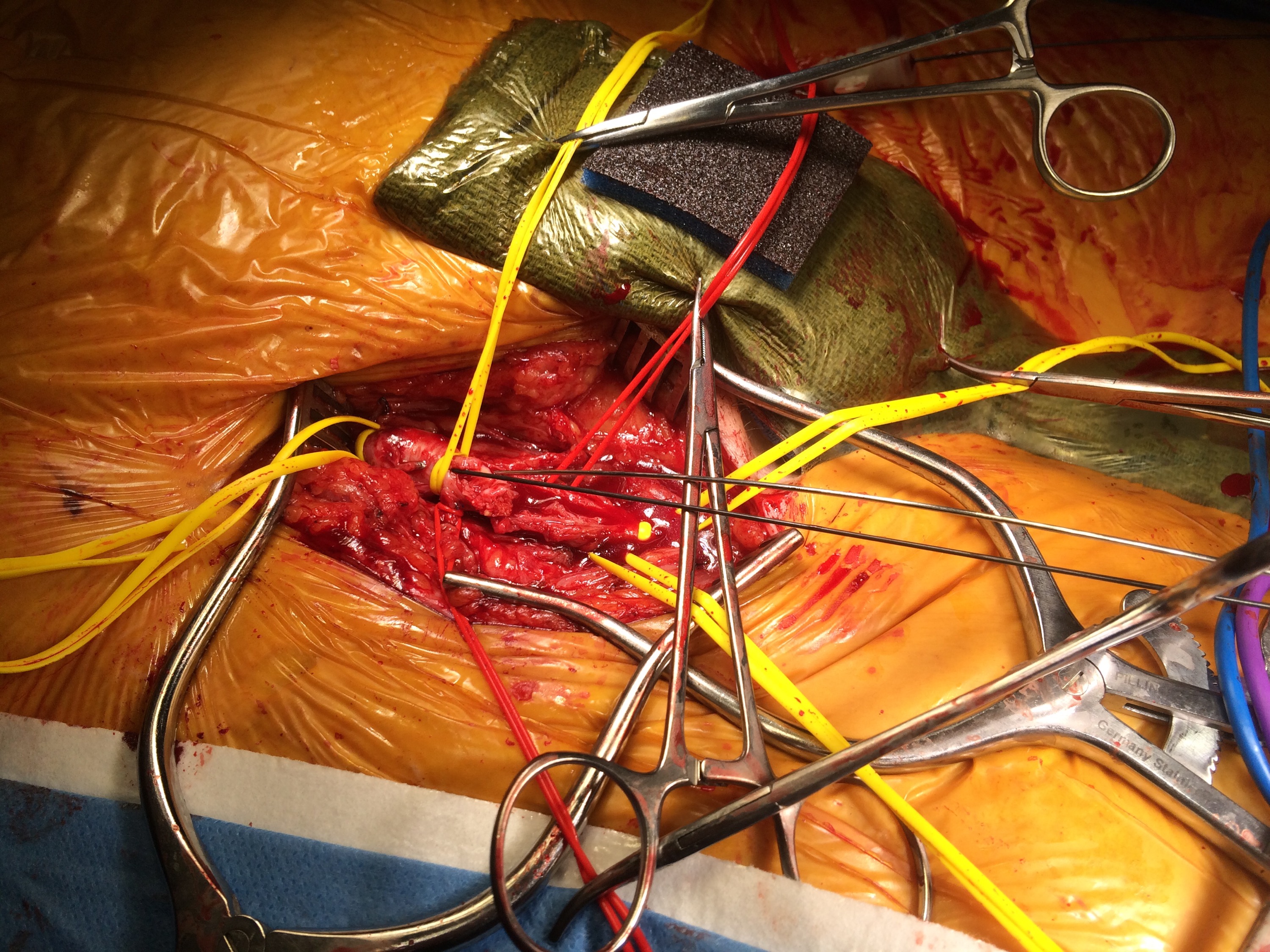
The operation went as planned. The external iliac plaque was removed in a single piece from the EIA origin.

IMG_1279

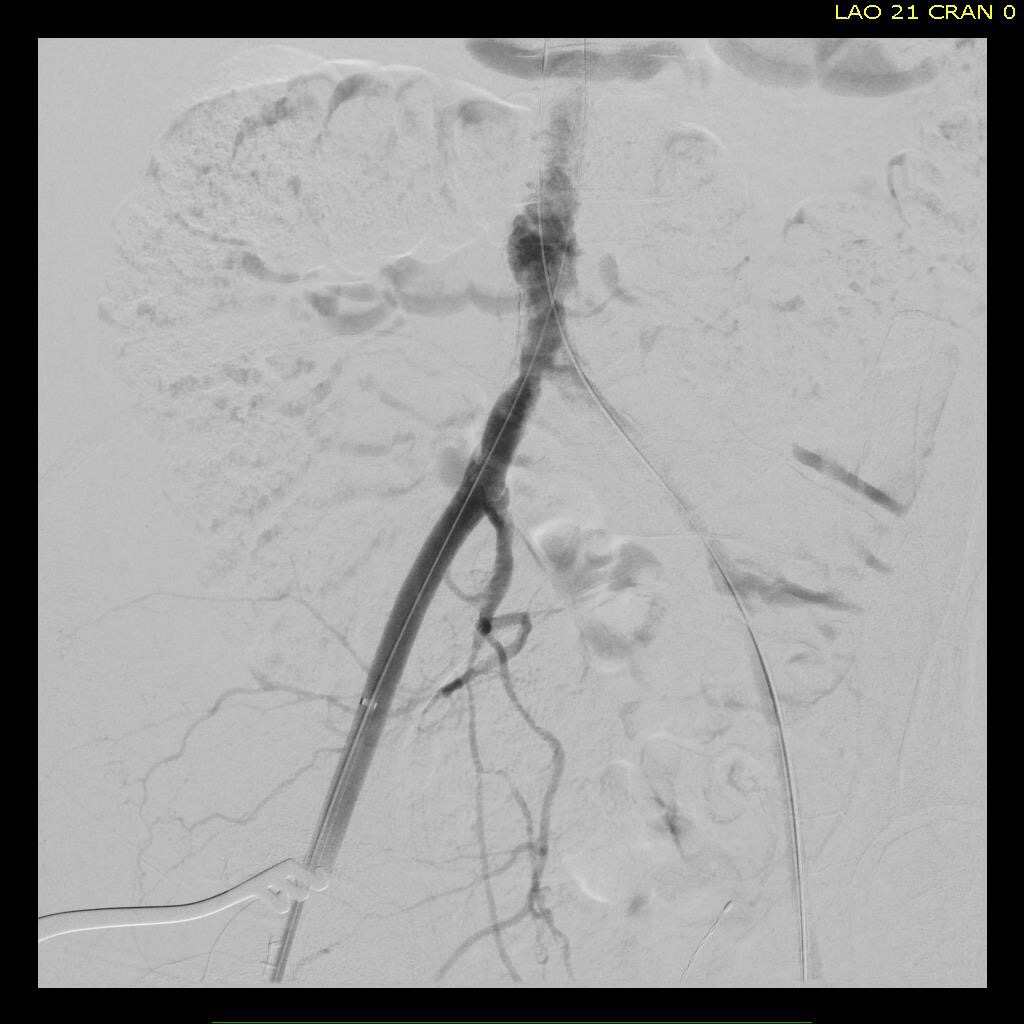
Arteriography showed the right EIA to be free of intimal disease, and dilators and ultimately the 22F sheath went in easily.

IMG_1276

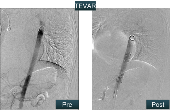
The TEVAR also went uneventully -the left subclavian which had a prior common carotid to subclavian bypass, was covered and the aneurysm and flap were excluded from the left CCA to the celiac axis.

IMG_1280

The most difficult part of the operation was removing the sheath, as is usually the case with a tight iliac, but the friction point was largely at the common iliac and not the external iliac. No artery could be seen extruding with the sheath at the groin while steady tension was applied to the sheath under fluoro. The aortic bifurcation was repaired with kissing iliac stent. The patient recovered well and her chest pain resolved.

I have done this for EVAR, including reopening occluded external iliac arteries, and even for a 26F access for TAVR, avoiding the need for placement of a conduit in selected patients.

Addendum: in followup, I had the chance to check up on the repair -the EIA remained large and patent.

before after

Categories
TEVAR

Aortic Zones

TEVAR zones

Categories
techniques TEVAR training

Kitchen-top Thoracic Stent Graft

IMG_1237

Before manufactured thoracic stent grafts were approved for use, you had to make your own. I think that even in theory you should have this in your mental locker, because it is pretty straight forward to accomplish. The patient was a homeless man who got struck by an SUV while crossing Broadway merely blocks from Columbia Presbyterian. The specifics are lost to time, but he was found to have among his multiple injuries a tear in his thoracic aorta at the ligamentum arteriosum. Cardiothoracic surgery felt that he was far too high a risk to undergo open repair. I was on call, and when I looked at this patient’s scans, I realized that he might survive with a stent graft across the tear, but the only suitable grafts were short aortic cuffs intended for infrarenal repair with short delivery systems. Being young staff, I called our site chief at that time, a grizzled veteran, for some advice about making stent grafts.

IMG_1232IMG_1234
The process is simple enough, and discovering it is like finding out that a seemingly complicated dish has an exceedingly simple recipe. The process starts with an iron and an ironing board, with which you press flat a Cooley graft of 32 or larger diameter. The Cooley graft is a fine weave graft that has pressed cylindrical folds that allow you to collapse it like a Slinky toy. Ironing between two sheets of paper allows you to avoid overheating the fabric.

IMG_1233
Once flattened and stretched, it is now ready for placement of stents. The stents shown here are Gianturco stents which typically are constrained with a monofilament and has barbs. The barbs are removed with needle nose pliers. 5-0 monofilament suture is used to secure the stents in the graft. More spacing allows for the graft to accommodate tortuosity, but the graft may bunch up in the sheath. The top and bottom stents should be within 5mm of the graft edge –this way you will remember that at deployment.

IMG_1235
For this case which required only one stent, three were made and they were autoclaved. Loading into a large sheath of 24F is done over a catheter to preserve a wire channel. The graft is pushed in using the umbilical tape or silk suture technique referenced in Oderich’s paper about reloading modified stent grafts.
Because of the large deliver system, a conduit was required and sutured end to end into the common iliac artery –I no longer do this unless there is a problem with severe plaque requiring endarterectomy. The graft was deployed by push-pull technique with the heart rate slowed pharmacologically. The patient stabilized from this, took several months to recover from his other injuries but was discharged and lost to followup.

IMG_1236IMG_1237
Is this knowledge helpful? In 2015, debatable, but in 2003, it saved a life.

Categories
TEVAR

A Fossil of the Kennedy Administration

From my archives, January 23, 2010,


A Fossil of the Kennedy Administration


The patient was sent to my clinic with the diagnosis of a saccular thoracic aortic aneurysm of 5x10mm. He is a 78 year old man who was working at restoring a vintage John Deere tractor when he developed chest pain. He underwent a cardiopulmonary workup which included a CT which showed a 5x10mm saccular aneurysm of the inner curve of his aortic arch opposite the origin of the left subclavian artery. This same CT showed no pulmonary embolism. His past medical history is significant for hypertension and he has never had surgery. His only hospitalization was in 1962 when he had a head-on collision which caused rib and collar bone fractures -he was the driver and he bent the steering column. 


His examination was normal. He had had a complete cardiac workup including a stress test which was also normal. He persisted on having this intermittent midsternal chest pain.


CTA images: above and below. The CT shows a small area of contrast fill consistent with a focal 5x10mm saccular aneurysm of the inner curve of the aortic arch. 


My impression is this is a residual saccular aneurysm of the thoracic aorta in the ligamentum arteriosum, related to the head on collision from time of the Kennedy administration. The injuries and the steering column damage suggest significant transfer of energy. It may have been the cause of his pain. We discussed surveillance with medical therapy (to include anti-inflammatories for any tractor lifting injuries) versus TEVAR. Ultimately we chose TEVAR.


This entry was posted on my old Medscape Blog, The Pipes Are Calling, with great comments and a poll. The poll results show a majority favoring medical therapy and watching. The only problem was that this patient was still having chest pain without a good explanation -pulmonary embolism and coronary artery disease were ruled out. The patient also lived two hours away from a hospital and would be lifting old tractors and their parts because that is what old farmers in Iowa do for fun –they also never truly retire. I think it also shows a general skepticism of surgical solutions –this feeling is based on historical bias with open surgery rather than contemporary results using percutaneous techniques.


The saccular aneurysm is an inherently unstable one as the angles on the surface of the aorta caused by a sac create areas of focused high wall stress. Rupture is usually fatal and the mortality rates for emergency surgery exceed 50%.


The other consideration was the relative risks of intervention have decreased with endovascular technique. Where mortality and morbidity of open repair on the proximal thoracic aorta ranged from 5 to 10%, with endovascular techniques, these fall to under 2-5%. I have been placing these thoracic endografts percutaneously and have had good results with low complication rates.


I performed percutaneous TEVAR of this saccular aortic aneurysm. The patient recovered well and went home after two days. Two years later, his CTA (below) shows complete resolution of the saccular aortic aneurysm. His chest pain never recurred.

Categories
imaging TEVAR

Suprising result from gunshot wound to chest


bullet CTA


The patient was shot in the right shoulder and had walked to the emergency room with some dyspnea and back pain. CXR showed a right sided pneumothorax but no bullet. The paper clip on the 3D VR view of the CTA shown above is the entry wound. The green line traces the centerline of the aorta, aortic arch, and the right carotid system. The patient’s assailant was shooting from a balcony of a movie theater. Vascular surgery was consulted for loss of pulse in left leg during trauma workup.

CT scan of the chest and abdomen showed blood in the mediastinum and haze around the distal thoracic aorta.

Remarkably the patient remained stable. My plan was to cover the aortic perforation with a stent graft, but an appropriately sized graft for patient’s size was not available at that time in 2009, so we used a Zenith RENU cuff. The patient on examination had an absent left femoral pulse. I chose to explore this and use it as the access site of the TEVAR. I also made sure the detectives put on scrubs to receive the bullet as US laws about evidence requires witnessed removal and acceptance of criminal evidence.

The cutdown revealed the bullet (9mm round) to be lodged in the common femoral artery. It was placed in a kidney basin with a loud clank and handed off to the peace officer for processing.

The bullet managed to miss the esophagus, heart, major pulmonary vessels, upper abdominal organs, and gently nestled in the aorta and embolized to the femoral artery in the emergency room.

The RENU cuff’s delivery system was long enough –at the time of this procedure, smaller diameter thoracic stent grafts were not available and in the setting of trauma with younger patients, particularly female patients, this was a problem. An aortic cutdown was sometimes necessary to deliver a 24mm aortic cuff up near a tear due to deceleration at the ligamentum arteriosum of the pulmonary artery and aorta. The patient recovered well and this case report was written up by Dr. Jared Kray who is now a vascular surgery fellow in Missouri –the article is in print for the January issue of American Surgeon.

Categories
EVAR TEVAR training

autoFestschrift for Dr. Matthew Eagleton

eagleton
Dr. Matthew Eagleton, picture from the internet

It’s Dr. Eagleton’s birthday. So in celebration, I recommend you read his authored or co-authored papers from this year. Link to MJE 2014 papers.


Categories
AAA EVAR techniques TEVAR

Percutaneous EVAR and TEVAR -how to get there if you aren’t doing it right now.

24F Access pEVAR
22 French Percutaneous Access circa 2008

Percutaneous access for EVAR and TEVAR does several things. First, the procedure becomes shorter by an hour or two, and (don’t discount not having nursing count instruments because the case was percutaneous). Second, the patients experience far less discomfort and it is easier to discharge them the next day when they have a bandaid versus an incision. And this leads to the third thing: not having an incision means it is far less likely that a groin infection will occur, especially in the obese.

There are three things which you must do before undertaking pEVAR. First, you have to become comfortable with using the Perclose S device in 6F-8F access -about 5 to 10 successful closures will do. You should become facile with the deployment of the sutures and closure of the access point. Avoid small arteries or heavily calcified arteries. This leads to the second point -all of your groin access should be ultrasound guided -this has been shown to improve results in pEVAR (Ref 1). I am a firm believer that the source of groin access complications starts with the initial needle stick. The 18g needle is basically a short 11 blade rolled up into a cylinder, and during groin access without ultrasound imaging, one can shear branch arteries, skewer arteries, dissect plaque, and access too proximally or distally, or into the profunda femoris.

needle is scalpel

The third need is access to 3D reconstruction software and multislice CTA. This gives you powerful ability to predict which patients are more suitable for a percutaneous approach, and which should have a cut down, and with 3D virtual reality reconstructions, you can plan where the incisions will be. In the skinny patient, this is not a pressing issue, but in the merely obese and the frankly obese, and the super obese, choosing to go percutaneous and avoiding a groin complication, which may be the one thing that debilitates the patient far more than a stent graft deployment, becomes an easy decision with experience.

As you build your 6-8F Perclose experience, you may notice that having too tight and subcutaneous tract can result in the suture catching on SQ fat, and not closing, or that bleeding won’t surface properly and create a hematoma under Scarpa’s fascia, often after the patient gets to the recovery room. Expanding on this principle, as you leap to 12F access and preclosure, I recommend you try this -make a 10mm incision, and using a tonsil clamp, pop through Scarpa’s fascia and seat the tips of the clamp under ultrasound on top of the soft part of the CFA that you intend to access. Gently spreading creates the space that you need to deploy the sutures and ensure that any bleeding will exit the skin and not dive under the fascia. It amounts to an ultrasound guided dissection of the common femoral artery. Before you remove the tonsil, you gently maneuver a micropuncture (always) access needle between the tines of the tonsil clamp until it gets to the artery -this keeps the eventual wire going through the tunnel you just made.

12F can usually close with a single Perclose, but start practicing by placing two Perclose sutures in a 10 oclock and 2 oclock orientation. Once the sutures are in, I make sure the  two ends of the suture are pulled out and the end loop of the suture is on the artery and I clamp these sutures to the drapes medially and laterally depending on how I deploy the two sutures. This also helps avoid catching the suture and driving it into the aorta.

After performing EVAR or TEVAR, I remove the sheath, leaving a wire -typically the stiff wire originally supporting the sheath and deploy one of the sutures. This first suture should cinch down onto the artery and substantially decrease the bleeding coming from the access site. I then deploy the second suture, and if the bleeding has stopped or is a steady dribble, I remove the wire. If pulsatile bleeding persists, I recinch the sutures using the knot pushers. If this decreases flow, I remove the wire, otherwise, I place a dilator, stop the bleeding and cut down. Cutting down after SQ dissection means merely dividing skin and tissues over the dilator, and the artery is easily visible for suture placement. If I remove the wire and there is still some bleeding, and usually there is, I place Gel-Foam soaked in diluted thrombin into the tract, reverse heparin, and hold pressure for 10-20minutes. It is very rare to have to convert after this is done.

thrombin gelfoam
thrombin-gelfoam into tract

The skin is closed with an absorbable 4-0 monofilament suture, and skin glue. I usually use the micropuncture needle to give an ilioinguinal field block with Marcaine. This gives 24hrs of pain relief.

bandaids

A note about incisions. Usually, with 3D VR imaging of CTA, the CFA and its quality (size and absence of plaque), and location relative to the inguinal crease can be ascertained. I try to make the access point at the inguinal crease or distally, as this goes under the subpannus of groin fat rather than through it.

groinaccess
3D-VR imaging can pinpoint optimal access

I sincerely believe sheath size is not the limiting factor to percutaneous access. Rather, it is the common femoral and iliac artery. Zakko et al at the University of Florida just published their experience on the obese with percutaneous TEVAR (ref 2), and found that while the arteries were deeper, the technical success rate of staying percutaneous (over 90%) was no different between their obese patients non-obese patients. The predictors of failure were poor access artery quality and size. I believe that you can select for patients most likely to succeed and greatly reduce failure. In this population, groin complications are potentially life threatening, and avoiding an open groin exposure is valuable.


References

1. J Vasc Surg 2012;55:1554-61 (ultrasound guided access)

2. J Vasc Surg 2014;60:921-928 (p-TEVAR and obesity)