Categories
AIOD CTA imaging techniques

3DVR -Very Helpful in Planning Open Surgical Cases

3DVR CIA Endart

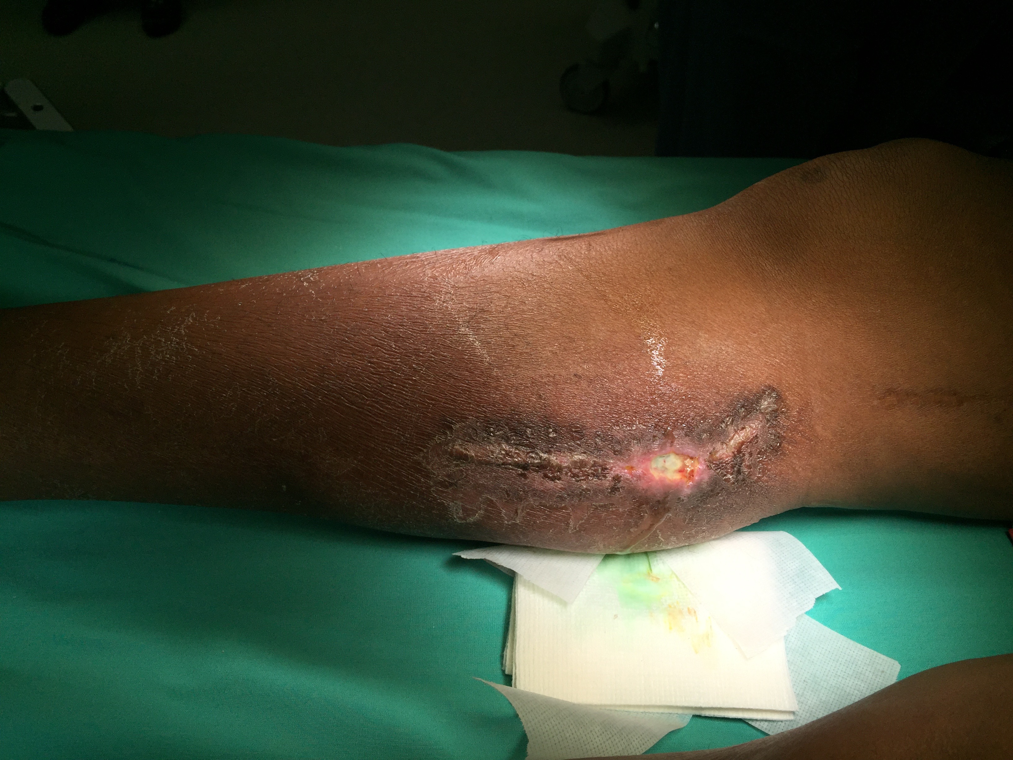
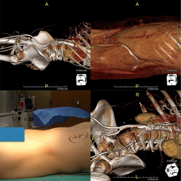
The images above show a patient with on isolated occlusion of his left common iliac artery. He was young, in his forties, but was a heavy smoker and suddenly developed claudication of his left leg which interfered with his work. He quit smoking and did not progress with exercise. Discussion involving possible stenting was made and initially offered but he turned it down because erroneously he assumed that his father’s coronary stents were the same as an iliac stent in terms of longevity. I do think that common iliac and aortoiliac occlusive disease is well treated with stents, but I felt it was possible to do a common iliac endarterectomy. We went over these images together and he settled on proceeding with endarterectomy.

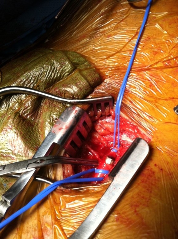
The images show how well the 3D Volume Rendering, which I mentally call Virtual Reality, of CTA makes it possible to plan out operations and exposures virtually. The bottom left image shows the surgeon’s eye view of the exposed vessel.

Below, the virtual and the actual are juxtaposed.

3DVR CIA Endart Exposure

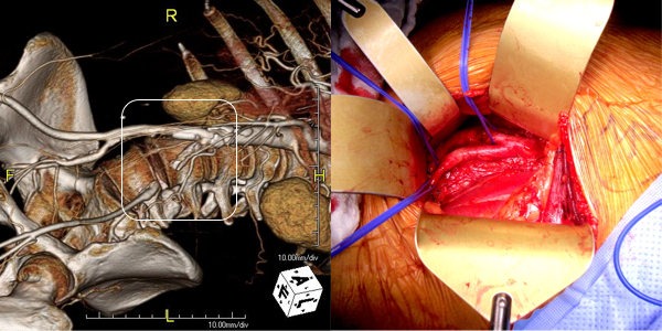
The outline on the virtual image (volume rendered) shows the areas of retraction -for the trainees, the retractor systems work to make quadrilaterals out of linear incisions, and as a rule, the incision should be twice the length of the square that you want to expose. The end points of the endarterectomy were at the aortic and iliac bifurcations.

SAMSUNG DIGITAL CAMERA

The arteriotomy was repaired with a patch at the iliac bifurcation -the common iliac was large and was repaired primarily.

SAMSUNG DIGITAL CAMERA

The specimen below was fibrocalcific. The thing about this disease is that the plaque truly has no endpoint -intimal thickening and mild plaque was present that could be taken all the way to the aortic root and to the feet on the other end!

SAMSUNG DIGITAL CAMERA

This patient did very well and had palpable pulses. He did not develop neointimal hyperplasia and successfully quit smoking.

One of the exciting developments is the ongoing development of wearable virtual reality and display solutions -particularly from the gaming industry. The gaming industry ironically drives all computer imaging because that is where the money is at. The advances in imaging trickle down to medicine -the VR images seen here are the result of the same algorithms that drive first person shooting games. It would be great to see this displayed intraop on a HoloLens, on a virtually positioned screen behind the assistant!

Categories
imaging Lymphatic training

Mind the Lymphatics: managing a persistent postoperative seroma

figure 1

The patient was referred from an outside institution for the development of a large tender mass in her below knee incision after a femoropopliteal bypass done with PTFE for ischemia after a aorto-bifemoral bypass. The patient reported swelling that grew in the months after the operation to the point that she was unable to walk without pain. On examination, she had a Nerf football sized swelling in her previous below knee incision without erythema. It was quite tender. CTA showed a patent aorto-bifemoral bypass and a femoropopliteal bypass to the below knee popliteal artery. Incidentally noted was the absence or occlusion of the profunda femoral artery. There was bland lymphedema below the knee.

Differential diagnosis included:

  1. Graft Infection
  2. Seroma from PTFE ultrafiltration leak
  3. Seroma from lymph leak.

Graft infections can present like this, but also drive local and systemic inflammation and in the absence of fevers and white counts, was highly unlikely. Occasionally, indolent infections with S. epidermidis will present with fluid collections but typically this is a late presentation. Ultrafiltration leaks from PTFE are fairly rare in my practice but can occur randomly. Most PTFE grafts nowadays come with an external wrap that acts as a seal against microporosity, but on occasion, I have seen protein rich fluid accumulated around PTFE grafts. This typically is not high pressure and accumulates along significant or whole length of graft. I used to treat that with graft excision and replacement, but I have had success with relining the graft with PTFE based stent grafts and externally draining the seroma.

I suspected this to be a seroma from lymphatic leak. The lymphatics are an unusual system of vessels in that they are remnants of an earlier circulatory system that was designed to move and mix nutrients and primitive phagocytic immune cells throughout the external compartment of an organism. They are diaphanous vessels that have smooth muscles that periodically contract like cardiac muscle, propelling fluid and cellular components past valves. Typically, cautery, suturing, and the inflammation of wound healing are sufficient to close lymphatics, but when there is potential space and a large lymphatic trunk that has been divided within it, that space will be filled with fluid, particularly with edema fluid that accumulates post surgically with dependency.

This patient was treated with I&D, but the lymphatic was identified by injection with Isosulfan blue in the subcutaneous space of the foot (between the toes). The dye is avidly taken up by the lymphatics and it can be used to identify the leak, allowing for extirpation and closure.

Isosulfan blue is injected into the subcutaneous spaces between the toes.

The vital dye will be cleared by the kidney -the pee will be greenish blue for a day or two. This is contraindicated in patients with known sulfa allergies.

The dye is seen in the wound within minutes without any added measures -no pumping or massaging was required. The patient had begun spontaneously draining the night before her operation.

The dye concentrates in the lymphatics which are easily identified.

IMG_5298

The lymphatics were ablated and a VAC dressing was applied. Two weeks later, there has been significant healing with complete resolution of the seroma.

IMG_5858

Loss of lymphatics at this level does not cause permanent injury but clearance of edema is slowed. Clearly, the avoidance of lymph leaks is the first step in preventing seromata, but when they occur, it is simple enough to identify and treat them using this technique.

They are one way self circulating pipes and therefore treating the afferent termini is all that is necessary.
Categories
Carotid imaging

The right and left carotid bifurcations usually look like this

IMG_1180.PNG

Maybe it’s the way the the bilateral dorsal aortas resolve into an asymmetric arch, but there is asymmetry in the carotid arteries which lets me look at a lateral carotid arteriogram and reliably predict which carotid artery it is. Image I is the usual conformation of the right ICA takeoff which may be tucked ,medially and posteriorly toward the pharynx. Image II is the typical forked appearance of the left ICA, and because it is longer and the plaque usually ends at the bend, this makes an eversion easier on that side. I began noticing this when I was on the CVRx trial applying stimulator leads on the carotids.

Categories
imaging TEVAR

Suprising result from gunshot wound to chest


bullet CTA


The patient was shot in the right shoulder and had walked to the emergency room with some dyspnea and back pain. CXR showed a right sided pneumothorax but no bullet. The paper clip on the 3D VR view of the CTA shown above is the entry wound. The green line traces the centerline of the aorta, aortic arch, and the right carotid system. The patient’s assailant was shooting from a balcony of a movie theater. Vascular surgery was consulted for loss of pulse in left leg during trauma workup.

CT scan of the chest and abdomen showed blood in the mediastinum and haze around the distal thoracic aorta.

Remarkably the patient remained stable. My plan was to cover the aortic perforation with a stent graft, but an appropriately sized graft for patient’s size was not available at that time in 2009, so we used a Zenith RENU cuff. The patient on examination had an absent left femoral pulse. I chose to explore this and use it as the access site of the TEVAR. I also made sure the detectives put on scrubs to receive the bullet as US laws about evidence requires witnessed removal and acceptance of criminal evidence.

The cutdown revealed the bullet (9mm round) to be lodged in the common femoral artery. It was placed in a kidney basin with a loud clank and handed off to the peace officer for processing.

The bullet managed to miss the esophagus, heart, major pulmonary vessels, upper abdominal organs, and gently nestled in the aorta and embolized to the femoral artery in the emergency room.

The RENU cuff’s delivery system was long enough –at the time of this procedure, smaller diameter thoracic stent grafts were not available and in the setting of trauma with younger patients, particularly female patients, this was a problem. An aortic cutdown was sometimes necessary to deliver a 24mm aortic cuff up near a tear due to deceleration at the ligamentum arteriosum of the pulmonary artery and aorta. The patient recovered well and this case report was written up by Dr. Jared Kray who is now a vascular surgery fellow in Missouri –the article is in print for the January issue of American Surgeon.

Categories
CTA imaging PAD techniques training

Intuition Aquarius (TeraRecon) Trick -Applying Virtual Reality to Operative Planning

I have used many different flavors of image post processing software including Osiris, Vitrea, and now Aquarius, aka TeraRecon. But I notice that outside of endovascular planning, people rarely use the virtual 3D reconstructed images (the pretty pictures) for anything other than posting images for publication in JVS, and even there I think we have reached saturation.

I have found 3D reconstruction to be especially useful for open surgical planning, and that is by doing two things. First, on viewing the 3DVR data, I reorient and center on the surgeon’s perspective, using left button to rotate the picture around the zero at the center of the screen, and the right mouse button to grab the whole image and recenter as necessary.

Window Leveling.001
Surgeon’s eye OR view

I then window-level in tissue density -this is done by pressing both the right and left mouse buttons, but you can choose this off the menu.

Window Leveling.002

I can plan the incisions and exposures from any angle -in this case, I can see the saphenous vein and its relative proximity to the CFA to perform an in site bypass to the AK POP. And I see the loci of the tributaries that I may need to ligate.

Window Leveling.004

This is a powerful tool that is often overlooked.