Categories
TEVAR

Aortic Zones

TEVAR zones

Categories
techniques TEVAR training

Kitchen-top Thoracic Stent Graft

IMG_1237

Before manufactured thoracic stent grafts were approved for use, you had to make your own. I think that even in theory you should have this in your mental locker, because it is pretty straight forward to accomplish. The patient was a homeless man who got struck by an SUV while crossing Broadway merely blocks from Columbia Presbyterian. The specifics are lost to time, but he was found to have among his multiple injuries a tear in his thoracic aorta at the ligamentum arteriosum. Cardiothoracic surgery felt that he was far too high a risk to undergo open repair. I was on call, and when I looked at this patient’s scans, I realized that he might survive with a stent graft across the tear, but the only suitable grafts were short aortic cuffs intended for infrarenal repair with short delivery systems. Being young staff, I called our site chief at that time, a grizzled veteran, for some advice about making stent grafts.

IMG_1232IMG_1234
The process is simple enough, and discovering it is like finding out that a seemingly complicated dish has an exceedingly simple recipe. The process starts with an iron and an ironing board, with which you press flat a Cooley graft of 32 or larger diameter. The Cooley graft is a fine weave graft that has pressed cylindrical folds that allow you to collapse it like a Slinky toy. Ironing between two sheets of paper allows you to avoid overheating the fabric.

IMG_1233
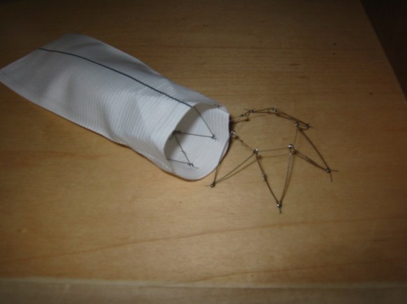
Once flattened and stretched, it is now ready for placement of stents. The stents shown here are Gianturco stents which typically are constrained with a monofilament and has barbs. The barbs are removed with needle nose pliers. 5-0 monofilament suture is used to secure the stents in the graft. More spacing allows for the graft to accommodate tortuosity, but the graft may bunch up in the sheath. The top and bottom stents should be within 5mm of the graft edge –this way you will remember that at deployment.

IMG_1235
For this case which required only one stent, three were made and they were autoclaved. Loading into a large sheath of 24F is done over a catheter to preserve a wire channel. The graft is pushed in using the umbilical tape or silk suture technique referenced in Oderich’s paper about reloading modified stent grafts.
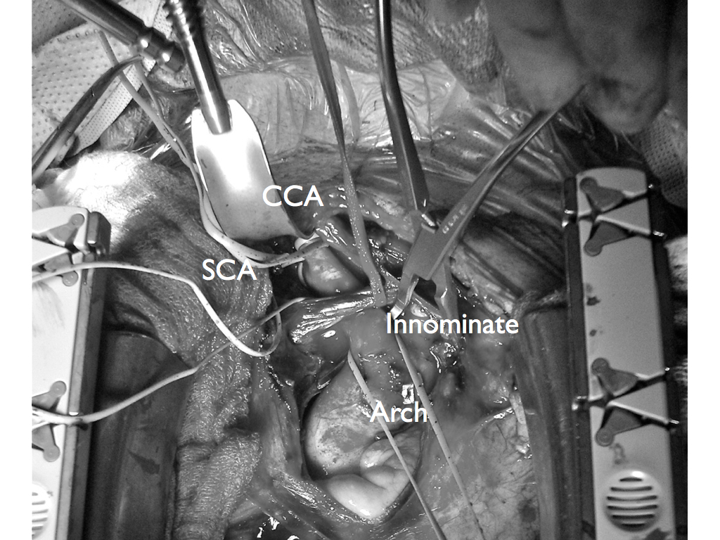
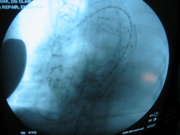
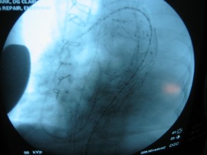
Because of the large deliver system, a conduit was required and sutured end to end into the common iliac artery –I no longer do this unless there is a problem with severe plaque requiring endarterectomy. The graft was deployed by push-pull technique with the heart rate slowed pharmacologically. The patient stabilized from this, took several months to recover from his other injuries but was discharged and lost to followup.

IMG_1236IMG_1237
Is this knowledge helpful? In 2015, debatable, but in 2003, it saved a life.

Categories
Lymphatic techniques Wounds

Extremity lymphatic leaks -a rare complication

2015/01/img_1225.jpg

This patient had had an endoscopic saphenectomy for coronary bypass at another institution and had developed a wound infection that eventually healed, but developed a pore at the scope site that continued to drain clear fluid soaking an ABD pad hourly. This had been cauterized and sutured without improvement.

2015/01/img_1227.jpg

The patient was taken to the operating room for dye lymphangiography and resection of the leak. The dye used was isosulfan blue which is lipophilic and is taken up by the lymphatics from the extra cellular space and pumped centrally within minutes of injection. The lymphatics are easily visible and the leak can be resected and the source vessels can be oversewn.

2015/01/img_1228.jpg

The suction cannula is on top of a lymphatic that was feeding the pore and was oversewn. I have had this occur in the forearm as well after a braciocephalic fistula creation.

2015/01/img_1229.jpg

The would was closed primarily. The lymphatics are evolutionarily the most primitive of our vasculature. They are gossamer threads often of single cell layer that have openings into the extracellular space and draw in fluid past bicuspid valves that bracket smooth muscle cells that periodically contract -a very primitive heart. The heart evolved from this mechanism.

It’s believed that multicellular organisms gained an advantage by maintaining an inner sea that was more organized and concentrated with nutrients than the surrounding sea, and to benefit all of the cells, it was necessary to circulate this inner ocean. The extracellular space is as porous as a sponge because that is what the ECM evolved from. Holding a wet sponge up in the air and watching the water go down with gravity is what happens when you stand up -edema. It is also why you can patiently and gently massage the extravasated saline from a displaced IV out of a hand and forearm. When the lymphatics stop pumping, the protein in the fluid eventually comes out of solution forming an insoluble solid -same thing that happens in cheese making when you add acid to milk -the hardened tissues of chronic lymphedema.

The pumping action is so efficient that injecting an amp of isosulfan blue into the web space between the toes subcutaneously will travel up the leg in about 5-10 minutes. There is a list of contraindications and precautions for its use, and hypersensitivity and anaphylaxis is reported to occur in up to 2 percent of patients.

This patient had primary closure and stopped leaking, after suffering from nearly a year of leakage.

Categories
AAA EVAR techniques training

Homemade laparoscopic suturing station -video

Tinkering in the basement, because bringing a laparoscopic tower and simulation setup into my house would be difficult.

Categories
Carotid PAD techniques

Innominate Endarterectomy -what the Satinsky clamp is for

Innominate.001

The patient, a 47 year old woman, was referred for syncope, but it was much worse than that. Excessive stress, standing, and thinking hard were described as causes of her syncope. Frequent headaches and lethargy lead to inability to keep a job. Several MVA’s resulted in revocation of her driver’s license. Added to that was a two to three pack a day cigarette habit. Her cardiac workup was negative, but it was noted that her carotid duplex was notable for a left ICA occlusion and left vertebral occlusion. Blood pressure in both arms was in the 70’s systolic while in her better thigh it was 90’s. She had an open right carotid system but the flows in the common carotid were attenuated. CTA of the arch revealed severe arch disease affecting the origins of her great vessels. The innominate artery was severely diseased to its bifurcation with a small <2mm channel of flow. She had a dominant right vertebral artery that was patent, and the right ICA had moderate disease at its origin. This was in 2009, and I entertained intervention, but wasn’t all that confident that kissing stents into the innominate was all that great of an option even though there are reports of innominate interventions in the literature. I had the opportunity to perform a handful of great vessel reconstruction with Ken Cherry during my fellowship and felt that this was an ideal case for an innominate endarterectomy.

This is one of those rare and infrequent cases from vascular surgery history. The exposures is one of the grand vistas of vascular surgery. The arch, thankfully, was only calcified at the origins of the vessel and clamped well. The endarterectomy was not that much different from an aortoiliac endarterectomy with a fibrocalcific plaque and was extended onto the common carotid while the origin plaque of the subclavian was plucked cleanly. The phrenic and vagus nerves were protected. The patient was centrally hypertensive as found by a long femoral arterial line and was kept that way for the duration of the clamp. A bovine pericardial patch was applied and the sternum was closed over a mediastinal chest tube.

The recovery was impressive for the patient’s immediately improved state of consciousness, lack of lethargy, and improved cognition. She was herself impressed enough to quit smoking during that admission for good. Her right brachial cuff pressure now correlated well. She went home POD 5. When I last saw her 2 years later, she was employed and symptom free with continued patency of her repair, consistent with the earlier reports of this operation (Cherry et al. J Vasc Surg; 989;9:718-14).

Categories
Journal Club

January 2015 Journal Club

Congratulations to Dr. Francisco Vargas for his December Journal Club presentation. January’s club meeting will be on the 20th.

Dr. Moqueet Qureshi: Open vs. Endo Popliteal Artery Aneurysm Repair

Dr. Deanna Nelson: VIASTAR Trial

Dr. Hazem El-Arousy: Cryografts in Infrainguinal Bypass