
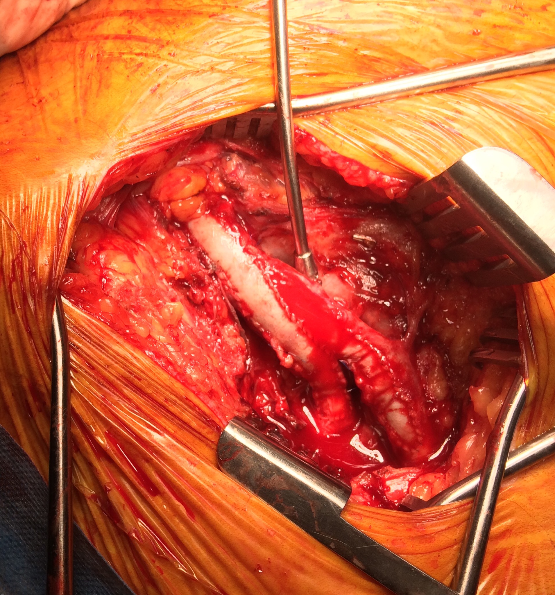
Dr. Teng, second from left, supported by her co-trainees, presented a poster at the International session on using cephalic vein transposition for SVC Syndrome, which was well received.
 A Surgeon's Notes
Dr. Teng, second from left, supported by her co-trainees, presented a poster at the International session on using cephalic vein transposition for SVC Syndrome, which was well received.
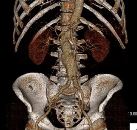
One of the many conversations I had with Dr. PJ O’Hara, who just recently retired, was about the place of traditional open vascular surgery. It is well known that many vascular surgeons are graduating with only a handful of open aortas. The idea of creating an open vascular (exovascular) fellowship was actually floated among the society leadership, but I suspect it was mainly the kind of idea that occurs when seasoned surgeons get together for a beer. This CTA above shows the kind of aneurysm that still benefits from open repair. It is a juxtarenal AAA with a highly angulated, short neck. This was one of the cases I did in my last practice. The patient did very well and went home on POD #5. A CT was done at a later date for possible dissection (there was none) but I got to check out my work (below). 
Unlike my patients who undergo EVAR, this patient won’t need intensive lifelong followup. While there is a small rate of complication in the mid and long term with open repair, these are infrequent and frankly rare. This is in contrast to the demands, often spelled out in the IFU’s, of stent grafts that require imaging and followup at 1 month, 6 month, 12 months, and annually for life. This is usually a CT scan and ultrasound, plus time and travel. While this is usually an agreeable tradeoff to most patients who are easily frightened by open aortic surgery, the cost to our healthcare system is not trivial. 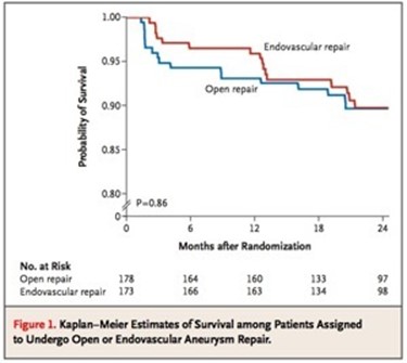
The DREAM trial showed that the benefit of EVAR versus open repair is lost after a year. Most of the benefit is in the short term –in hospital stay, complication rates, and recovery time. EVAR also allows more surgeons and even nonsurgeons to treat aneurysms. Clearly, for the higher risk patients, the extra year or two of complication free life is worthwhile and avoiding possible death from open aortic surgery may be a good thing, but for the majority of patients, we still have to ask far more and far better from the technology. This starts with getting reintervention rates closer to zero and significantly diminished followup protocol printed on the IFU. It also means allowing it to cost the same as open surgery. It probably means changing the way we assess and approve medical devices.
My personal journey through the past twenty years since I started my career (I was an intern in 1994!) has been a witness to vast changes in how we treat vascular diseases. Vascular surgery was more art than science, and many fellowships were indentured servitude to a famous surgeon, usually with a fearsome reputation. There was selection pressure in the process for certain personality types – fanatically committed to received wisdom and suspicious of change. Frankly, I was not a good fit, being relatively open minded, suspicious of dogmatism, and always looking for a better way.
There was an industry sales representative back those Manhattan days. David Hunt took an interest in the peons and always brought in the best food and swag –I know this is verboten these days, but I’m talking about history here. He brought in needle holders and sutures, and trays for holding grafts for practicing anastomoses, which I did practice on those interminable weekend calls up in our Stuyvesant 10 call rooms. When you ran out of grafts, a call to Dave, and he’d bring in several bags of graft (PTFE). Clearly, the crack dealers and lobbyists didn’t have a monopoly on this sort of sales technique.
One day, he brought in stent grafts –something out of the labs, and mockups of AAA’s. My mind reeled playing with EVAR. This was the future. That fall, I pulled some favors and left work early, and got to the Hilton wearing a blazer over my scrubs. Sneaking into the Veith Symposium, I was shopping my own future. I walked around the halls laden with all kinds of stent grafts and mock ups of space age operating rooms with fluoroscopy built into them. Dodging the security, I snuck into lectures and listened to the early data, and the resounding condemnation of the angry old men. I had to be part of this. This was supercool. This was the future.
I wasn’t alone in this endo-enthusiasm. A whole generation of vascular surgeons fell into its spell. There was the urgency of training to avoid the perceived obsolescence of not being able to perform endovascular procedures. Back then, it was very difficult to get training and subsequently privileging to do endovascular. I had the great fortune of working with Dan Clair while we were at Columbia in the early 00’s.
Even back then, I also perceived a rush to cast off open surgery, even in myself. There was a thrill at crossing a long SFA occlusion with a wire. It felt like victory sparing someone a long vein bypass operation. Every year, new gadgets came along to make the crossing and opening of closed arteries slicker and easier. But the truth echoed in the condemnations of the angry old men, many who were at this point retiring en mass, was that most of these procedures weren’t very durable. Where a vein bypass would be good for years, these interventions were sometimes only good for a few months. Many practitioners, usually not vascular surgeons, vocalized that two or three of these procedures was better for the patient than any single huge operation, and (sotto voce) was better for the revenue stream. Procedural failures definitely put water on the fire, especially after the news that many of the investigators for these new devices were also investors.
Endo-enthusiasm grew into eventually a more mature perspective. Which brings me to the point of this meandering entry. I see the best results when the range of potential therapies are tailored for the needs of the patient. A frank discussion about the short, mid, and long term outcomes of any approach allows for a deliberative planning discussion that many patients, especially those who come with literature and research in hand, appreciate. This can only be possible with a practitioner who has mastered both endo and exo– vascular techniques.
I tell people who are applying for vascular surgery training that one of the best metrics for judging a program is the volume of hybrid procedures being performed. It speaks to an ease with all possible techniques and a philosophy based on imagining the best possible path for treating a patient. While there is nothing wrong with seeing programs and practices with an “open guy” or “gal” and the “wire guy” or “gal,” there will be an abundance of both endo and exo-enthusiasm, that is bias.
I believe that this secures the future of vascular surgery. Other specialties can generate unlimited number of endovascular specialists, but only vascular surgery can produce the individual who can perform a redo common femoral endarterectomy, profundaplasty, endovascular aortoiliac recanalization, and infrageniculate vein bypass to save a leg and a life. Only vascular surgery can produce the individual who can judge that with any credibility that limb salvage attempts are likely to fail and recommend primary leg amputation and rehab and have the patient walking on a modern prosthetic limb within a month, and maybe even running within a year. And that same surgeon can perform a tibial intervention to heal an ulcer, and understand that it only needs to stay open 6-12 months and forebear reintervention when the artery is closed and the patient healed and asymptomatic.
So I head to the VEITH symposium looking forward to seeing what’s next. I have a badge this time.
JULY 30, 2008 10:55 PM
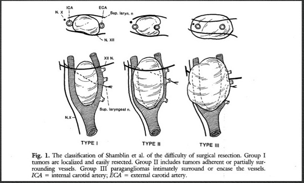
Group I: small tumors, minimal attachment to carotid vessels, easily resected
Group II: larger with moderate vascular attachments
Group III: large tumors encasing carotid artery, often requiring resection of tumor and carotid artery
This was in contrast to what I was taught as a resident in New York City, that carotid body tumors were once a decade or career occurrence. Most of the cases found in my practice at that time were diagnosed by ultrasound, often on carotid screening. No one knows why they cluster in the prairie.
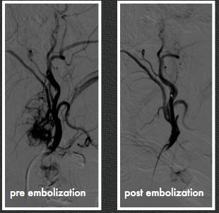
The case in the top movie had bilateral tumors; this occurs in 5%. The bottom movie shows a highly vascularized lesion with a branch coming from the external carotid.
This was embolized on the morning of surgery [figure below], and the tumor was avascular and shrivelled up at extraction.
Generally they are not malignant but are characterized by steady growth, mass effect, and eventual unresectability. Some are malignant and will metastasize, generally to lymph nodes. It is believed that malignant carotid body tumors are in the minority.
The use of embolization in larger tumors is a topic that has its proponents and detractors. I have found it useful in larger tumors, but unnecessary in the smaller ones. There is some risk to the patient and these can be technically challenging cases for the neuroradiologists, as these tumors take multiple sources from both internal and external carotid arteries.
References
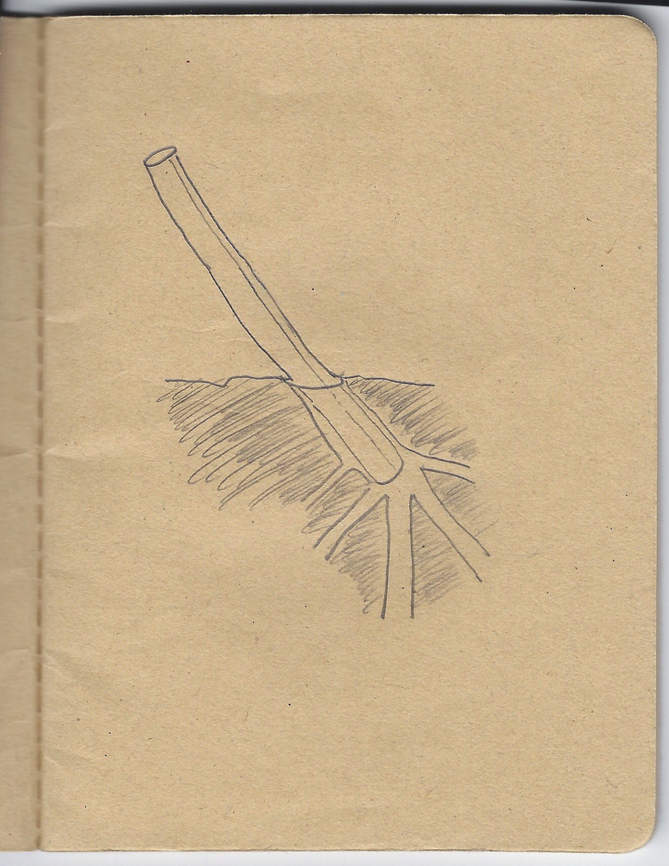
The usual situation is a multiple redo or infected groin with heavy scarring. Woody is the proper adjective. The common femoral artery may be obliterated or buried in the scar but a small profunda or its major branch may be accessible. Or you just run into it and get bleeding. Rather than bemoan your fate, you may be able to make a purse out of sow’s ear by exposing the artery and sliding in an appropriately sized Argyll shunt. Backbleeding into the shunt means that you haven’t dissected (hopefully) the artery, and now you have control over the surface edge of an artery. You can then clamp the shunt. You can assess your situation and decide that spending another two hours digging out two centimeters of 3mm artery may not be worthwhile, but you also decide that it is important to preserve this vessel.
It is straightforward to anastomose graft to the arterial stump. The shunt keeps you from narrowing the anastomosis, as you are well aware from carotid shunting. While you are doing this, if you have a Rummel tourniquet or vessel loop around the distal external iliac, you can feed the leg via this shunt as long as you remember to have the graft over the shunt. And remove it when you are done.
Another scenario is in revascularizing an intercostal, lumbar artery, or backbleeding posterior origin accessor renal but don’t want to do it right away.

Thousands of filters were placed over the past decade and the cows are coming home to roost. My feeling is that if a removable filter goes in, there must be an appointment or mechanisms in place to arrange for it to come out, anticoagulation must be started as soon as feasible, and kept on as long as possible if the filter is to remain in. Failure is infrequent for the conical designs, and not an issue if the filter is removed. How long after implant that a filter can be removed seems to be a moving target. In my personal experience, I have safely removed them out to two years, but I have partners who have gone beyond that by multiples. Two rare late failure modes of IVC filters can be devastating and life threatening.
IVC Perforation

This patient developed a vague upper abdominal pain and plain radiography showed the filter on a tilt. CT shows the legs of this Simon Nitinol filter extending into the right kidney and duodenum.

The 3 D VR images assisted in operative planning –as is my habit, it rotate the image into the surgeons-eye perspective to plan the incision.


The green arrows point to the exposed legs of the filter once the right colon and duodenum were rotated out of the way. The duodenum required only a serosal suture. The vena cava above and below the filter and both renal veins had to be controlled to remove the filter which was extirpated in pieces. I have had to do this about once a year or two. The youngest patient I operated on was a 20 year old who had a filter placed after a car accident at 17, but never had it removed. The legs of the filter had eroded into his duodenum causing an abscess.
Iliocaval Thrombosis
The figure below shows two panels with a Trapease filter associated with an iliocaval thrombosis. This patient had cardiovascular collapse and severe bilateral lower extremity edema after a long car ride.
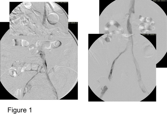
Venography showed iliocaval thrombosis. Thrombolysis was started and the second panel on right of Figure 1 shows the result.
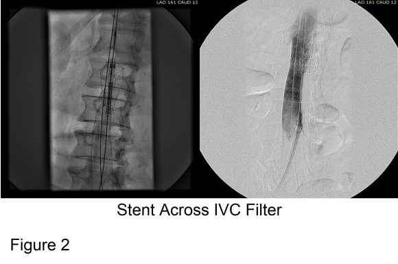
Large WallStents were used to support the recanalized iliocaval system from the common femoral veins to the filter. A Palmaz stent was deployed across the filter (Figure 2).

Figure 3 shows the final result. Interestingly, stents placed across the inguinal ligament into the common femoral vein seem to do fine in contrast to those placed in the artery. IVUS is necessary to confirm good results. Acceptable short term, and durable mid to long term results are reported.
Remove Them While You Can
Filters should be considered a short term therapy to decrease the risk of pulmonary embolism, and should be removed as soon as it is safe. There seems to be no magic time interval beyond which removal cannot be attempted. If permanent filter placement is planned, it should only be for established indications.
There are several areas yet to be satisfactorily traversed by endovascular technology and the common femoral artery is one area. While not completely a no stent zone, stents and interventions in the CFA do poorly compared to the open surgical alternative. As vascular surgeons we know that the key to inflow problems is the produnda femoris arteria and she does not tolerate being ignored, stented across, or ballooned too much. I have tabulated some areas that are still in the purview of open surgery in no particular order :
1: systemic infection
2: failure of stent grafts
3: rupture/hemorrhage/trauma
4: thoracic outlet obstructions
5: cancer
6: SVC syndrome after failure of interventions
7: MALS
8: popliteal entrapment
9: hypothenar hammer syndrome
10: very large thoracoabdominal or juxtarenal aortic aneurysms (until we get FDA approved off the shelf devices)
11: dialysis access
12: extreme limb salvage
13: severe aortic occlusive disease
14: CKD on the cusp of dialysis
15: congenital vascular disease
17: trauma/contaminated fields
18: low risk patients
19: common femoral artery
20: subclavian artery/innominate artery
21: carotid endarterectomy -for now
The list is open ended and you may add in the comments below, but the list in some parts is esoteric. The data is sobering if you read “Predicted shortfall in open aneurysm experience for vascular surgery trainees,” by Dua et al in the 10/2014 JVS. When I trained, I graduated with about 50 open AAA under my belt. Dua et al are predicting 10 per trainee in 2015, and 5 per trainee by 2020.
Who will do my open AAA?


The NYT reports the increasing use of hospital EMRs and registries to help make clinical decisions based on experience not yet published. Of course we must use all the tools available within our databases which is an extension of our knowledge. But I also get the other side of the argument.

The graphic is a painting I did several years ago meant to be a market sign for a vascular surgeon in some faraway place. I get no proceeds from their sale, but if I do, they will be turned over to a vascular related charity or foundation.
