
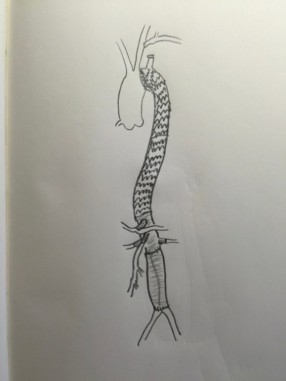
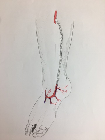
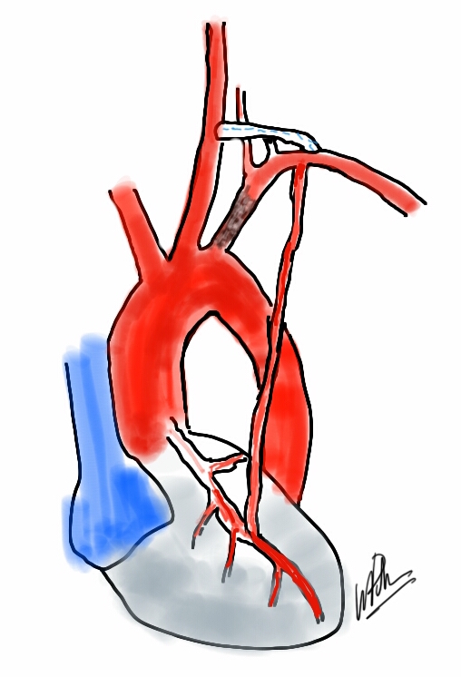
I taught myself to draw during medical school when I couldn’t figure out the three dimensional relations of structures. I discovered that if you just draw the shading of an object, it pops out in three dimensions. Over the years, I took to carrying little notebooks to sketch out anatomy and proposed operations for patients through this medium. While I found this to be a handy tool that I used only occasionally, since moving to Abu Dhabi, where much of my communicating is done through an interpreter, my drawings carry a much greater weight as direct communication of my thoughts and intentions.
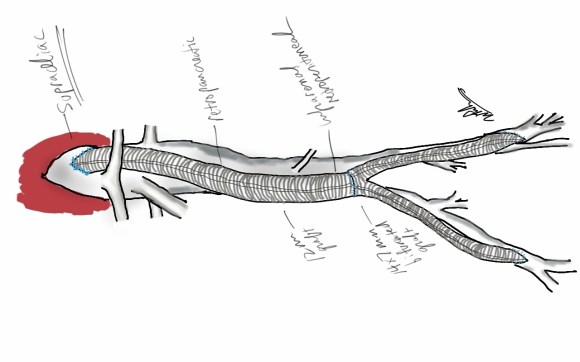
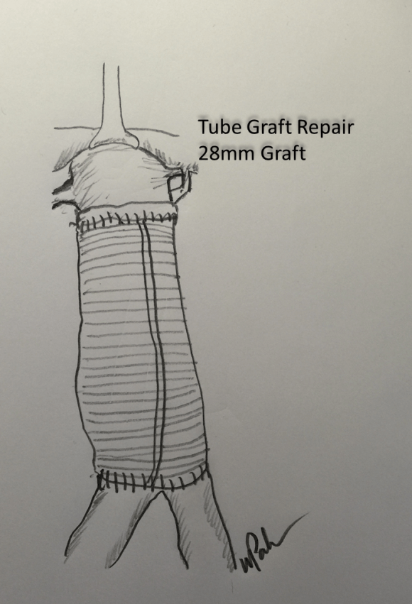
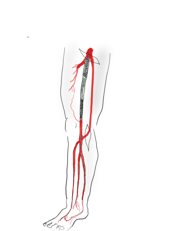
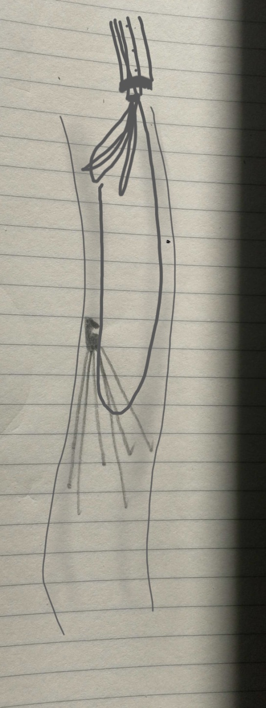
Drawing helps the patient and family understand the unseeable. It gives form to words that are often confused like blood vessel, graft, stent, artery, and vein.

What is informed consent when patient’s cannot describe their problems to their friends and relatives what the problem is and what is going to be done about it?

I usually draw with the pen in my shirt pocket and some copier paper, but sitting down and doing a proper sketch is soothing and very helpful for me as the surgeon to previsualize the goals that I have to reach during an operation to take the patient across the finish line. During meetings and conferences, I sketch into one of those fancy bound notebooks that I collect.

While pencil and markers do a fine job, the real magic is in using tablet based sketching software, using layers, to build serial images of the steps of an operation.

I am increasingly tempted to use these images as my operative note, but understanding that words are needeed for billing, I comply. Even so, I find it helpful to put these illustrations on my EMR notes, because it allows everyone to see and understand what I saw and what I did. I leave you with some of my illustrations with attached comments.

Our best shot


One reply on “At the Intersection of Art and Science”
Hi Dr.Park, your illustrations are great, explanations of anatomy also. Can you sketch your explanation of nutcracker ie. the small intestine hanging off of the sma? I could never figure out why the angle was so steep, it’s always just described as the artery pulsating. I e read through you many of your texts and tweets and really appreciate the detail.
LikeLike