Categories
SVC SVC Syndrome Venous

SVC Syndrome: Right Internal Jugular Vein Pressure is Intracranial Venous Pressure

preintervention venogram.png


The patient is a young father in his early thirties who complains of severe headaches, dyspnea, and inability to function or interact with his toddler. He complains of visual disturbancers when bending over and has dilated neck veins when recumbent. He was born with aortic stenosis and has undergone three aortic valve replacements and has had pacemakers since childhood. His most current pacemaker is a DDDR pacemaker in his left subclavian venous position, but he has two dead leads in his right subclavian vein. His symptoms started in his twenties but has worsened sharply over the past year. An effort was made to remove his dead leads at a tertiary referral center, but they couldn’t.

On examination, he had significant findings from dilated neck and supraclavicular fossa veins, a left chest pacemaker, and scars from his sternotomies. Duplex showed a patent bilateral internal jugular veins (IJV) with minimal respirophasic variability.

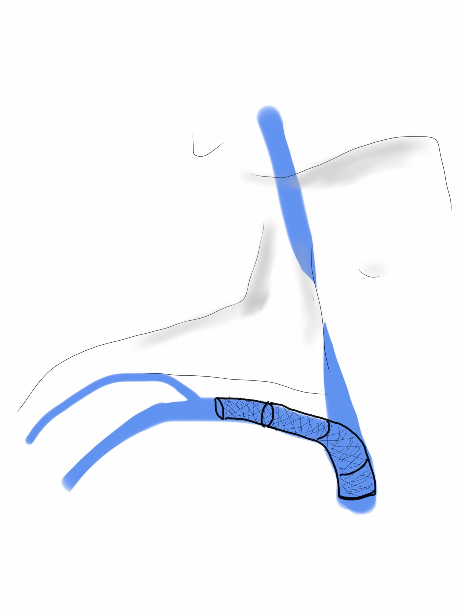
Venography (composite, above) showed superior vena cava occlusion consistent with his diagnosis of SVC Syndrome. I measured venous pressures, which we normally don’t do, because I was curious about them. Right IJ access with ultrasound with a sheath revealed pressures on the monitor in CVP mode of 22mmHg. Access from the groin into the heart revealed a right atrial (RA) pressure of 14mmHg. The SVC was crossed from below into the right IJV, and the SVC was dilated with a balloon -no stenting was possible because of the presence of the leads. His pacemaker did not fail during the procedure. After balloon dilatation, contrast passed easily from his IJV into his RA (below). His pressure in the R. IJV was 17mmHg and in the RA was 16mmHg.

post intervention venogram.png

He immediately felt relief and in 1 month followup had sustained symptom relief with patent flows seen in right IJV. He understood it was likely he would need regular reintervention when symptoms returned.

Discussion:

While IVUS is usually the way to check on patency after venous intervention, the presence of permanent pacemaker leads, including a set of nonfunctioning leads from the right SCV, made its use moot -venoplasty to 12mm was done in multiple stations and there was to be no stenting which is the usual next step if IVUS found residual stenosis. The more interesting finding here is the pressures measured in the IJV. The symptoms of SVC syndrome come from venous hypertension, but we rarely if ever measure pressure. Intracranial pressure (ICP) which is used to monitor for critical hypertension in the cranium typically after surgery or trauma, is considered high if over 15mmHg, moderately elevated over 20mmHg, and severe when over 40mmHg. ICP is greater than or equal to intracranial venous pressure, and in patients with SVC occlusion, this pressure is equal to the IJV pressure. With an IJV pressure of 22, this patient’s ICP was likely over 20mmHg, explaining his incapacitation.

IJV pressure is easily obtained and may justify urgent intervention in acute SVC syndrome, although more data needs to be obtained. This patient will benefit most from a more durable solution, but for now, he is very pleased to be able to take part in the care of his young child.

Categories
SVC Venous

A Palma Procedure for SVC Syndrome

IMG_0252.JPG

The enthusiasm for stenting has driven the misplacement of many well intended stents. The problem with stents in the central veins is three fold. First, the typically large stent chosen for placement in the SVC, the Wall Stent, by design expands while shortening but maintains a uniform diameter. If deployed partially in the subclavian vein, it remains constrained at the smaller diameter and far longer than intended. The other problem is that while ballooning can be done repeatedly, once they are in, stents limit how much can be ballooned as material grows and accumulates rapidly in the stent. There are no FDA approved devices for debulking this material on the venous side (I have asked the laser folks if anyone has used -no). The third challenge is deploying or embolizing into the heart, and this often requires a sternotomy or thoracotomy to retrieve the wayward stent. Unfortunately, you can’t compress the head like you can the legs, and these patients have overloaded their remaining drainage even with 24 hr upright posture. Spandex Lucha Libre masks would not treat the cerebral edema that causes intense headaches and neurotic symptoms.

This patient began his problems with effort thrombosis and hypercoagulability, found and treated in his home institution. He underwent first rib resection and stenting, but he rapidly thrombosed his stent despite anticoagulation, and this resulted in more stents until he had stents deployed across the confluence of the left and right brachiocephalic veins. This inevitably occluded and he developed SVC syndrome. He underwent two open bypasses first with vein then with PTFE by his local surgeons but these occluded. When he came, he had the swollen face and conjunctival edema of someone suffering from SVC syndrome. He had been told that there was no more that could be done and he would likely die within the year. He decided to seek a second opinion and made the long trip to the Clinic.

The ultrasound and CT showed his stents to be closed, but even after I opened his stents basically by ballooning and putting in more stents (10mm), he still had symptoms. This required an imaginative solution. IVUS by the way is important in these procedures.

The procedure to open the subclavian to SVC stents was done via the cephalic vein which was large and patent. Duplex of his neck revealed dilated internal jugular veins and it struck me that I had a good a chance at draining the head with a transposition of the cephalic vein to the IJV.

The challenge was how to tunnel this -above or below the clavicle. It was not entirely obvious because the superficial tunnel would be subject to compression while the subclavicular route was likely heavily scarred and subject to compression and kinking after turning upward.

I chose to tunnel over the clavicle and confirm a good turn of the vein by sending a catheter and wire through it and shooting venograms. The vein was taken from the antecubital fossa up to the shoulder. It was exposed, marked in situ, mobilized, flipped and tunneled to the neck where the IJV was dissected. It was anastomosed to a generous venotomy created with multiple applications of a 5mm aortic punch.

IMG_0251-0.JPG

This immediately relieved his symptoms, and he did well for about 6 months when he called urgently and drove in because his symptoms had returned. I thought he had closed his transposed vein, but duplex showed that it was his stents that had closed, and that his vein had stayed open. I reintervened on the stent via the brachia veins and his symptoms resolved again, and he remains happy and providing for his family. He will be due for his 1 year followup soon.