Categories
Venous vte

The student is now the master: IVC filter removal is easy until it is not, then it is very difficult


The inferior vena cava filter when it first invented by Dr. Greenfield was a minimally invasive solution that offered continued caval patency. The options up to then were ligation of the inferior vena cava using sutures or with an implantable plastic clip. The use of these filters exploded over the past 15 years corresponding to increasing recognition of venous thromboembolism as a morbid complication, the increasing numbers of implanters, and the introduction of retrievability.

Removing filters is a serious business because leaving them in for life is not inconsequential. Typically, the period of time that the filter are required for protection exceeds the limits of retrievability recommended on the instructions for use. It is not generally understood that many filters can be retrieved years after implantation, but it is not as simple as retrieval within a few months of implantation which can be done in under 30 minutes. In patients like the one in the illustrations, several years after implantation, the filter comes out only with some patience and a little help from friends.

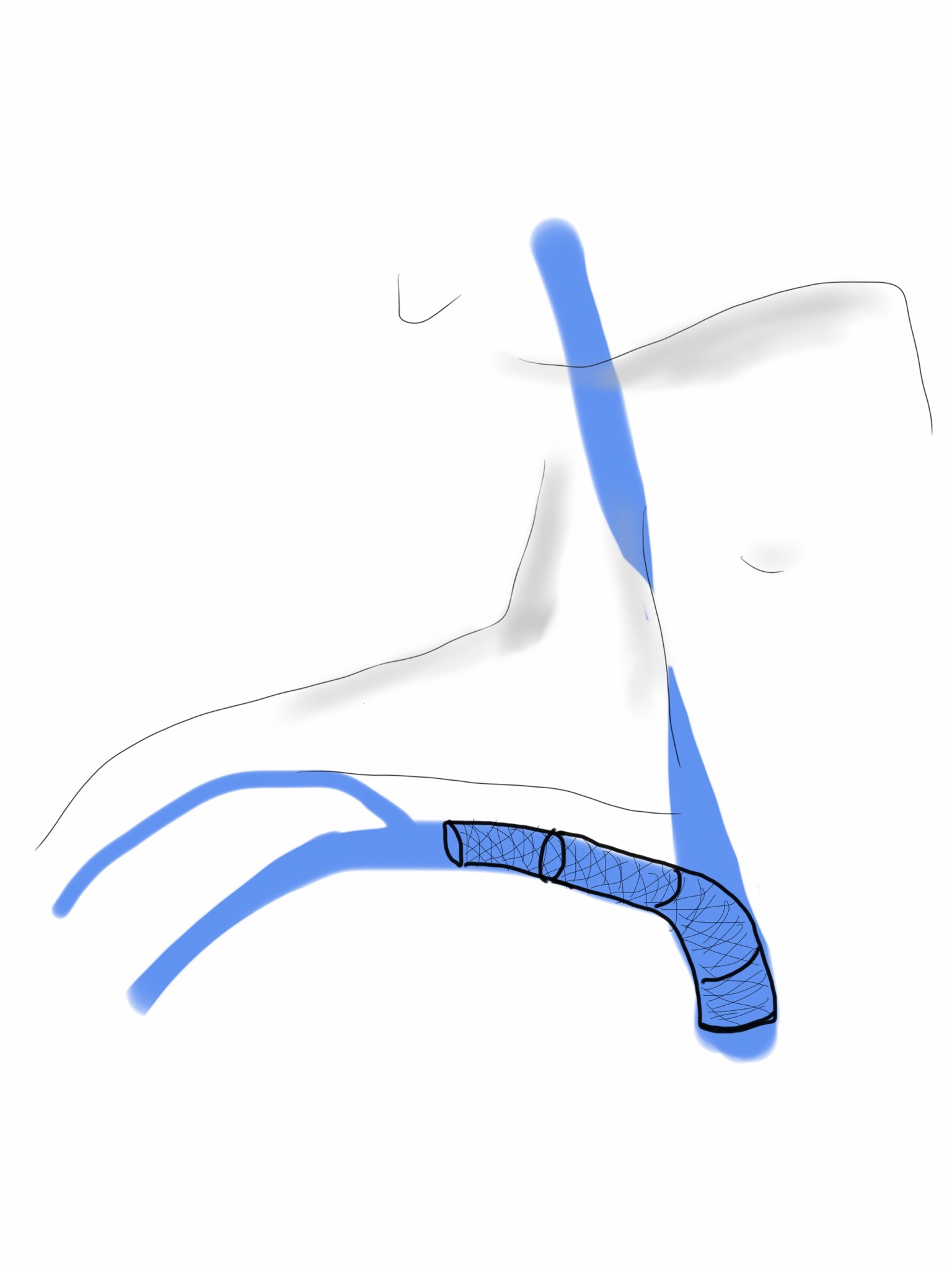
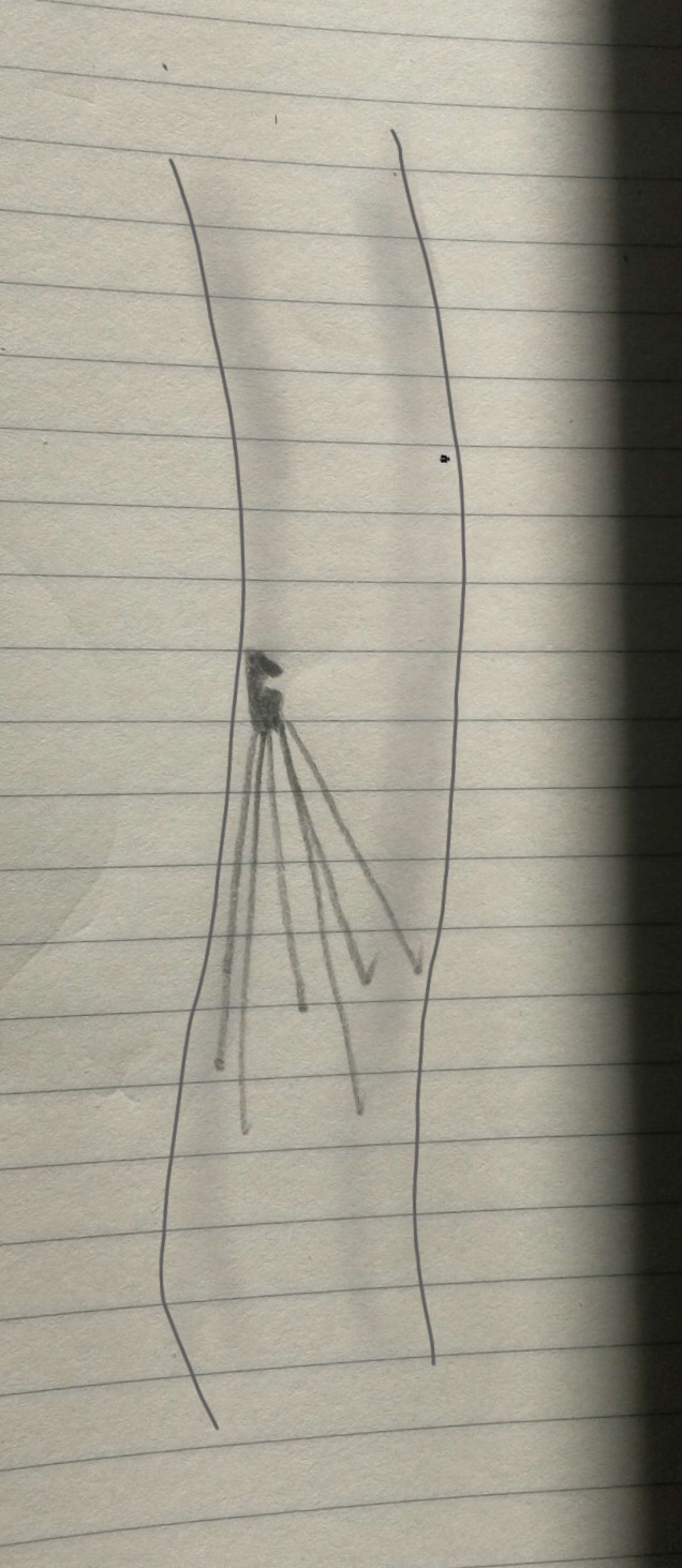
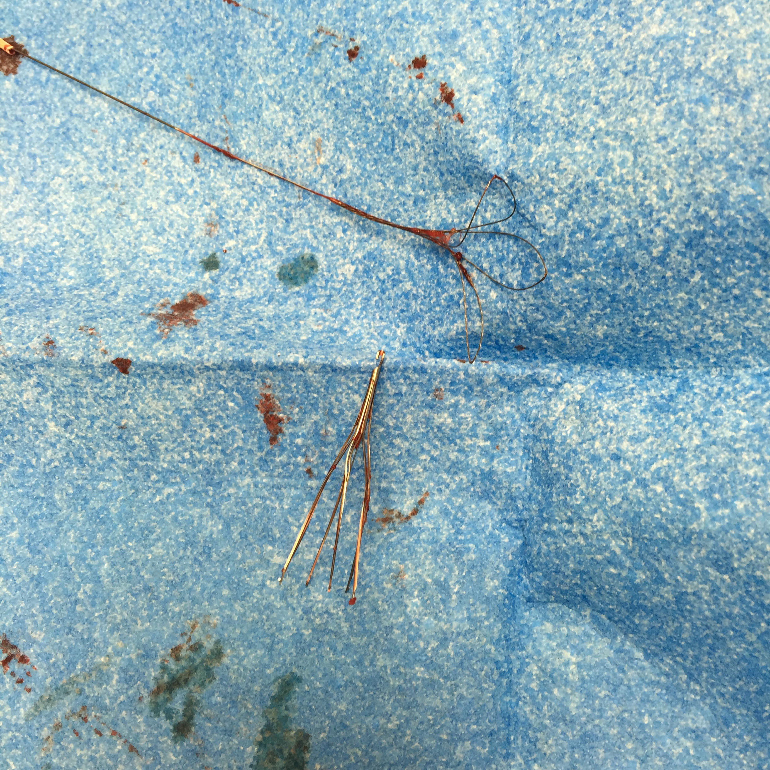
The IVC filter is embedded in the right sided wall of the vena cava and the hook would not engage. From a right internal jugular vein, wire access to the filter was achieved and an 18F x40cm sheath was placed through which a 12Fx50cm sheath was placed. Through this, a floppy glide wire was directed above the struts of the filter, and it curved around and snaked under one of the far struts.

This allowed me to snare the wire and bring that out.


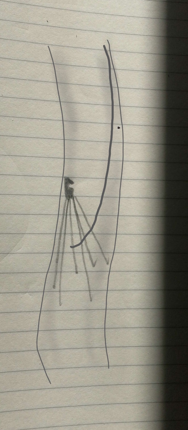
I placed another wire through the sheaths and removed the sheaths which were around both the Glidewire which was wrapped, and the second wire which was through. The sheaths were then replaced over that second wire, giving me room to maneuver I inside the 12F sheath. The first wire was then retracted with modest tension and it succeeded in lifting the hook away from the wall, allowing me to snare the hook through the sheaths.


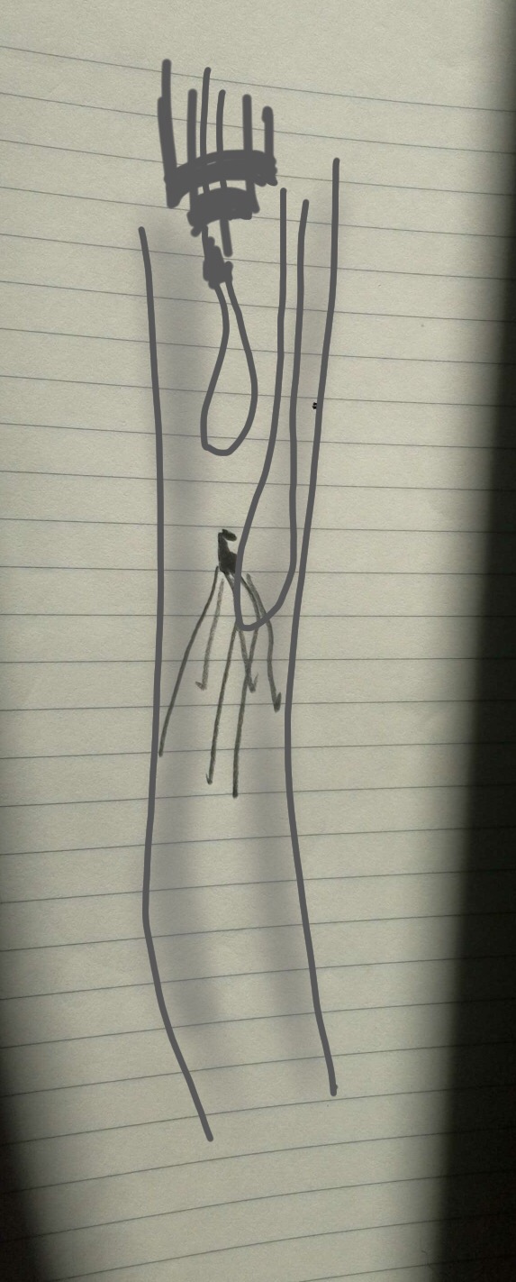
Once the top of the device was securely in the 12F sheath, the first wire was removed and the filter was removed.


The retrieval of an IVC filter device within the parameters of IFU (instructions for use) is like level one of a video game. Challenging for the novice, but eminently doable. The retrieval of these filters left in for years is more like level 25 of the same video game. The nice thing is having friends who can give you tips on defeating that level.

At VEITH symposium a couple of years ago, Dr. Paul Foley presented data and technical details on removing these filters, and this has been enthusiastically taken up by my partner Dr. Christopher Smolock who happened to be walking the halls the day of that case. His tip: “18F Sheath over 12F sheath, tilt the filter, and capture,” he said. “Wasn’t Foley your resident back at Columbia (in 2004)?” he added.

“Yes…” I replied.

“Now the student has become the master,” quoting Darth Vader. Which was fine with me because that made me Obi Wan Kenobi, which isn’t too bad. The great privilege of being a teacher is having that go around full circle. Or as Vader said, “The circle is now complete.”


Categories
SVC Venous

A Palma Procedure for SVC Syndrome

IMG_0252.JPG

The enthusiasm for stenting has driven the misplacement of many well intended stents. The problem with stents in the central veins is three fold. First, the typically large stent chosen for placement in the SVC, the Wall Stent, by design expands while shortening but maintains a uniform diameter. If deployed partially in the subclavian vein, it remains constrained at the smaller diameter and far longer than intended. The other problem is that while ballooning can be done repeatedly, once they are in, stents limit how much can be ballooned as material grows and accumulates rapidly in the stent. There are no FDA approved devices for debulking this material on the venous side (I have asked the laser folks if anyone has used -no). The third challenge is deploying or embolizing into the heart, and this often requires a sternotomy or thoracotomy to retrieve the wayward stent. Unfortunately, you can’t compress the head like you can the legs, and these patients have overloaded their remaining drainage even with 24 hr upright posture. Spandex Lucha Libre masks would not treat the cerebral edema that causes intense headaches and neurotic symptoms.

This patient began his problems with effort thrombosis and hypercoagulability, found and treated in his home institution. He underwent first rib resection and stenting, but he rapidly thrombosed his stent despite anticoagulation, and this resulted in more stents until he had stents deployed across the confluence of the left and right brachiocephalic veins. This inevitably occluded and he developed SVC syndrome. He underwent two open bypasses first with vein then with PTFE by his local surgeons but these occluded. When he came, he had the swollen face and conjunctival edema of someone suffering from SVC syndrome. He had been told that there was no more that could be done and he would likely die within the year. He decided to seek a second opinion and made the long trip to the Clinic.

The ultrasound and CT showed his stents to be closed, but even after I opened his stents basically by ballooning and putting in more stents (10mm), he still had symptoms. This required an imaginative solution. IVUS by the way is important in these procedures.

The procedure to open the subclavian to SVC stents was done via the cephalic vein which was large and patent. Duplex of his neck revealed dilated internal jugular veins and it struck me that I had a good a chance at draining the head with a transposition of the cephalic vein to the IJV.

The challenge was how to tunnel this -above or below the clavicle. It was not entirely obvious because the superficial tunnel would be subject to compression while the subclavicular route was likely heavily scarred and subject to compression and kinking after turning upward.

I chose to tunnel over the clavicle and confirm a good turn of the vein by sending a catheter and wire through it and shooting venograms. The vein was taken from the antecubital fossa up to the shoulder. It was exposed, marked in situ, mobilized, flipped and tunneled to the neck where the IJV was dissected. It was anastomosed to a generous venotomy created with multiple applications of a 5mm aortic punch.

IMG_0251-0.JPG

This immediately relieved his symptoms, and he did well for about 6 months when he called urgently and drove in because his symptoms had returned. I thought he had closed his transposed vein, but duplex showed that it was his stents that had closed, and that his vein had stayed open. I reintervened on the stent via the brachia veins and his symptoms resolved again, and he remains happy and providing for his family. He will be due for his 1 year followup soon.